Category Archives: Current projects

Scavengers in the ocean

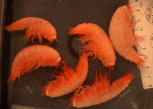
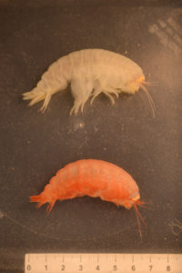
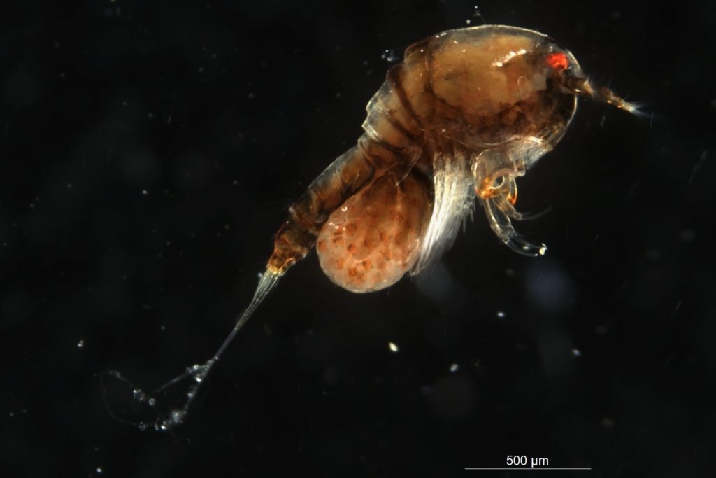
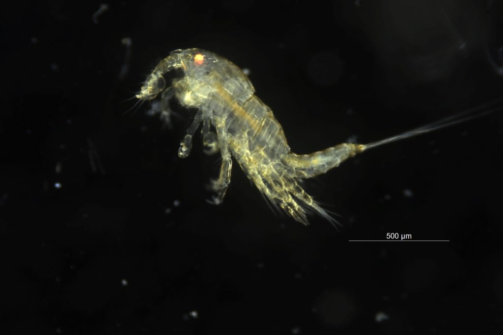
Lysianassoid amphipods from a trap in Raudfjorden, Svalbard. Photo: AHS Tandberg

Most animals are sloppy eaters. They have their favourite piece of food that they go for, and then they leave the rest. This allows for others to pick up where others leave. One of the laws of ecology is that “there is no such thing as an empty ecological niche”. That can be translated to “where there is a food-source (or a place to live) someone or something will use that food-source (or place to live). And that gets us to the sloppy eaters out there, and not least the animals picking up after all the sloppy eaters.

From the pigeons crowding under your cafe-table for your panini-crumbs to the rats in our sewers, our “local scavengers” tend to be animals we feel slightly uncomfortable around. Is it different with the scavengers we dont see so often? It does not seem that way. Vultures  are not the most popular birds, even the word “vulture” has a negative connotation – and we mainly use it in its non-bird meaning.

How about the scavengers of the sea? As on land, we have many different animal-groups that can be classified as scavengers. Many of the marine scavengers are invertebrates (even if some fishes also scavenge). Let us look at the scavenging Lysianassoid amphipods. Are these as little loved in our world as the rats and vultures seem to be?

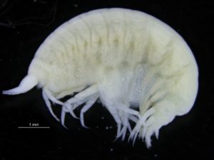
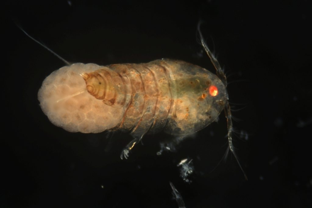
A typical lysianassoid amphipod. Photo: AHS Tandberg

Lysianassoid amphipods can mostly be distinguished from other amphipods by their “telescope-like” antennae: a very fat inner article with the two next looking like a collapsed old fashioned radio-antenna; two short rings. We know that the antennae of crustaceans are often used to “smell” things in the water – food or mates or possibly even enemies. It is not thought that the radio-antenna-shape of the Lysianassoid antenna specifically has to do with being a scavenger, as other amphipods and indeed several other crustaceans not having such an antenna are also scavengers. But most Lysianassoids have that antennae, and it makes for an easy first-sorting for the scientist. (Getting further – towards a genus, or even species name on the other hand, is not so easy).

Other general traits in most Lysianassoids, are the smooth exterior, and their high swimming abilities. Both are good if you need to get to some leftover food-source fast, and to “dive” into the food-source while not getting stuck through the entry.

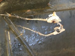
Leftovers of bait (polarcod) after 24 hrs in the trap. Not much left for dinner… Photo: AHS Tandberg

And this is where many Lysianassoids loose out when it comes to human appreciation. They seem to love to scavenge on fish caught in fishnets and traps, and both professional and hobby fishers don’t like to share their catch. We dont think it is very appetising to find our fish-dinner “infested” by non-fish. I am quite sure the scavengers being pulled up with their lovely find of dead or dying fish also are not pleased with having to share their dinner with us.

Lysianassoid scavenging amphipods are the focus of our NBIC-financed project NorAmph2. Here, we will collect and register what different species are present in Norway, and we will try to barcode them. These are quite tricky animals to identify properly, but luckily we have teamed up with the best lysianassoid-expert we know – Tammy Horton from the National Oceanography Centre in Southampton, UK.

We use baited traps to collect: put some lovely, smelly fish out there and see who comes to dine. So far, we have collected from Svalbard in the north to Kong Haakon VIIs Hav in the south, and from the intertidal to the deep. They are often many, and the size-variation is great. We look forward to continuing finding out what species we have, and to see if what morphologically seems one species really is (only) one species genetically. (This previous blog-post (in Norwegian) tells the story about one scavenging amphipod that turned out to be 15 (or maybe even more!) separate species)

Anne Helene

Sea slugs of Southern Norway; farewell but not goodbye!

A note from the Norwegian Taxonomy Initiative project (artsprosjekt) “Sea Slugs of Southern Norway” (project home page), which ran from 2018 to the end of April 2020.

Dear all,

The Sea slugs of Southern Norway project reached its terminus at the end of April, with sending the last reports of our collection and research efforts to Artsdatabanken (the Norwegian Biodiversity Information Centre).

What we have been able to build up these last two years is of immense importance for the scientific collections of the Natural History Museum of Bergen (University of Bergen)  and for (Norwegian) biodiversity research.

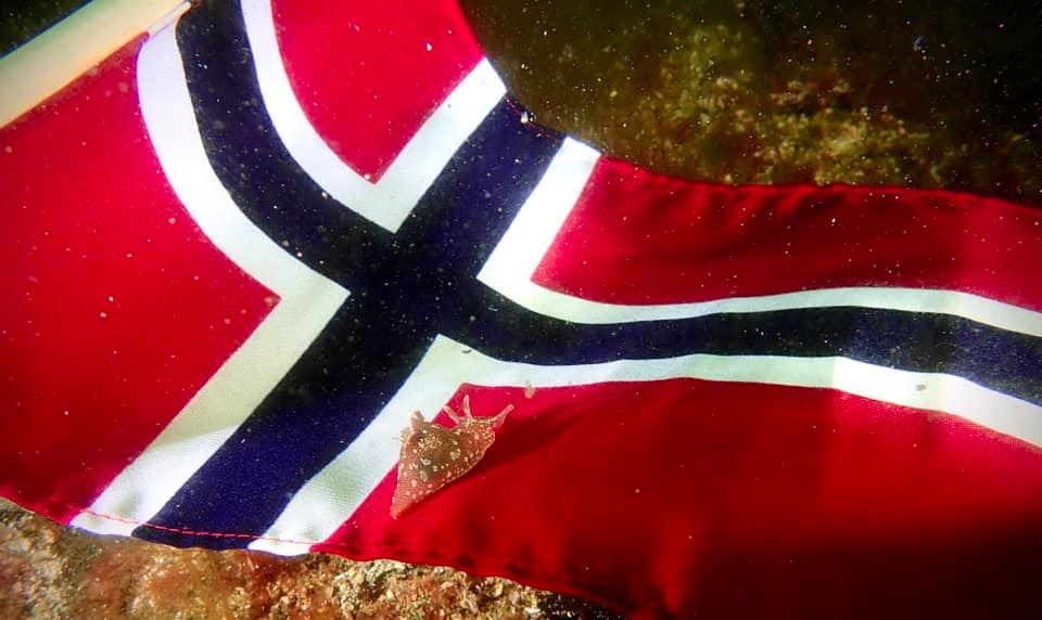
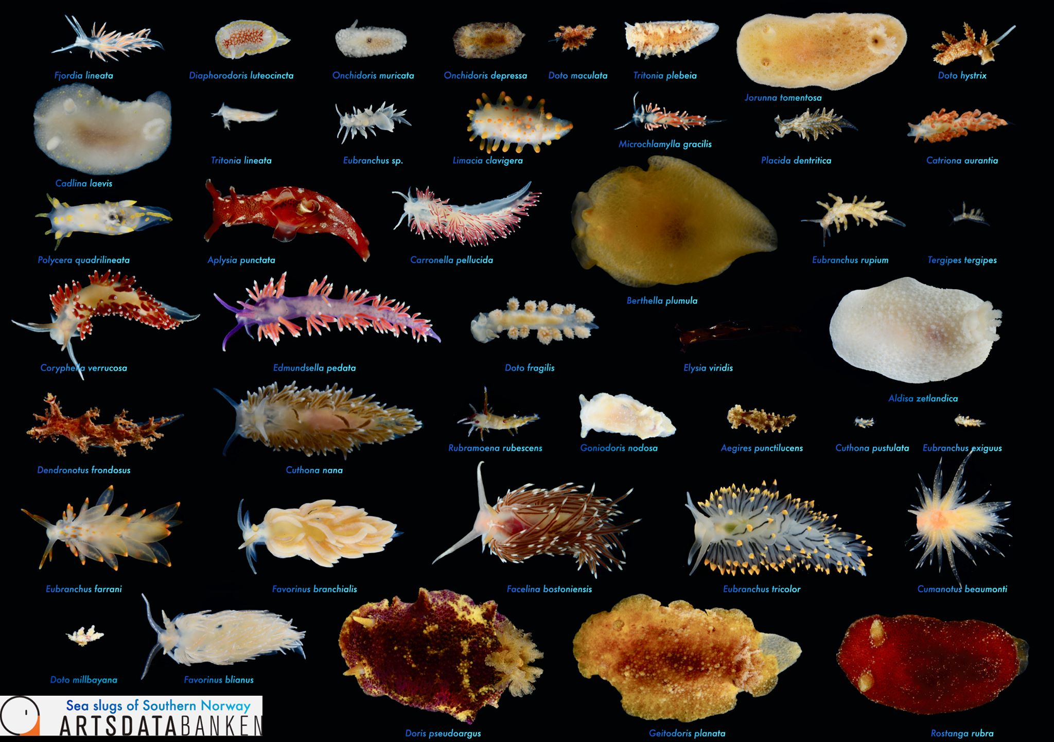
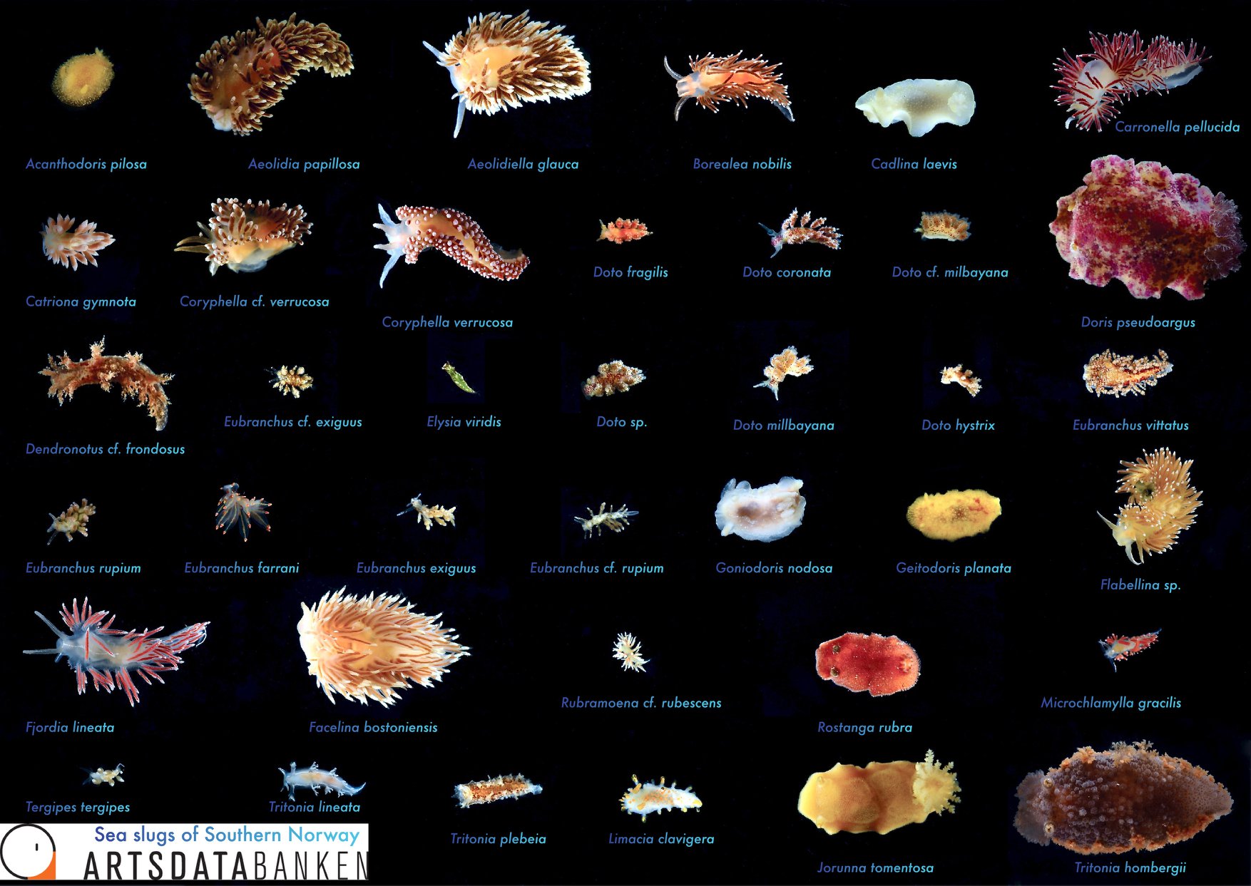
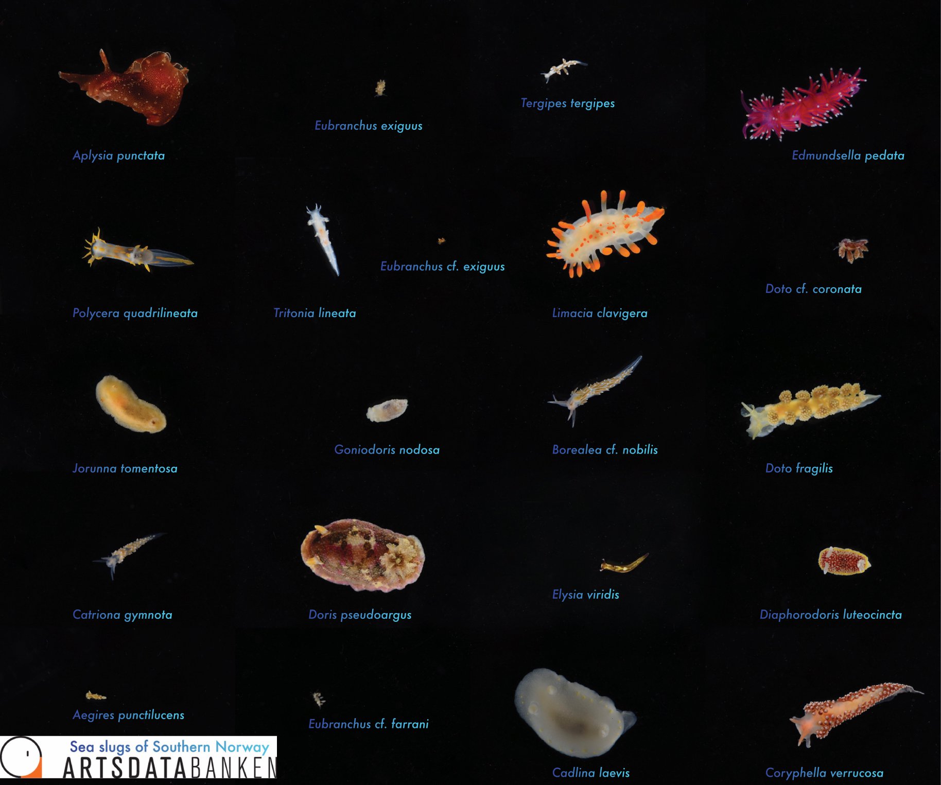
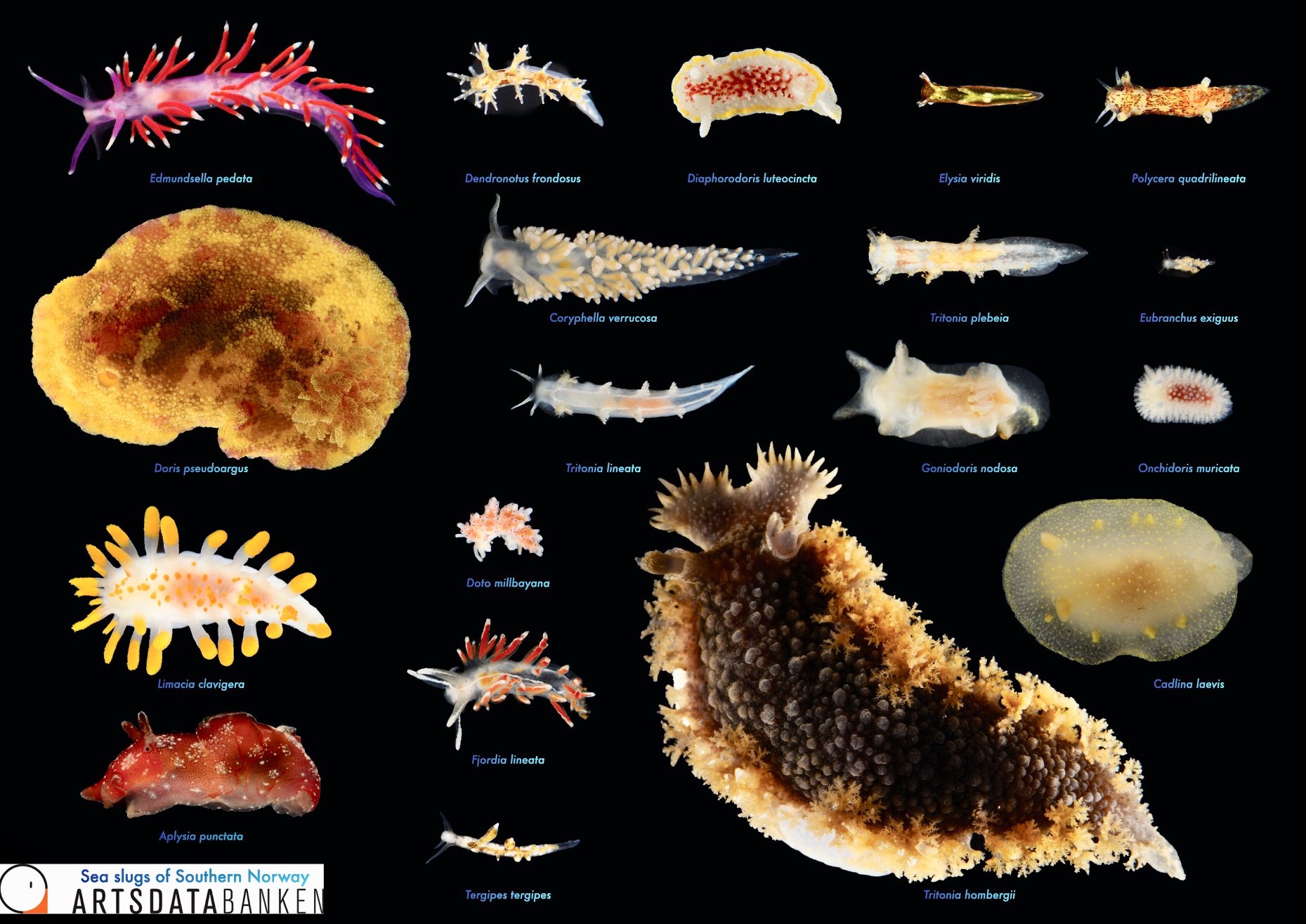
Sea slugs of Southern Norway managed to collect over 1000 lots covering 93 different sea slug species, of which 19 are new for Norway and a few new to science (we are working on it!).

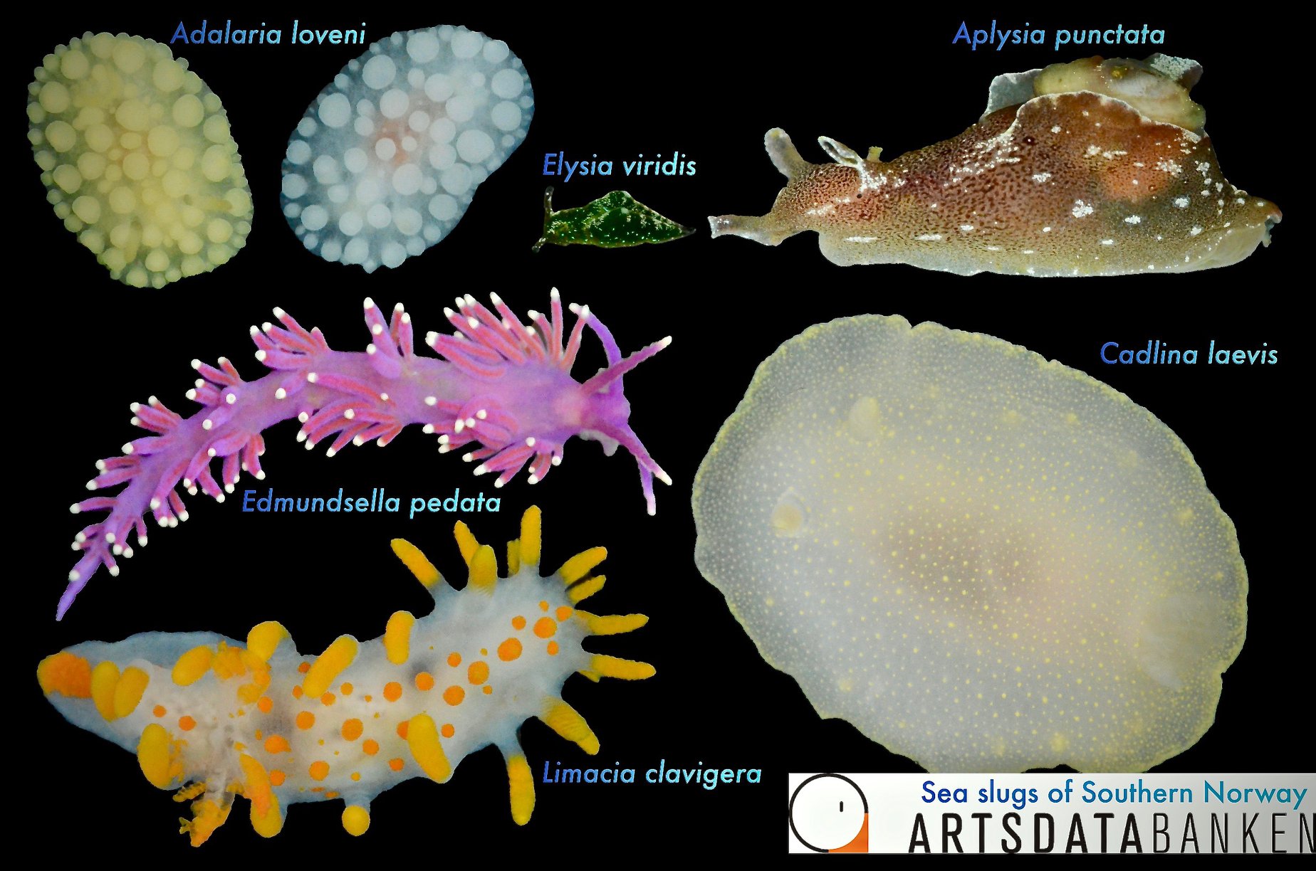
Below are photos of the species that were collected at different sampling events.  The photos are made either by the researchers associated with the project, or by the amazing team of citizen scientists.

Look at these beauties!

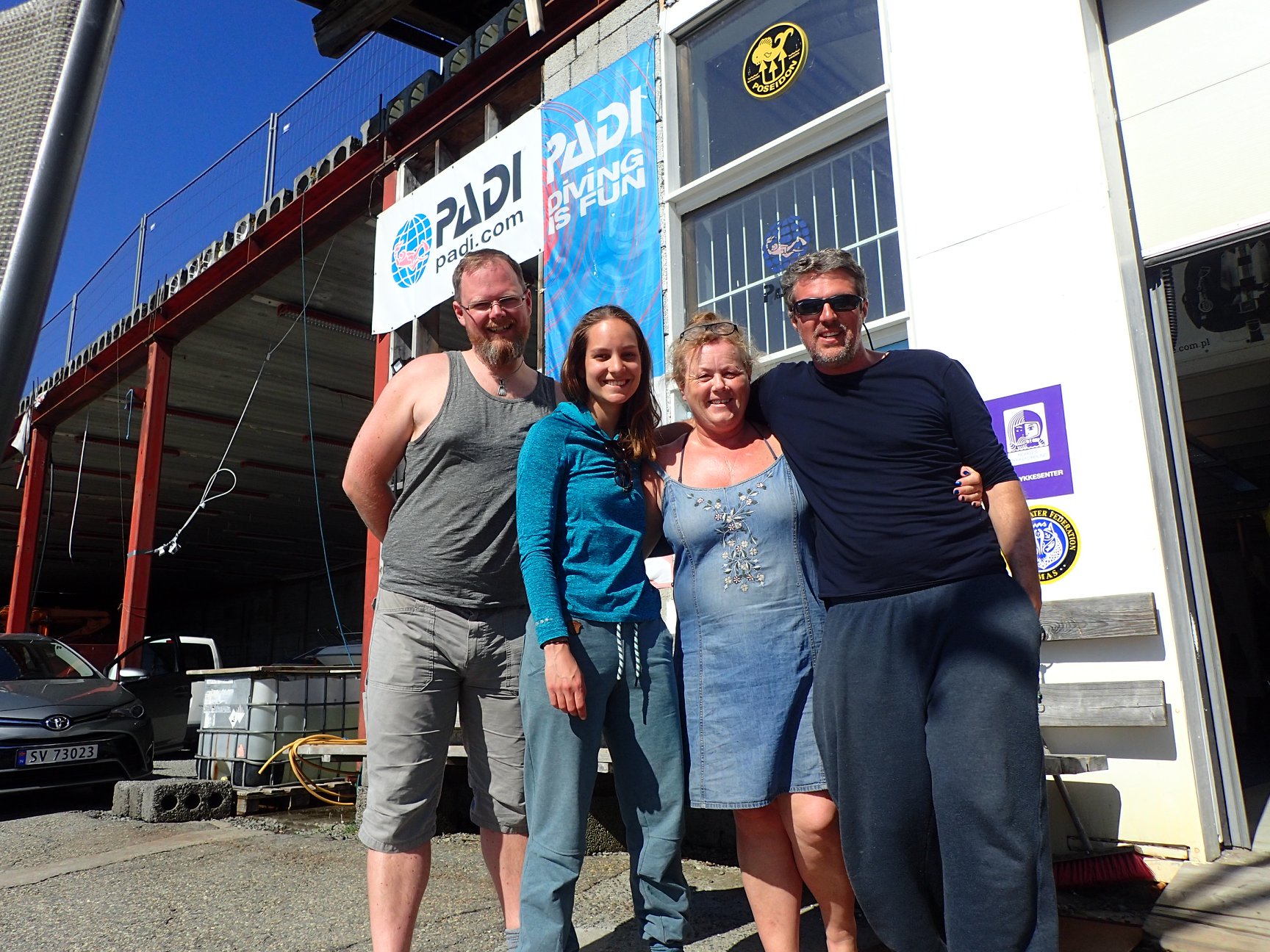
This would absolutely not have been possible without the special effort of our knowledgeable citizen scientists, and we would like to use this opportunity to name a few that were extraordinarily productive during the last years and provided the project and the Museum with valuable samples; Nils Aukan, Roy Dahl, Viktor Grøtan, Heine Jensen, Tine Kinn Kvamme, Runa Lutnæs, Ole Christian Meldahl, Jenny Neuhaus, Bjørnar Nygård, Anders Schouw, Erling Svensen, Cecilie Sørensen, Mona Susanne Tetlie, Anne Mari with Ottesen, Mandal Dykkerklub, Hemne Dykkerklubb, Slettaa Dykkerklubb, SUB-Studentes Undervannsklubb Bergen, Larvik Dykkerklubb, Sandefjord Dykkerklubb, and all the others that made big and small contributions.

A big thank-you to all contributors!

Would you like to know more about the citizen scientists part of the project? Check out this paper (starts on page 23) by Cessa and Manuel: Sea Slugs of Southern Norway: an example of citizens contributing to science.

Mandal team

One of the core components of the projects success was our outreach effort on all kind of social media platforms. During these two years these platforms got much more traffic than we initially thought; apparently we have many Norwegian sea slug fans, within and outside of Norway!

Therefore we decided to continue with our outreach efforts to keep everyone engaged and up to date about these wonderful animals in our ocean backyard, but with some minor adjustments. Some of you might have already noticed a few changes during the last days on the Facebook page  and our Instagram account. From today onward, the social media pages will cover sea slugs of all of Norway, and is now named accordingly. We also welcome a new admin to the facebook group: Torkild Bakken of NTNU University Museum. Welcome Torkild, the more expertise the better, so we are very happy to have you onboard!

We encourage everyone in this community to continue to be active and share your findings and knowledge with other.

Let’s carry on enjoying the wonderful world of sea slugs of Norway!


-Cessa & Manuel
 

HYPCOP kickoff!

Follow us at @planetcopepod!

Tuesday may 19 was the first fieldwork of the new project called Hyperbenthic Copepoda (HYPCOP). You can read more about the field work and see some photos and videos from the field in the previous blog post. 

Copepoda are small crustaceans that are found all over the world in both marine and freshwater habitats. Species can be planktonic (drifting in the sea water) or can be parasitic and a large diverse group of them live on algae in the hyperbenthic (living near the bottom) zone. Copepoda are very important food source for many organisms like small fish, they are on the bottom of the food pyramid, together with other zooplankton. Without copepods, a lot of bigger animals would no exists. Despite being so important, not much is known about the biodiversity and taxonomy of these animals, especially that of the species that live near the bottom.Some species like Calanus finmarchicus are the main nutritional basis for many fish species, and therefore of great importance for the Norwegian fish strains.  Therefore Artsdatabanken is funding the new project HYPCOP in order to unravel the biodiversity and taxonomy of hyperbenthos copepods. With special focus on the species in the group Harpacticoida that live in the water masses just above or near the bottom. Copepods from shallow water will be collected in coastal areas, in deep fjords and on the continental shelf.

The Institute of Marine Research (IMR), Natural history museum of Bergen (UiB), Norwegian Institute for water research (NIVA) and the Norwegian Barcode of Life (NorBoL) are working together to survey the diversity of marine copepods in Norwegian waters and expect to find and describe species that are new to science and new for Norway! Currently some of the taxonomic competence in Norway is lacking, but through collaboration with foreign experts this knowledge will increase among Norwegian researchers and students!

The projects duration will run from 2020 until 2023 and last week was the official kickoff with some fieldwork to get fresh material to work with! Together with the project Hardbunnsfauna we drove to a local favorite collection site of us; Biskopshavn; very close to Bergen city center. Around the hard substrate we found lots of freshly grown algae that contained many small animals for us to collect! In order to get good quality samples we needed to be in the water and snorkel. With plankton nets we collected algae and sieved the water column catching the smallest of the animals; copepods!

And even though this was just a first test of equipment and collection methods, it was not without success. Back in the lab the microscope revealed the beautiful and diverse world one drop of seawater contains. A lot off small crustaceans and of course the copepods were omnipresent.

Our findings had to be shared and especially for #InternationalDayForBiologicalDiversity the copepods cannot be left out as they from such an important group! See for yourself the beauty of our copepod planet!

-Cessa

Field work in Biskopshavn

Happy International Day for Biological Diversity 2020!
On this day, we wanted to share a few glimpses of our most recent field work:

We were finally able to – with some precautions in place – resume our field activities again this Tuesday; we had a lovely day trip in the sun to Biskopshavn, a locality just a few minutes drive away from the lab.

Here we collected animals from the shallow sub-littoral (from just below the tide mark to ~3 m depth) for the new project on Copepoda (see more about that here), and for Invertebrate fauna of marine rocky shallow-water habitats (Hardbunnsfauna).

Below is a short video from the field & lab (including the inevitable Littorina on the lam!), and a few of our findings from the day.

This is a polychaete in the family Syllidae. If you look at the tail end on the top image, you can see that it is about to breed: these animals can do so with schizogamy, which is the production of stolons (enlarged in lower image) which are budded off and become pelagic, swimming away to breed. The stolons form complete new animals, but differ from the stock animal in a number of respects, such enlargement of the eyes, reduction of the gut, and different musculature. The stolons die after breeding.

One of the animal groups Hardbunnsfauna is focusing on is the Bryozoa, or moss animals. Pictured is a Bowebankia spp. Due to COVID we haven’t been able to host our international specialists here this spring. We are amassing a nice collection of animals, and do our best to identify them – we will  begin preparing plates for DNA barcoding soon, and then involve the taxonomists once we have the results.

-Katrine, Cessa & Jon

Meet ZMBN 130407!

How much information do you think we have on the animals in our collections? 🤔

Quite a lot more than you might think, and here to help us show you, we have a small snail from the shore. Meet specimen #ZMBN 130407, a Littorina saxatilis 🐌 (rough periwinkle/spiss strandsnegl).

We collected it one year ago on our fieldwork up North, in Tendringsvika near Tjeldsund (Troms): our northernmost station on the trip.

Tendringsvika in Troms

Here’s a short video of the habitat: notice how the sea urchins dominate once we get below the intertidal zone!

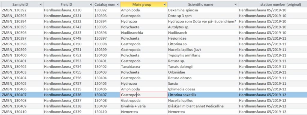
To be able to use the Invertebrate Collections for research, we need to know quite a lot about each animal (“specimen”). Standard information would be where, when and how it was collected, who collected it, who identified it (and revisions), notes about the habitat, images if any, and the museum number that it is registered within our database.

A screenshot of how it may look when a specimen is registered in our database

If there is genetic data – like here, a DNA barcode as part of NorBOL – we also need the genetic information. This information is stored in the international barcode library BOLD (BOLDSystems.org), where it is organised in projects containing information linked to the physical specimen, and to the DNA.

Small snail, much data!

If you look at the lower right corner, you will find information about specimens that have identical DNA sequences, and who are therefore grouped together in what is called a BIN in BOLD (/OTU). Most of the other specimens with identical DNA barcode have also been identified to Littorina saxatilis, but not all…that’s one reason to keep the animals in museum collections, so that identifications can be re-checked if needed 🔬.

Through our project (hardbunnsfauna) on shallow water hard bottom fauna from Norway, we are helping build a good DNA barcode library of species that occur in Norway – with reference (“voucher”) specimens in the scientific collections of the University Museum of Bergen, and with our partner, NTNU University Museum.

-Katrine

Science Communication – Creating Scientific Illustrations

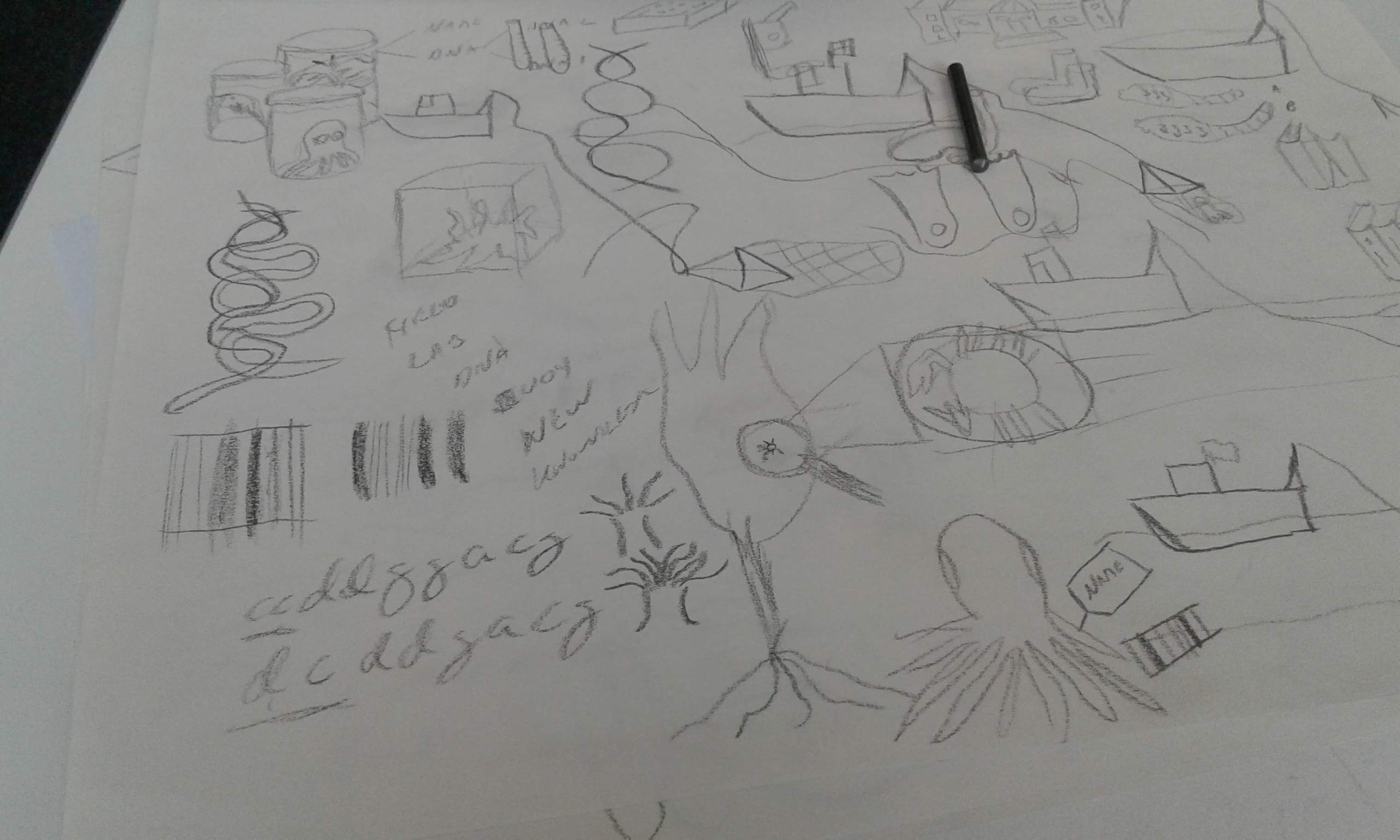
What on earth is this going to become?

I (Katrine) recently attended a course on how we can use illustrations to (better) communicate our science.

The course was offered as a joint effort of four Norwegian research schools: CHESS, DEEP, ForBio and IBA, and I got my spot through ForBio (Research School in Biosystematics).

The course was taught by Pina Kingman, and covered a lot of different topics in four days, from messy drawing with charcoal to using graphic software for digital illustrations:

  • Principles of design and visual communication
  • How to apply these principles to illustration and graphic design, which in turn will inform all visual material you might want to create, including; graphical abstracts, presentation slides, poster presentations, journal articles, graphs, data visualisation, project logos, animations and outreach material.
  • Best practices for poster and slide presentation design
  • Step by step method on how to draw your own research
  • Introduction to sketching by hand
  • Crash course in digital illustration with mandatory pre-course digital tutorials

Now, we were sternly told on day 1 that we were not allowed to say that we could not draw…but let’s say that some people have more of an affinity for it than I do – see above for proof! None the less, a concept was to be developed, discussed and improved during group work, and ultimately transformed into a digital illustration by the end of day 4.

Most of my fellow students were creating something related to their ongoing research, such as an illustration to be used in a paper of their PhD. On the last day we presented our work for the class, and got the final feedback from the group. Spending a whole day looking at cool graphics and learning about people’s work on such varied topics as water flow in magma, colour patterns on Arctic rays, better diagnosis of tuberculosis, and ecosystem modelling was really enjoyable, and the feedback I got was very helpful.

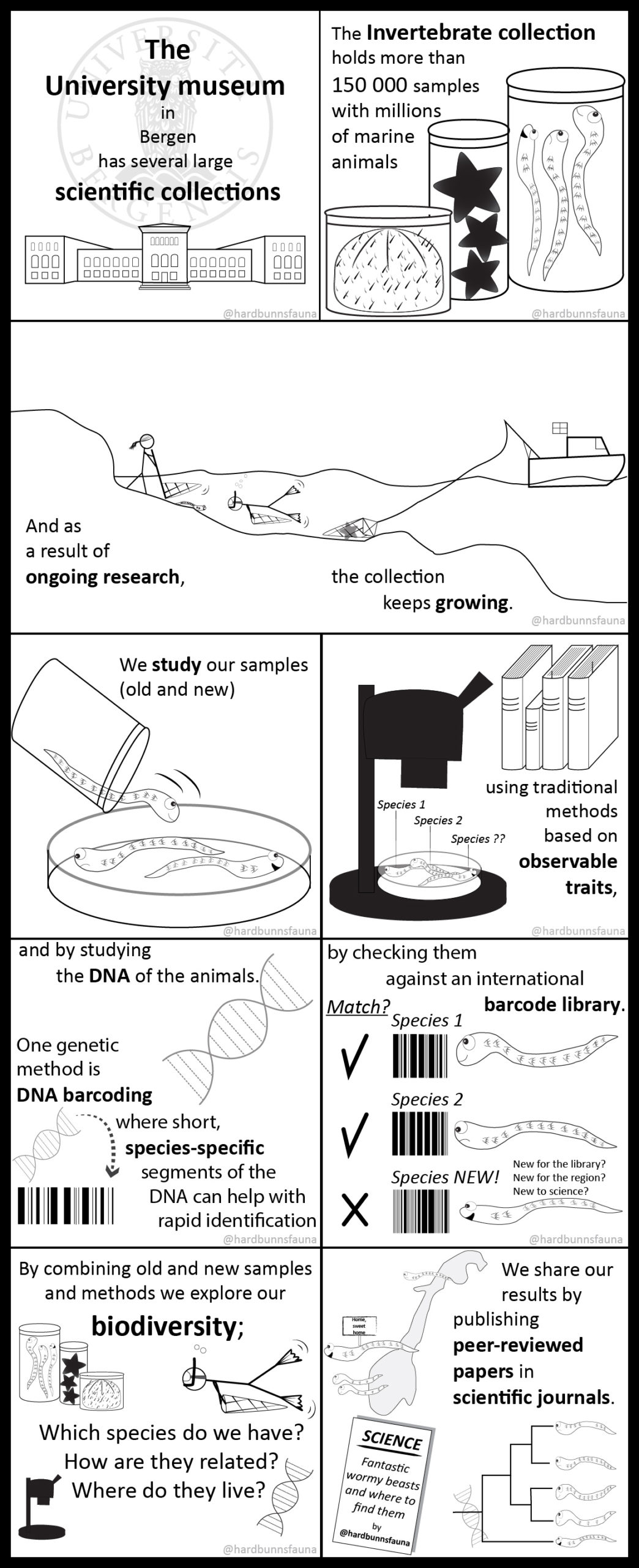
I opted for an outreach-approach, creating a lot of small illustrations that will be individually useful in future presentations and such, and which could be combined into a small comic about our scientific collections. The comic has been shared on Twitter and Instagram (do follow @hardbunnsfauna!), and now here:

The end product of the course; a short introduction to our scientific collections, how we work, and how we integrate data such as DNA-barcodes and morphological traits of the animals to do our research!

Thank you to Pina, Mandy (& the other arrangers), and the class for a wonderful learning environment and a fun couple of days!

-Katrine

NorHydro in South Africa: the 6th International Jellyfish Blooms Symposium

The 6th International Jellyfish Blooms Symposium was the last big academic event in 2019 attended by team NorHydro, and we were very happy to have presented our project in such a relevant meeting!

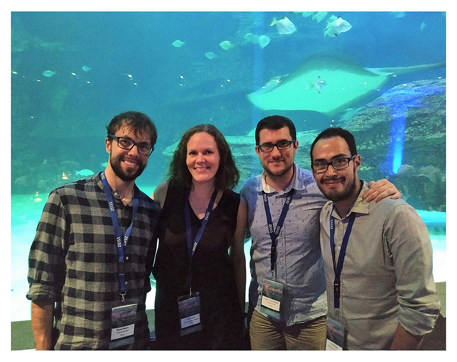
Team NorHydro at the 6th Jellyfish Blooms Symposium, from left to right: Maciej Mańko (University of Gdansk – Poland), Aino Hosia (UiB – UMB), Joan J. Soto (UiB – Sars Center), and Luis Martell (UiB – UMB).

In many ways, the Jellyfish Bloom Symposium (JBS) is the most important meeting of scientists working with medusozoans in the world. Professionals from different countries, backgrounds and lines of research meet every 3 years in this symposium to present their results, discuss new findings, and chat with colleagues about the state of knowledge in the group. So of course our Artsdatabanken project NorHydro had to be present, especially after a session focused on polyps was announced for this particular edition. The importance of polyp stages – the object of study of NorHydro – is now widely recognized in jellyfish biology, and understanding the ecology and diversity of polyps has become a key point in the study of jellyfish blooms.

This time, it was the turn of the University of the Western Cape, Iziko South African Museum and the Two Oceans Aquarium to host the JBS, which was held in Africa for the first time.

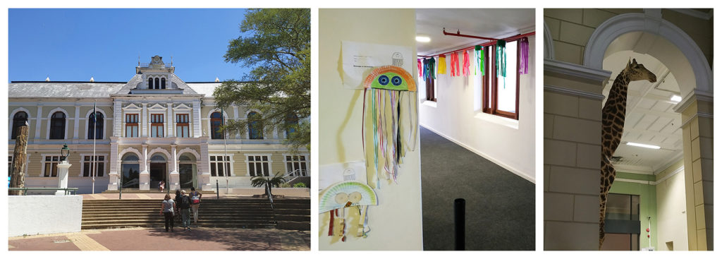
Iziko Museum was the venue for the oral presentations and poster sessions. The way to the auditorium was marked by jellyfish but we still had to pass under the vigilant eyes of a giraffe.

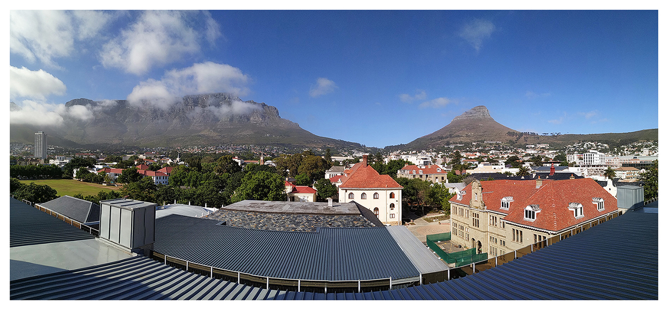
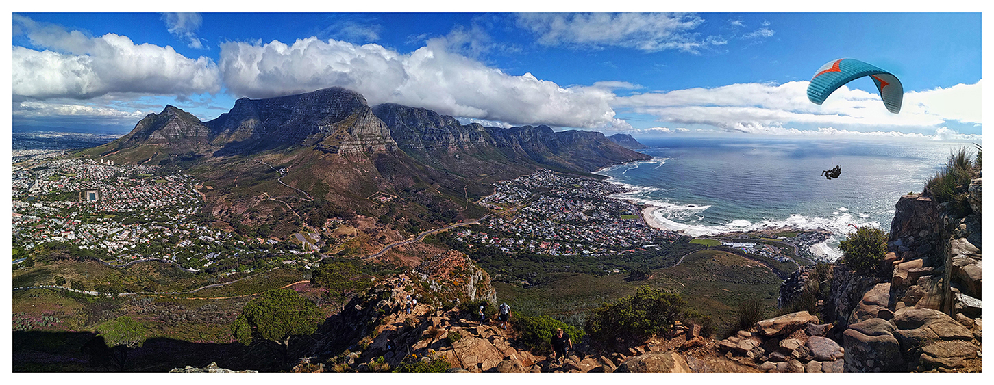
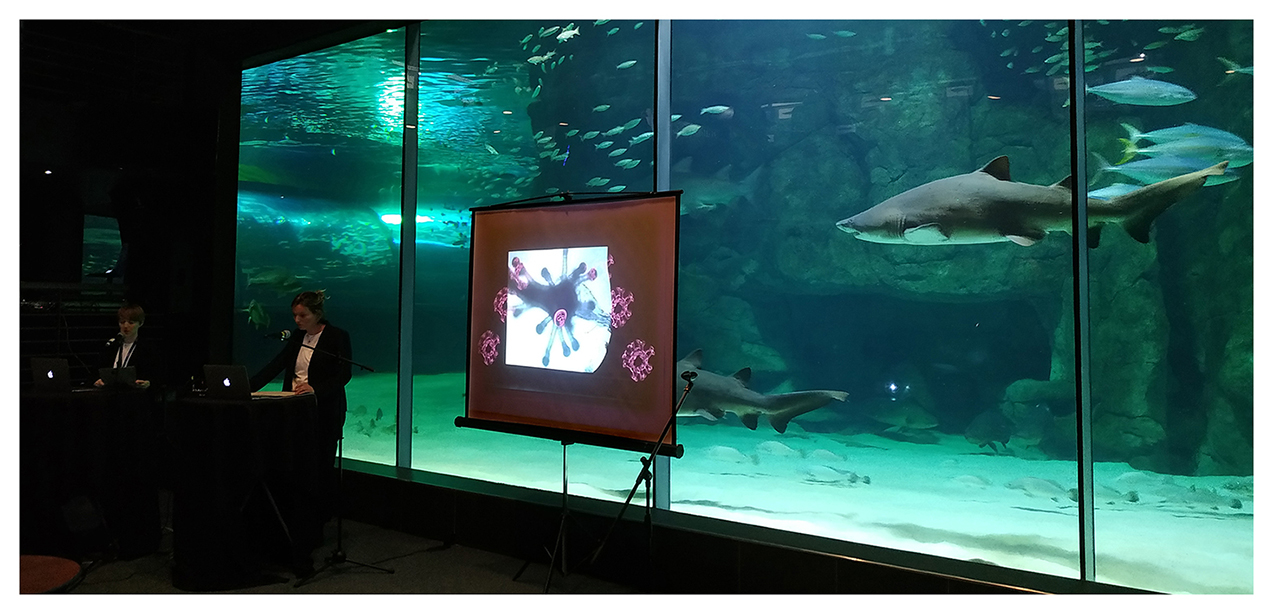
The city of Cape Town provided a beautiful setting for discussions on gelatinous matters and sharing of jellyfish-related stories, and we even got to see some of the local hydrozoans from the surroundings, both in the aquarium and in the sea.

Not everyone was happy with the high concentration of jellyfish researchers in Table Mountain. Photo: Joan J. Soto Àngel

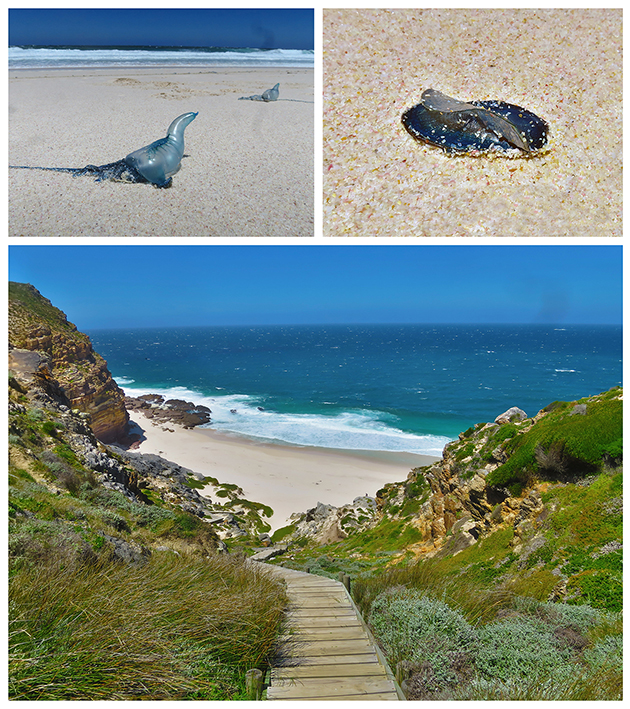
We were lucky to see some hydrozoans by the sea. Several specimens of the siphonophore Physalia physalis (top left) and the anthoathecate Velella velella (top right) were stranded in Diaz Beach, near the Cape of Good Hope (bottom).

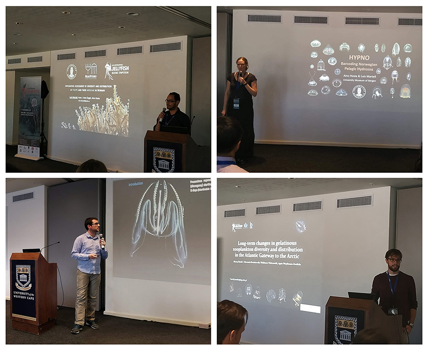
The oral presentations and poster sessions covered many subjects on jellyfish biology, not only the dynamics of polyps but also the relationship between jellyfish and humans, the role of jellyfish in the ecosystem, and the diversity of medusa, ctenophores and salps.

Overall, the participation of NorHydro in the JBS was very succesful. We received positive feedback about the results presented, and people were very interested in our upcoming activities, particularly the course on hydrozoan biology and diversity (read more and apply here).

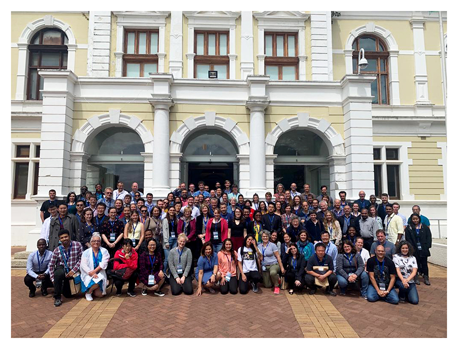
All the participants of the 6th Jellyfish Blooms Symposium

NorHydro was so warmly welcomed that we are already looking forward to sharing more about the hydrozoans of Norway in the next JBS in 2022!

– Luis

Keep up with NorHydro in Facebook and in Twitter with hashtag #NorHydro.

Research Internship – Francesco

In the last part of 2019 Francesco Golin collaborated with us as an intern in project NorHydro. Francesco is a student at the University of Algarve, where he is enrolled in the International Master of Science in Marine Biological Resources (IMBRSea). We asked him about his internship and this is what he told us:

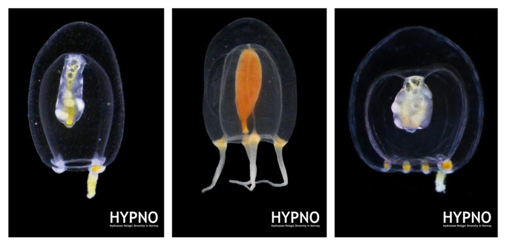
During the 2019 autumn semester I joined Luis Martell and Aino Hosia in project NorHydro as a research intern. My research contribution was aimed at finding how many species of the hydrozoan genus Euphysa are present in Norwegian waters, and how to define them morphologically and genetically. Euphysa is a common genus with 22 accepted species, but many of them are not easy to tell apart from each other, which is why we decided to implement an integrative approach for species delimitation including morphological and molecular analyses.

Some of the species of Euphysa occurring in Norway. From left to right: Euphysa aurata, Euphysa flammea, and Euphysa sp

Working on board during the cruise

My first mission as an intern was collecting some samples of Euphysa and other gelatinous organisms. Luckily, the opportunity to do so presented itself during the student cruise associated to BIO325, a course in which I participated as part of my studies at UiB.

During this cruise I used a light table to spot the tiny jellyfishes brought on board by the Multinet, then I placed them on a Petri dish and took pictures of them with a camera attached to a stereomicroscope, before transferring them to an Eppendorf tube filled with ethanol.

All these elements (the pictures of each organism, the associated sampling data, and the samples themselves) are needed for species delimitation of hydromedusae. The pictures are used to compare the morphology of different individuals and to identify important diagnostic characters (unfortunately, ethanol-fixed jellyfish are not useful for morphological analysis), while the ethanol-preserved samples are used to obtain DNA sequences.

The light table used to spot the gelatinous zooplankton

Some siphonophore parts are very transparent, and thus they are some of the most difficult animals to spot in plankton samples.

The hydrozoan Aglantha digitale (left) was very abundant in all my samples. Other cnidarians, such as this anthozoan larva (right) were also present.

My second mission consisted on gathering the original descriptions of the different species of Euphysa. This information is necessary if we want to understand what makes each species different, and will come handy when analyzing the individuals and their pictures collected on the field. Talking about species boundaries, I had the opportunity to attend a course on “Molecular Species Delimitation” offered by the University Museum. In this course I learned how to perform the analysis of DNA sequences for species delimitation, using some common software (MEGA and R) for this purpose. These are important tools that will allow us to assess the diversity of Euphysa in Norway, and together with the morphological analyses these data will help us determine if new species have to be described.

Now the semester has ended and my internship is over. Nevertheless, I hope my help was meaningful, as I want to continue being a part of this research project in the future. I will keep myself updated with the changes in the taxonomy of Euphysa, so I’m sure I will be able to join NorHydro again when I’ll come back to Bergen!

-Francesco

CT scan all the things!

Oslo Natural History Museum 02.12.2019 – 06.12.2019

Literature for the week of our CT scanning course


From December 2 till December 6 Justine Siegwald, Manuel Malaquias and I (Cessa) attended a CT scanning course in Oslo at the Natural History Museum.

The course was organized by the Research School in Biosystematics (ForBio) and Transmitting Science.

The group attending the CT scanning course in Oslo at the Natural History Museum

The three of us all work on mollusks and one of the main reasons to deepen ourselves in CT scanning techniques is because of studying the internal anatomy of these often small and delicate specimens.

Species descriptions are not only based on external morphology and/or DNA barcoding, a lot of the species differences are small details within the internal structures. Like the reproductive organs and digestive system (e.g. radular teeth) of the animals. This is very laborious work, and can be challenging especially with the smaller specimens of only a few millimeters long. Besides some species are difficult to collect and can be a rare collection item of the museum. Once you cut these animals to study them, there is no turning back and the specimen is basically destroyed. Being able to see the insides of the animal without touching it would therefore be ideal. CT scanning comes very close to that and with powerful X-ray we can almost see every detail of the insides of the animals without the need to cut them open

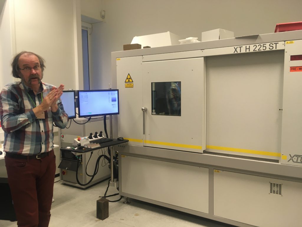
The CT scan in the Natural History Museum of Oslo with one of the teachers in front (Øyvind Hammer)

Lecture about the technology of the CT scan and X-ray

However, how easy as this sounds, CT scanning soft tissue comes with some challenges. Soft tissue means that the x-ray contrast is often very low. Even with modern good x-ray detectors it is still difficult to detect the different internal structures. Therefore, during the course, we were taught how to artificially increase the contrast of the tissue by staining the specimens before mounting them in the CT scan machine.

Coating a specimen with a metal (e.g. gold or platinum) is only useful if you want to see external details of the study object, coating will not help revealing internal structures. For radio contrasting the internal anatomy, you can stain specimens with high density liquids like iodine to enhance the x-ray contrast. So, we went for this option and left our specimens in iodine ethanol solution overnight


After the staining process, we needed the wash out any extra iodine before mounting the specimen into the CT scan. A good scan is not simply pressing the button of the machine; there are a bunch of settings that can be adjusted accordingly (e.g. tube voltage, tube current, filament current, spot size, exposure time, magnification, shading correction, etc.). The scan itself can take hours and file sizes of 24GB for a single object are not uncommon, which means you need a powerful computer with decent software to process this information. Part of the course was also about how to visualize and analyze the files of the CT scan with software like Avizo

Video 1. Software Avizo has a user-friendly interface for analyzing all sorts of CT scanning files.

The week gave us some very promising first results (se video below), and also new insights about how to increase the X-ray contrast of our sea slug samples (lots of hours of staining and very short washing steps).

Video 2. 3D reconstruction of one of Justine’s shell samples

It was a very fruitful week and besides interesting new ideas for scanning our museum specimens, we also met many old and new friends during the week that was as inspiring. ForBio and Transmitting Science did a really great job with setting up this course and I can highly recommend it to anyone who is interested in CT scanning.

Explore the world, read the invertebrate blogs!

-Cessa

Field season’s end

Sletvik field station, October 15th-23rd 2019

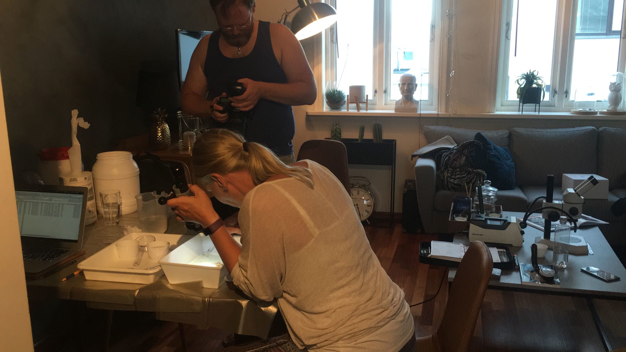
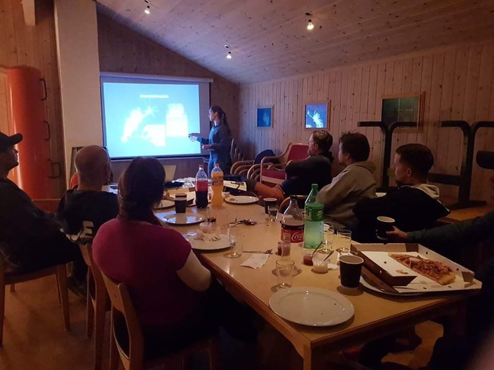
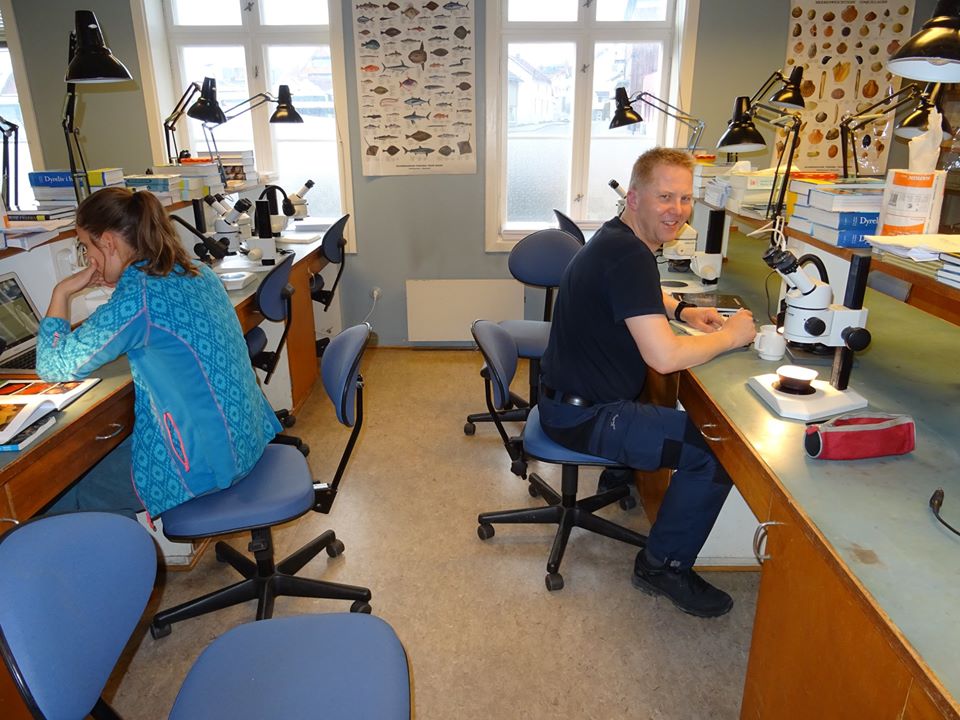
We wanted to make a write-up of the last combined fieldwork/workshop we had in 2019, which was a trip to the marine field station of NTNU, Sletvik in Trøndelag, in late October. From Bergen, Luis (NorHydro), Jon, Tom, and Katrine (Hardbunnsfauna) stuffed a car full of material, microscopes, and drove the ~12 hours up to the field station that we last visited in 2016.Beautiful fall in Trøndelag

There we joined up with Torkild, Aina, Karstein, and Tuva from NTNU university museum, students August and Marte, and Eivind from NIVA. We also had some visitors; Hauk and Stine from Artsdatabanken came by to visit (if you read Norwegian, there’s a feature about it here), and Per Gätzschmann from NTNU UM dropped by for a day to photograph people in the field.

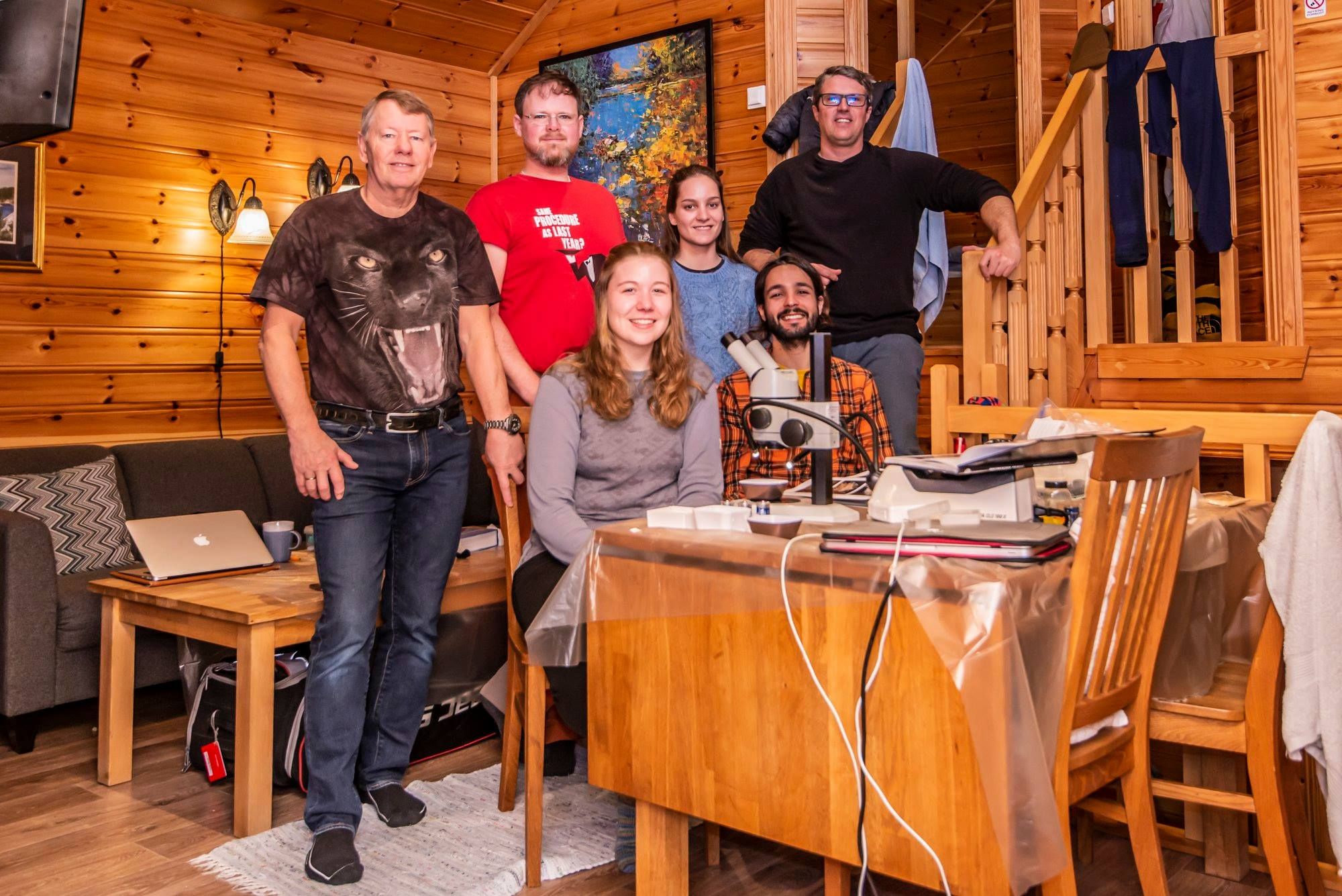
Most of the workshop participants lined up Photo: Hauk Liebe, Artsdatabanken

During a productive week the plan was to work through as much as possible of the material that we and our collaborators had collected from Kristiansand in the South to Svalbard in the North. Some of us went out every day to collect fresh material in the field close to the station.The Artsprosjekts #Sneglebuss, Hardbunnsfauna, NorHydro, and PolyPort gathered at Sletvik, and with that also the University museums of Trondheim and Bergen. Of course we were also collecting for the other projects, and the museum collections.

One of the things Hardbunnsfauna wanted to do whilst in Sletvik was to pick out interesting specimens to submit for DNA barcoding. This means that the animals need to be sorted from the sediment, the specimens identified, and the ones destined to become barcode vouchers must be photographed and tissue sampled, and the data uploaded to the BOLD database. We managed to complete three plates of gastropods, select specimens for one with bivalves, and begin on a plate of echinoderms, as well as sort through and select quite a few crustaceans and ascidians for further study.

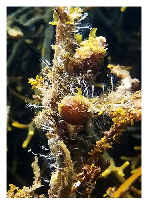
Collecting some fresh material was particularly important for NorHydro because the hydroids from the coasts of Trøndelag have not been thoroughly studied in recent years, and therefore we expected some interesting findings in the six sites we managed to sample. We selected over 40 hydrozoan specimens for DNA barcoding, including some common and widespread hydroids (e.g. Dynamena pumila), some locally abundant species (e.g. Sarsia lovenii) and exceptionally rare taxa, such as the northernmost record ever for a crawling medusa (Eleutheria dichotoma). We also used a small plankton net to catch some of the local hydromedusae, and found many baby jellyfish belonging to genus Clytia swimming around the field station.
Plan B when the animals (in this case Leuckartiara octona) won’t cooperate and be documented with the fancy camera; bring out the cell phones!

It was a busy week, but combining several projects, bringing together material spanning all of Norway, and working together like this made it extremely productive!

Thank you  very much to all the participants, and to all the people who have helped us gather material so far!

-Katrine & Luis