Category Archives: Student Projects

ForBio Annual Meeting 2023 at the University Museum of Bergen

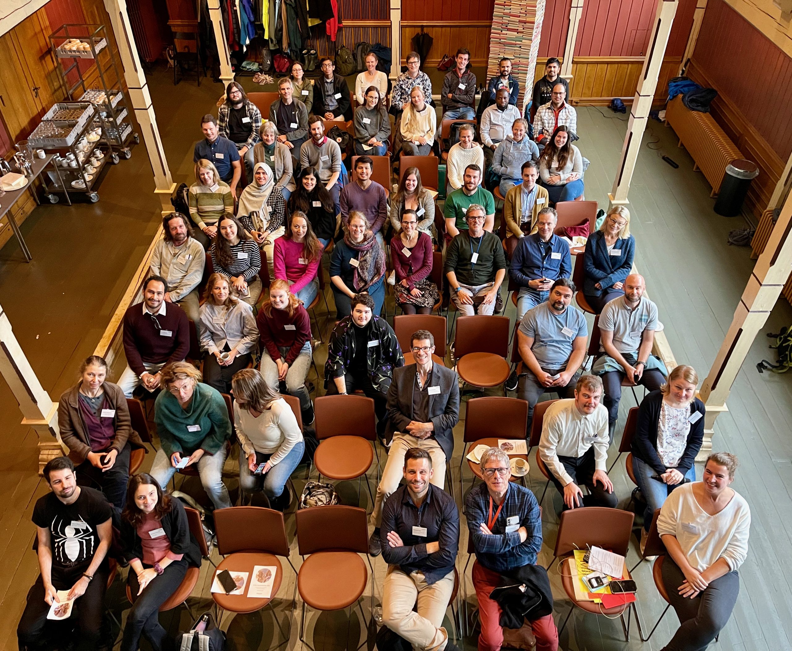
ForBio – Research School in Biosystematics held its 12th Annual Meeting at the University Museum of Bergen on September 19-21st.Sixty participants from Norway, Sweden, Iceland, Czech Republic, Poland, Germany and Peru presented their research results in various fields of biosystematics.

The main day of the meeting took place in the Tårnsalen – a beautiful venue at the top of the museum building and was opened by a talk by Endre Willassen on the history of marine research at UiB.

The participants - 60 of them - in the historical Tårnsalen. The photo is taken from up on a gallery in the room, so it's a bird's eye view

The participants in Tårnsalen. Photo: ForBio

Four keynote speakers gave lectures on general biodiversity research topics of high interest to a wide audience.
Michael Bok from Lund University talked about the evolution of visual systems in marine annelids. Lovisa Gustafsson from the Stavanger Botanic Garden talked about the evolution of reproductive isolation in Arctic plants. Tomas Roslin from the Swedish University of Agricultural Sciences discussed the ways to assess enormous diversity of insects using DNA barcoding, and Agnes Weiner from NORCE gave a lecture on using ancient DNA in the past marine biodiversity assessments.

ForBio [LINK to homepage] is a cooperation between the main Natural History Museums in Norway: Natural History Museum, UiO; University Museum of Berge, UiB; NTNU University Museum; and the Arctic University Museum of Norway, UiT and supported by the Norwegian Biodiversity Information Centre.

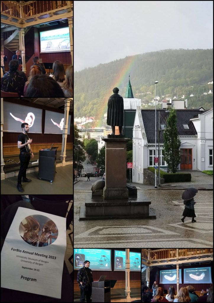
Collage showing people presenting, and a statue of Christie with a rainbow over it.

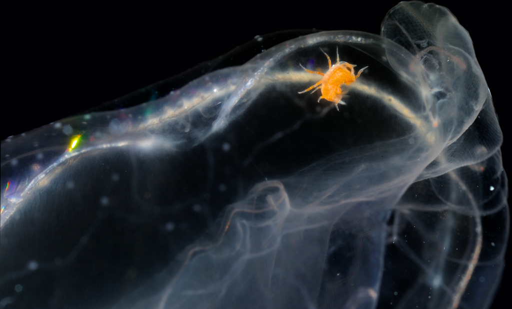
From top left: Endre Willassen presenting his talk on the history of marine research at UiB, Christie (the statue) standing outside the Museum in quite typical Bergen weather, Miguel Meca on the systematic revision of orbiniid polychaetes, the result of his PhD project, a snapshot of the program, Vincent McDaniel gives his speed talk about the parasites in jellyfishes (ParaZoo project), and Martha Everett gives her talk on the diversity of scale worms in the Arctic, the results of her PhD project

ForBio aims to advance biosystematics education for students and postdocs working in the fields of botany and zoology. The annual meetings is a great arena for the students to meet their peers and mor senior scientists and to present their research project results, exchange ideas and expand their networks.

You can find upcoming courses and events here: link

-Nataliya Budaeva
ForBio-coordinator

The Cnidaria+Ctenophora Research group at the PRIMALearning Jellyfish Workshop in South Africa

On the 12th of February at the crack of dawn, we had the amazing opportunity to go to Cape Town to attend a Jellyfish workshop. The “we” in question are the three authors of this blog post: Vincent, Vetle, and Håvard. We are master students all working with jellyfish-related topics, and some would go as far as to call us jellyfish enthusiasts. Our work is part of the museum’s Artsprosjekter NorHydro and ParaZoo, and we were happy to represent the invertebrate collections and UMB at this event.

The workshop, held at the Iziko South African Museum, was organized by PRIMALearning and was a collaborative initiative between the University of Bergen and the University of Western Cape. This gave the three of us, accompanied by a few other UiB students not affiliated with the University Museum of Bergen, the chance to visit Cape Town. Some of us for the first time.

On the first day of the workshop, we were greeted outside the museum by Mark Gibbons and his PhD student Michael Brown who was the representative from UWC and would be teaching parts of the workshop. With them was also Anne Gro Vea Salvanes as the representative for PRIMALearning and the University of Bergen, she was also joined by our own UMB scientists Aino Hosia and Luis Martell, who were also part of the teaching team.

The first day included introductions from all the participants, and we got to know each other bit better. We also got a brief introduction to the world of jellyfish and their taxonomy before the night ended with a delicious dinner together with all the participants.

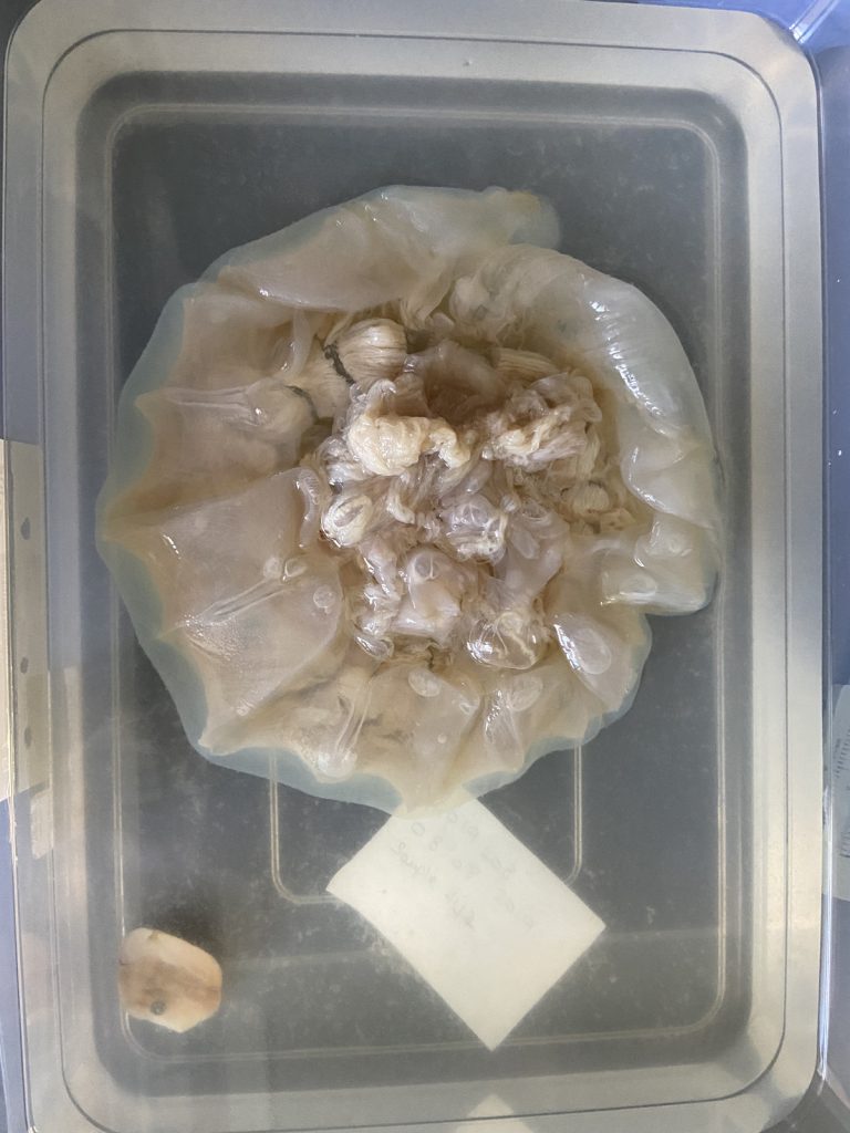
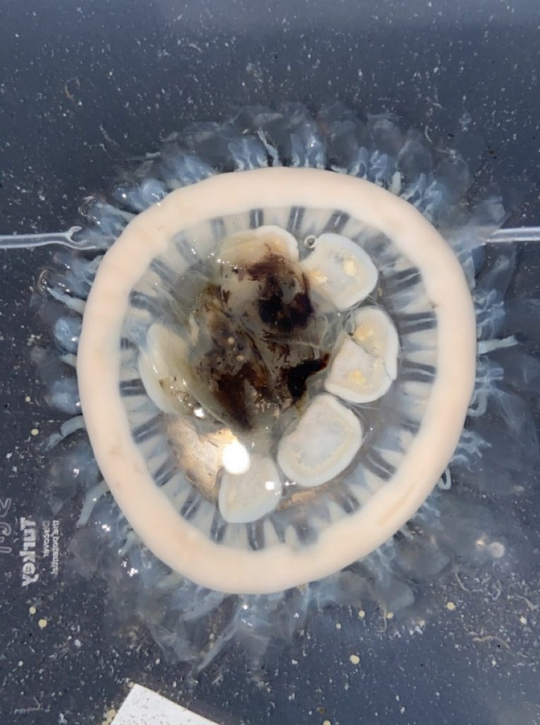
The second day started with lectures about the large or ‘true’ jellies (Scyphozoa), before we got to get our hands dirty looking at preserved samples of jellyfish. We were met with a broad diversity of scyphozoans that was passed around between the students so we would get a shot at identifying them.

We examined fixed material of scyphozoan jellies representative of the three major groups within the class:


The rest of the day after the workshop was spent at the beach, enjoying the sun and local wine. Cape Town is called the windy city, and it did deliver on its name, but nothing could stop the sun-deprived Norwegian students from going outside to soak up some rays.

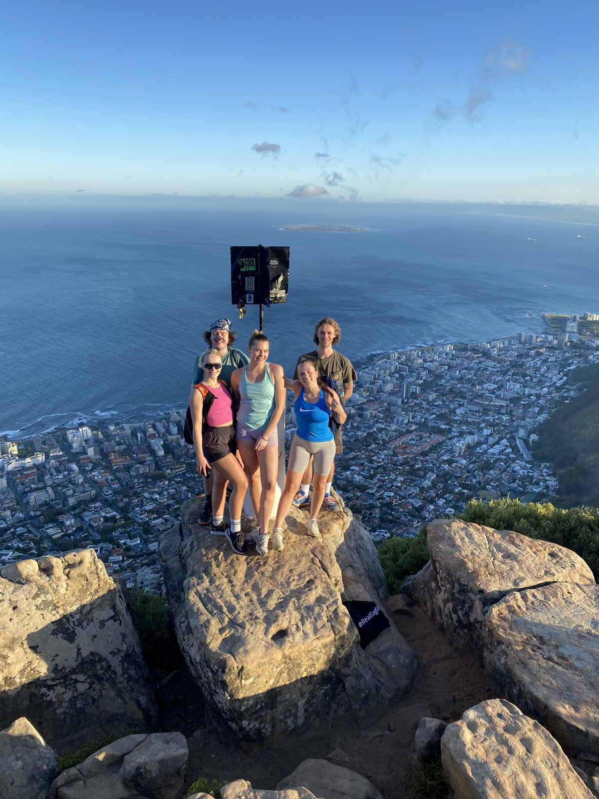
The third day was Hydrozoa day 1, a topic dear to our hearts and it was taught by our own MSc supervisor Luis Martell. Some of us were a bit tired this day because we decided to climb Lion’s head mountain before the workshop started to see the sunrise.

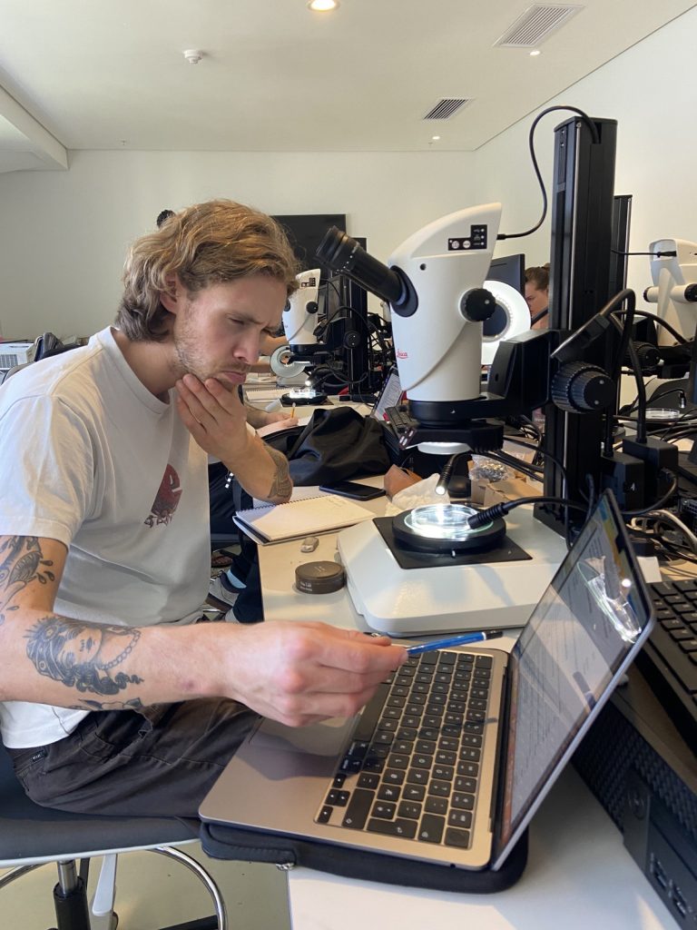
But this did not stop us from eagerly working with the preserved hydrozoan samples we got to look at. We identified all hydromedusae and siphonophores with the help of a stereomicroscope:

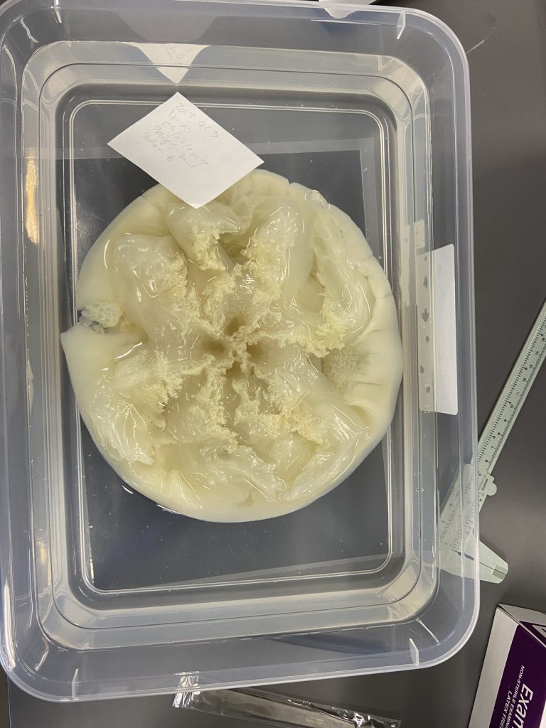
 A dissected carybdeid cubozoan jellyfish. IC: Håvard Vrålstad


Day four was box jellies (Cubozoa) day, a class neither of us was very familiar with. So we were excited to learn about these unknown and sometimes dangerous animals.

Luckily the animals were less deadly when preserved and we could therefore touch them while identifying them.


This day ended early so we took the opportunity to get down to Simon’s Town were we spent the rest of the day looking at the local penguins and wildlife at the beach.

A snapshot of the fauna we observed at Simon’s Town. IC: Vincent McDaniel

On our last day, we were introduced to the alien world of siphonophores by our own Aino Hosia. These animals are close to Håvards heart (if you ask him on a good day).

They were a nice ending to an awesome workshop, and we can honestly say now that our interest for jellyfish has grown, and we look forward to seeing even more of them in the future.

On February 19th the vacation/business trip was unfortunately over for Vetle and Vincent and they had to pack their bags and prepare to leave Cape Town and head home to Bergen, while the “slightly” more fortunate Håvard stayed behind in the windy city to enjoy a few more days of leisure.

Participants and teachers at the jellyfish workshop. IC: Anne Gro Vea Salvanes

We want to thank PRIMALearning for arranging the workshop, the University of the Western Cape for hosting and providing us with samples to work with, the Iziko Museum of South Africa for the location as well as providing refreshments, and to the University of Bergen for arranging accommodations. Also, a very special thanks to all the lecturers who presented and were very patient with us during the lab work.

-Vetle, Vincent, and Håvard


Are you interested in becoming a master student in marine biology at the University Museum of Bergen? Information about available projects can be found here (more will be added soon!):

Marine Masters at the University Museum of Bergen
– available thesis topics in marine biodiversity

Hydrozoan team at ForBio 2022 annual Meeting

Do you remember that feeling of dread before you must present in class about a topic you didn’t really study for? Your mind racing, trying to scramble a coherent story to tell the sea of eyes fixed expressionless on you and your powerpoint? We believe we all have at least one memory of this from our days in college.

That was a similar feeling to what we felt on November 28th, 2022, when we had just landed in Trondheim and were on our way to the Norwegian University of Science and Technology (NTNU) for this year’s ForBio Meeting, a 3-day gauntlet where we will present the Master’s projects we’re currently working on. Except this time, we weren’t presenting in a small classroom full of uninterested teenagers thinking about tik tok dances, but in an auditorium full of fellow researchers who work on your same field, and who will probably have extremely difficult questions at the end of your talk. Being the first time we present in an environment like this, one can’t help but imagine all the worst outcomes

And so we’re sitting there, watching the hours and the talks go by, thinking to ourselves “I should/shouldn’t try that for my talk!”. At lunch, all we think about is our talks. When we’re doing some light sightseeing in Trondheim, all we think about is our talks. We’re laying in our beds the night before, and all we think about is our talks. “How will I make my topic sound as professional and knowledgeable as I want to?”, we think to ourselves.

The day arrives, and again the dread starts setting in, right until the moment they call each of our names. At our every turn, we walk down the steps, grab the microphone, and start talking. Except for some reason, this time it feels like we’re in control. The words flow effortlessly; we even crack a joke or two, and the audience laughs. There’s no stumbling around the words like those college days.

Because this time, we’re not the average seat-warming student. This time, we’re the ones that have spent cold and raining hours on a research vessel or diving to get our samples; we’re the ones who have spent hours working together with our supervisors, reading and learning about our topics; we’re the ones who have spent hours looking down a microscope trying to identify our organisms. This time, we’re the ones who know what we’re talking about, and the audience is there to learn from us.

After each of our talks, we give our acknowledgements, and everyone claps. The questions thrown at you are answered effortlessly, and the moment is finished with a thumbs up to our supervisors, returning to our seats smiling.

The most exciting part for us was showing to experts what we do with our favorite invertebrates: Hydrozoans. These organisms are an inconspicuous class in the phylum Cnidaria that most people ignore. Our job was not only to present what we do with them, but to show what they are, what they do, how fascinating they can be and why they are important. And we think that we did a good job.

Now we’re finally free to enjoy the world famous Trondheim bike lift (and the Nidaros Cathedral too), closing the night with burgers, beer and friends.

Everything has been great on this trip and we will always remember the advice from other professionals; how different or similar the work of each one is; and the feeling that you are part of a group of specialists who are excited to share their knowledge with others. But, above all, we have learned why these conferences are important: Knowing what other researchers are doing gives us the chance to collaborate together and help each other, because working as a team is how science moves forward.

The hydrozoa group (and friends) from UiB participating at the ForBio meeting

Pedro, Ana & Håvard

Student visit – Ana González

MSc student Ana González visited the collections last month as part of project NorHydro, where she spent some weeks in the lab working with her samples. Here is an account of her experience:

The challenge of identifying benthic hydrozoans
Hydrozoa is a fascinating but poorly understood group of invertebrates, in part because their identification is not always an easy task. I have been studying benthic hydrozoan communities for over a year now, in particular those living in the shallow waters of Mallorca (Spain), and I have realized that the diversity of forms and structures in the group is higher than I had imagined at the beginning of my studies, and their identification is more difficult than I expected. The assemblages of hydrozoans in the Mediterranean are of course very different from the ones that occur in Norway, but something that both communities have in common is that morphological identification of the animals (i.e. telling which species is present based only on the characteristics we can observe) is challenging, which is why one of the aims of my visit to the University Museum of Bergen last December was to learn a different technique (DNA barcoding) that can help me improve the identification of my samples in cases when the morphology of the specimens is not good enough.

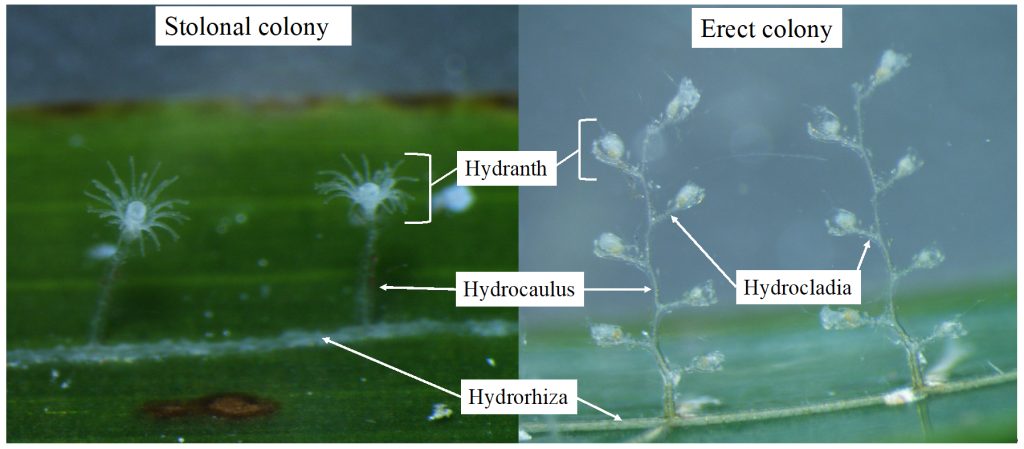
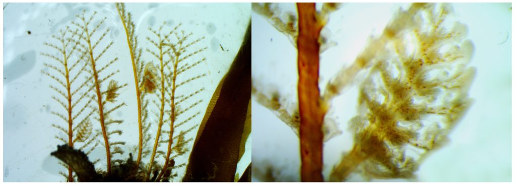
Some of the morphological characters that are used to identify benthic hydrozoans. On the left side a member of Campanulariidae, with a stolonal colony, and on the right side Monotheca obliqua with an erect colony.

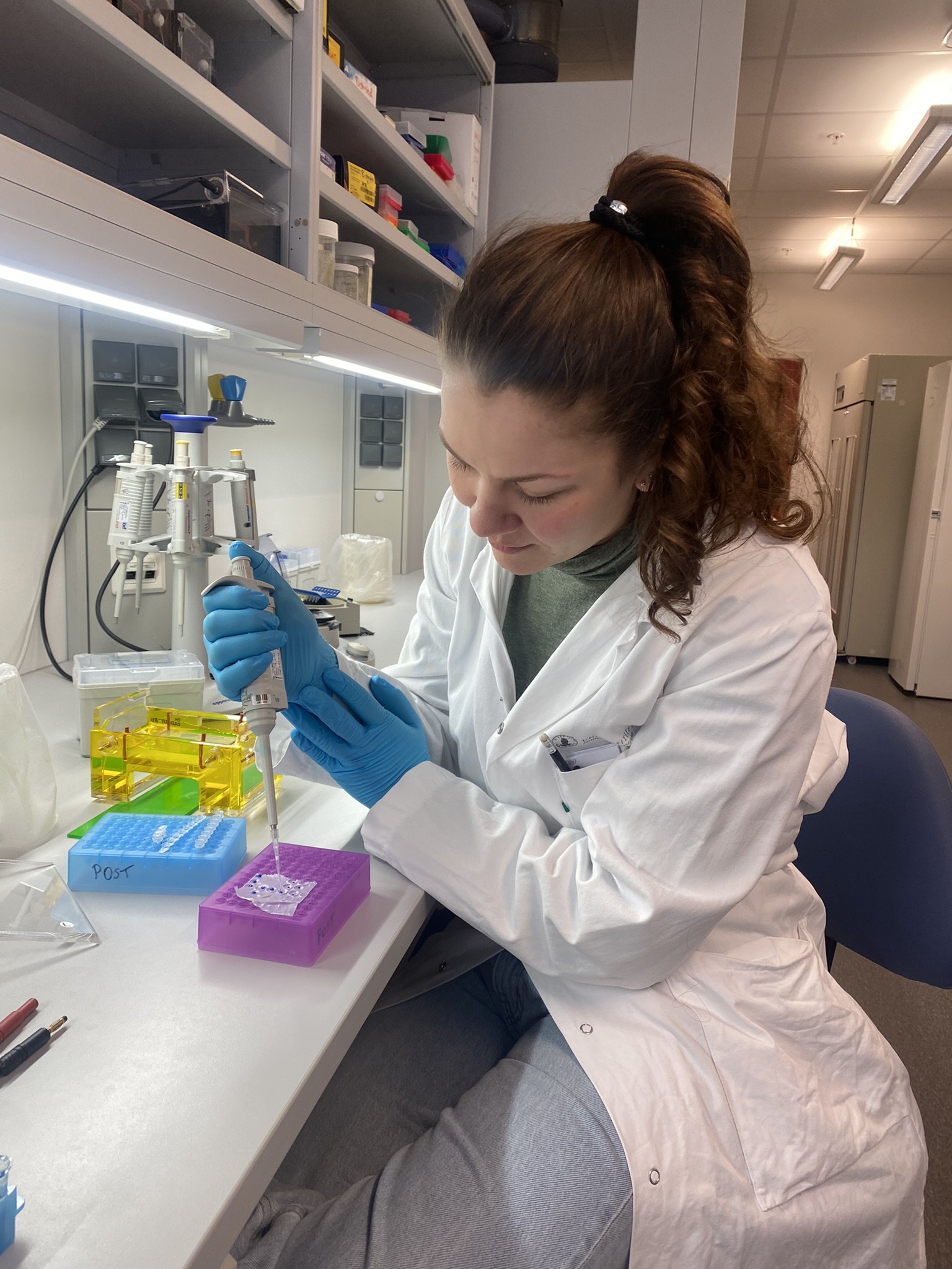
DNA barcoding consists in finding a short DNA sequence (the barcode) that is similar for all members of one species but different from all other species. It is a relatively recent tool that –among other things– has helped the scientific community identify specimens that for one reason or the other cannot be identified based on how they look. In some groups, such as many colonial invertebrates, this technique has become a key asset because the colonies are often too young or not reproductive, or the important characters for identifications may be found only in one stage of the life cycle and not in others. For this visit I had the chance to bring all my samples from Mallorca to Bergen and I set to extracting the DNA of selected specimens, amplifying two different barcode genes (COI and 16S), and obtaining clean sequences for them. I discovered that, when it comes to DNA barcoding, every step of the process is important, and being patient and careful is essential.

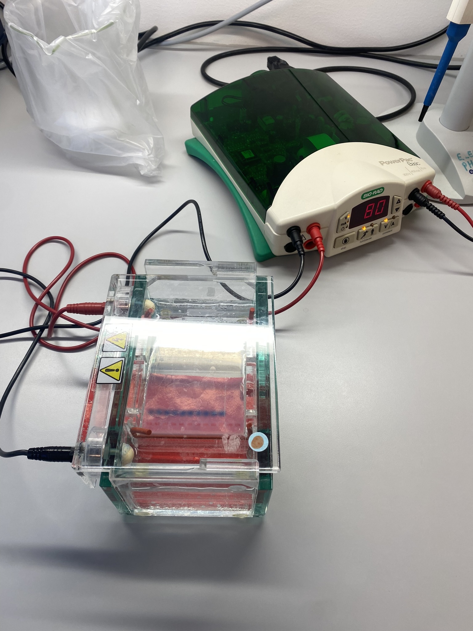
Me at the DNA lab, running the electrophoresis for my samples.

Getting good results in the DNA lab depends on several factors like not forgetting any step and avoiding contamination as far as possible, but the work does not end there: once you have your sequences they have to be cleaned, quality-checked, and finally compared with others. This means that having a complete and trustworthy database of DNA barcodes is necessary, especially if you want to use the sequence to help you corroborate the identification of a specimen. When done right and with a good database, the DNA barcodes can be useful to detect differences between hydrozoan assemblages growing in different parts of the world or between different substrates and levels of anthropogenic impact, which is what I am doing in my MSc project.

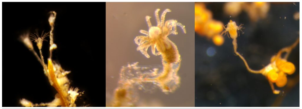
Left: Clytia sp growing on the marine plant Posidonia oceanica. Center: A polyp of Halecium sp, one of the most difficult genera of Hydrozoa to identify based only in morphology, especially when the colony is not reproductive. Right: Eudendrium sp., found in harbours in Mallorca in high abundances.

The analysis of DNA sequences is a powerful tool to compare specimens of distinct populations and in some cases animals that apparently belong to the same species turn out to be completely different (e.g. cryptic species). This is not uncommon for benthic hydrozoans, which have high morphological diversity but also high levels of plasticity, resulting in colonies from different species sometimes being very similar to each other when they grow in similar substrates. As useful as DNA analyses are, however, it is also important to consider their limitations. For example, while the abundance of each species in a given community is important to describe the ecological status of a habitat, estimating abundance is still not always possible from sequence reads in DNA analyses.

Many cryptic species have been discovered in Aglaopheniidae thanks to the combination of DNA barcoding and morphological analysis

The use of DNA barcodes in my work is not limited to my current project, as I hope my identifications and sequences will help a little bit to improve the databases for future studies of hydrozoan communities in the Mediterranean Sea, and maybe even allow other researchers to compare their samples with the species found on other parts of the world. I think that looking closely at each specimen is the best way to truly know variation, so both morphology observations and DNA analyses should be combined to obtain good estimates of the diversity of a taxon in any locality. For example, whenever the DNA analyses reveal differences in two clades that were thought to be the same species, it is time to search for new taxonomic characters that we might have missed before, and for that reason it is also important to have a good knowledge of the morphology of each species. Both morphological and DNA-based identifications have limitations and advantages so, if you have the opportunity to use both, why choose only one?

Ana

2021 in review for Hardbunnsfauna

Another year of our “Hardbunnsfauna”-project;  Invertebrate fauna of marine rocky shallow-water habitats: species mapping and DNA barcoding (funded by the Norwegian taxonomy Initiative) is coming to an end.

I opted for an easy way to show some of the activities we’ve had on our by selecting a post from each month on our Instagram account to share.

Do give us a follow, if you aren’t already: we are @hardbunnsfauna on both Instagram and Twitter!

Click on the images to expand them

January: Field work on R/V Hans Brattstrøm in gorgeous (but FREEZING) weather

February: our report from field work in Saltstraumen got published

March: Workshop at Espegrend field station together with the projects HypCop and NorChitons

April: results are coming in on some of the DNA barcoding we are doing. Sponges (like the blue one here) are tricky to barcode, but we are getting some interesting results!

May: we have also barcoded a lot of other groups, including a substantial amount of microgastropods (tiny snails)

June: The first master student from the project successfully presented his thesis

July: We played marine invertebrate bingo (did you get a full set..?)

August: Fieldwork in the neighborhood; we sampled invertebrates from the fjord clean-up SUB was doing in Puddefjorden

September: We participated at an event at Os together with Havkollektivet, introducing the invertebrate and vertebrate locals to each other

October: Katrine was on a research cruise with limited internet, but did sample many interesting critters for the project

November: Field work in Haugesund, where Slettaa Dykkerklubb arranged a course on marine biology for divers

December: Pre-end-of-year-hectic-season, but we are enjoying the contributions coming in (physical and electronic) from our wonderful citizen scientists!

Then we wish you all some very
-Katrine

Research internship – Carla García Carrancio

On summer 2021 I had the opportunity to conduct a research visit at the University Museum of Bergen under project NorHydro. Concretely, I was working with the hydrozoan collections, where I got to know first-hand the role played by the curators and the importance of the collections. I examined several specimens and digitalized their associated data creating e-vouchers. Having all the information of specimens in a digital format is very important because it allows other researchers to access the material without having to come to the museum and helps to make the inventory more accessible and organised. I also improved my knowledge of hydrozoan diversity by identifying numerous specimens deposited at the museum. For that, I used some keys for both thecate and athecate hydroids from North—West European waters as well as the guidance of my MSc supervisor Luis Martell.

The main difference between leptothecate and anthoathecate hydroids is the lack of theca in the latter (the theca is a cup-like structure that protects the polyps), but some of them can be very tricky to identify since anthoathecate hydroids may have theca-like structures, and the theca of lepthothecate polyps may be difficult to see at first sight. Also, when you look at a sample, you may found several hydrozoans growing on the same substrate all together, making identification even more difficult. One of the characteristics used to differentiate species is the presence and the shape of the reproductive structures (gonophores), but they are not always present if the polyps are not reproductive.

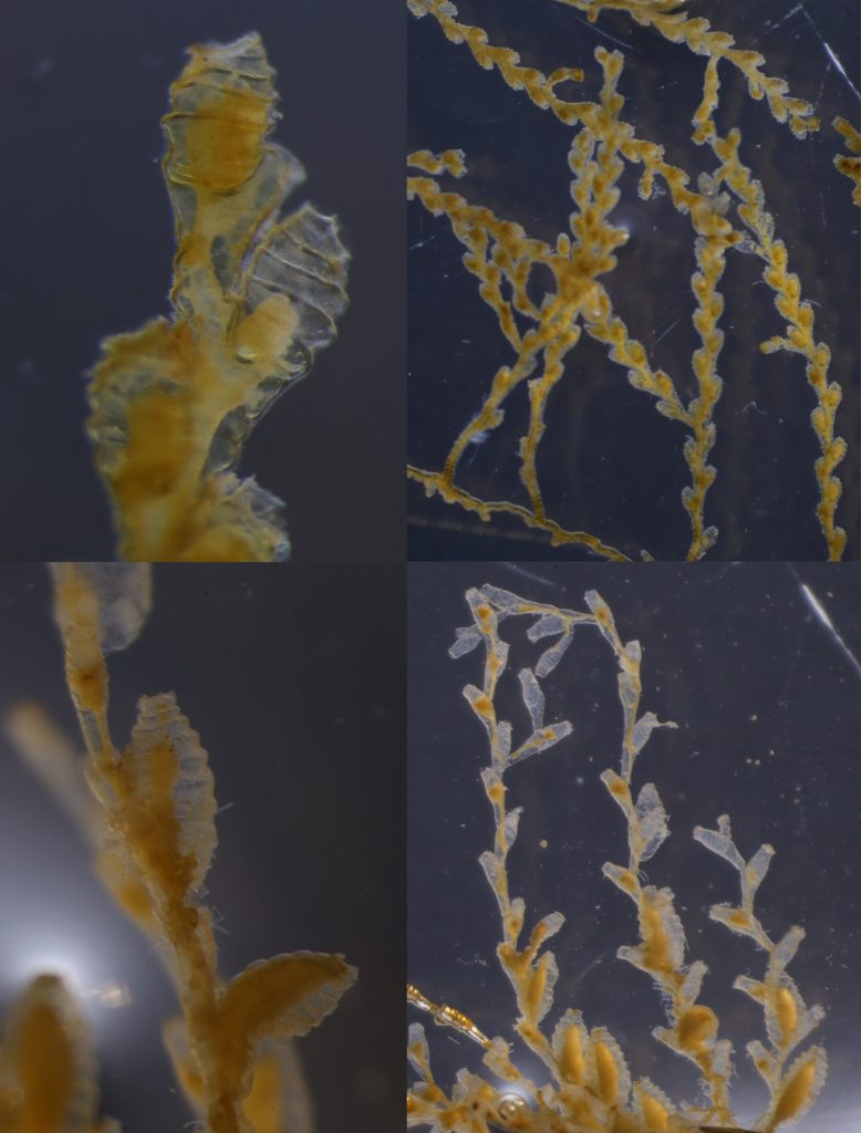
Sertularella rugosa (top row) is without a doubt one of my favourite hydrozoans. The hydrothecae resemble a bee hive and the colony has a zig-zag appearance. However, it is easy to confuse it with Sertularella tenella (bottom row). Pictures credits: Carla García.

Polyps of the family Campanulariidae. This common family is characterized by the presence of a bell-shaped theca. Pictures credits: Carla García.

During my stay, I also had the chance to go sampling on a research boat, which helped me to understand better the procedures and requirements that are necessary to collect hydrozoans. We used a wide-mouthed plankton net that went up and down at a constant speed to avoid damaging the jellies and other gelatinous organisms from the plankton. After sampling, we took the cod-end to the laboratory. There, the content of the cod-end was poured on a light table. Then, we selected interesting specimens (including hydromedusae belonging to genus Euphysa) with wide mouthed pipettes and transferred them to Petri dishes filled with fresh seawater to observe them better under a microscope.

Towing the plankton-net which went down to 650m to capture some gelatinous organisms. As you can imagine, going up and down such a long distance takes a lot of time, but it is never boring with colleagues like Aino Hosia (right). Picture credits: Carla García.

I was lucky enough to get samples of Euphysa aurata and Euphysa sp., but they did not want to pose for my photo and kept moving around. Picture credits: Carla García.

We took the opportunity to collect some shallow-water benthic hydroids just in front of the Marine Station. Picture credit: Carla García.

Last but not least, I worked at the DNA lab, which allowed me to gain experience in new molecular techniques that I had not used before and to adapt myself to different (and very modern) facilities.

This experience has been simply great for me. I loved the working environment and the fact that everybody was always there to give me a hand. I have learned a lot and I am taking with me many friends that I hope to meet again when I come back to Bergen.

If you want to know more about projects of NorHydro and HYPNO, visit NorHydro’s home page and Facebook page, and check the hashtags #HYPNO and #NorHydro inTwitter.


-Carla García-Carrancio

Hello Jorunna artsdatabankia; new sea slug for Norway and to the World!
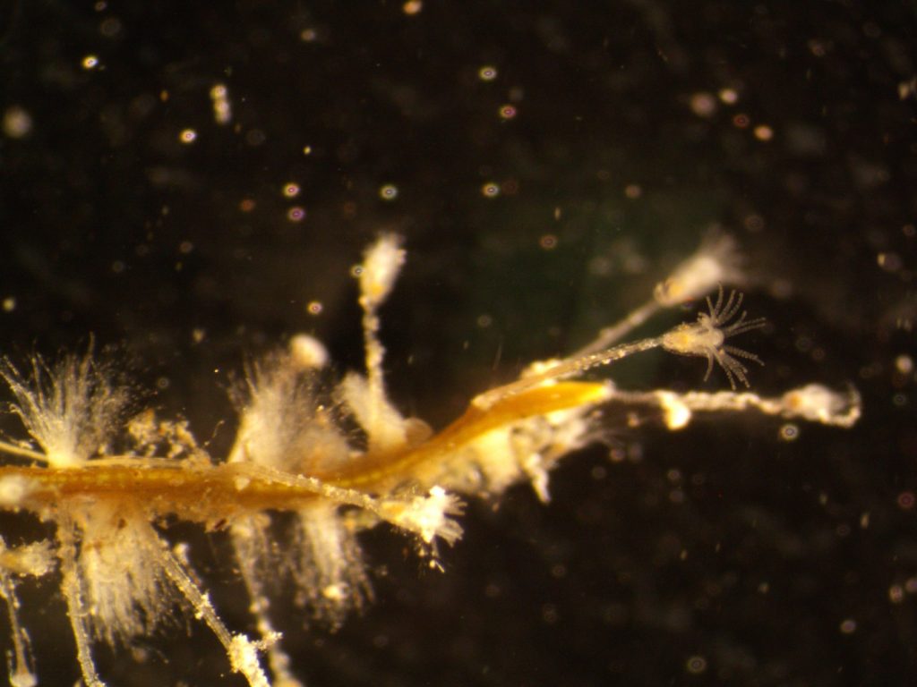
In 2018 former master student Jenny Neuhaus started working under supervision of Manuel Malaquias and Cessa Rauch on the sea slug species Jorunna tomentosa.

Jenny presenting her work on Jorunna tomentosa on the world malacology conference in the USA

It was known already for some time that this sea slug occurs in a wide variety of colour patterns (morphotypes). With the increased discovery of cryptic species due to improved molecular techniques we wondered if we were dealing with a single species or several cryptic lineages.

For a long time the different colours and patterns were regarded as natural variation within the species, consisting of shades of grey-white, cream-yellow, pale orange and either plain of blotched with light brown or chocolate brown spots of various sizes, distributed either irregularly or in lines, or combination of both!
But it was this variety that tossed up the question eventually whether we are dealing with a single species after all.

The diversity within Jorunna tomentosa

The nudibranch genus Jorunna consisted of eleven recognized species occurring in European waters. At that time, Jorunna tomentosa (Cuvier, 1804) was the only known species of this genus to be found along the Norwegian coastline. Prior to the study, the northernmost record of J. tomentosa was listed from Vestvågøy, Lofoten, Nordland. Today we know that the species is found at least 550 km further North in the Magerøysundet, Troms og Finnmark.

Jorunna tomentosa has an oval-elongate body shape with different colours varying from grey-white to cream-yellow and pale orange. They can reach a size up to 55 mm and occur at depths from a few meters down to more than 400m. They feed on sponges of the species Halichondria panicea, Haliclona oculata and Haliclona cinerea. J. tomentosa can be found from Finnmark in northern Norway, southwards along the European Atlantic coastline, the British Isles, the French coast, Iberian Peninsula, Mediterranean Sea up to Turkey, and the Azores and Canary Islands. Besides the species has even been recorded from South Africa.

Jenny Neuhaus in the lab of Prof. Marta Pola in Spain dissecting specimens for anatomical studies

Jenny compared specimens from different parts of the world, including Norway, Ireland, Spain, France, Portugal including the Azores and South Africa. She took tissue samples for genetic studies and dissected them for their anatomy.

For the genetic studies we selected three different gene markers called COI, 16S and H3 to check how these morphotypes compare with each other and evaluate the meaning of genetic distances.

From the genetic distance analyses, it became clear that we were dealing with a “cryptic species complex”, as a clade of three specimens showed substantial genetic difference compared to J. tomentosa but seemed morphologically indistinguishable from another at first glance.

As sea slug anatomy is a matter of complexity, especially since each animal possesses both male and female reproductive organs (hermaphrodite), the expertise of Prof. Marta Pola from the University of Madrid was essential to conduct detailed morpho-anatomical studies. We were able to detect structural differences in the rasping tongue (radula) and parts of the reproductive apparatus.

Meet Jorunna artsdatabankia!

Jenny sequenced the DNA of 78 specimens of which 60 where successful for using in the final phylogenetic analyses. Her results supported the presence of a new Jorunna species, and in addition a possible case of incipient speciation in J. tomentosa with two genetic lineages morphologically undistinguishable. The new Jorunna species was based on material collected from Norway (Kristiansund, Frøya & the North Sea).

Jorunna artsdatabankia

J. artsdatabankia has a plain white to yellow background colour accompanied by small brownish spots irregularly placed on the body surface. Its distributional range is so far restricted to Norway, being recorded from Skogsøya, Frøya (Trøndelag), Brattøya, Kristiansund (Møre og Romsdal), and a North Sea plateau (60.726944 0.505371) with a depth range from 27 to 350 meters, suggesting a sympatric occurrence with J. tomentosa.

Jorunna artsdatabankia in comparison to Jorunna tomentosa

The name attributed to this new species was chosen to recognize the work of the Norwegian Biodiversity Information Centre (Artsdatabanken) for their instrumental role promoting and supporting biodiversity research in Norway.

– Cessa Rauch, Jenny Neuhaus, Manuel Malaquias


Sea slugs of Norway Instagram: @seaslugsofnorway

Sea slugs of Norway Facebook: www.facebook.com/seaslugsofnorway


The paper can be found here:

The genus Jorunna (Nudibranchia: Discodorididae) in Europe: a new species and a possible case of incipient speciation. Jenny Neuhaus, Cessa Rauch, Torkild Bakken, Bernard Picton, Marta Pola, Manuel António E Malaquias (2021), Journal of Molluscan Studies, Volume 87, Issue 4, December 2021, eyab028, https://doi.org/10.1093/mollus/eyab028

On the study of the diversity of Euphysa in Norwegian Waters

NorHydro’s student and collaborator Carla García has just defended her MSc thesis! We asked her to share with us a little bit about her project and this is what she had to say:

During summer 2020 I started learning about hydrozoans through online chats with my supervisor, Luis Martell. At that time, I was not familiar with these curious animals but they caught my attention and by September that year I had already fallen in love with them so I moved to Mallorca to pursue a Master’s Degree in Marine Ecology and work with hydrozoan diversity. For my master’s thesis I worked in a case that was part of project NorHydro (Norwegian Marine Benthic Hydrozoa), led by Luis at the University Museum of Bergen. The global pandemic has put many limits and obstacles in the way of my project, but I have been able to cope with them successfully by combining presential lessons in Mallorca with online courses and meetings with my fellows in Norway.

Me in Mallorca sampling my first benthic hydrozoans (Pennaria disticha and Eudendrium sp.) Picture credits: María Capa and Carla García.

My research was focused in the diversity of genus Euphysa (family Corymorphidae), which had not been assessed in Norway before. I wanted to determine which and how many species of this genus are present in Norwegian and adjacent waters and to characterise them morphologically and genetically. The problem with these animals is that many Euphysa species are not easy to tell apart from each other and there is a big taxonomic controversy within this group. For example, Euphysa aurata is one of the most frequently reported species in Norway, but are we sure that all the records belong to the same species?

Detailed observations of the samples raised my suspicions about the possibility that a complex of species could be hidden within this species name. To try to solve this problem I used an integrative approach combining molecular (DNA barcoding) and morphological analyses (documenting diagnostic characters). The result of this was that we discovered a new species of Euphysa in the waters outside Bergen and we are in the process to describe it!

Medusae of Euphysa aurata (top row) and Euphysa sp. nov. (bottom row), the main taxa covered in my project and two easily confused species. Picture credits: NorHydro and HYPNO.

The first step in the approach I followed was the field work. I gathered samples of hydromedusae and polyps from different localities across the Northeast Atlantic waters and also used some preserved specimens from the University Museum of Bergen. Because last year’s restrictions during the pandemic, part of my samples had to be sent by post to Mallorca, where I carried out the second step: laboratory work. This part included all the steps that go from the DNA extraction to the sequencing of three specific DNA fragments (called barcodes), which allow us to identify and distinguish between species based on similarity with reference sequences from online databases. Then, I applied some phylogenetic analyses and species delimitation tools. What these techniques do is to infer the evolutionary history of the three DNA markers that I sequenced (COI, 16S, and ITS). The results are visualized in the form of phylogenetic trees that we have to interpret and to decide if they are reliable or not. Then, I used species delimitation software to delineate species boundaries. The last step was to look again at the morphology and search for diagnostic characters that allow us to differentiate the species of Euphysa.

Sampling with my lab partners Mariana Strauss and Raúl González. We were looking for hydroids, small worms, and goose barnacles for our respective research projects. Picture credits: María Capa and Carla García.

Each and every life form that make up our planet, no matter how small it is, is a key component to the functioning of the Earth as a whole, and for that they deserve to be studied. Knowing how many species inhabit the Earth has always been one of the main questions of science, so I am really happy to contribute to a better understanding of ocean biodiversity and concretely, to hydrozoan systematics.

If like me, hydrozoans have piqued your curiosity and you want to know more on the activities of NorHydro and my results, you can check NorHydro’s home page and Facebook page, as well as the hashtags #HYPNO and #NorHydro inTwitter.

-Carla García-Carrancio

On the Hunt for Tiny Polyps

Two weeks ago I had the chance to go field-sampling on the research vessel Hans Brattström. The sampling this time was focused on a broad range of marine invertebrates ranging from Hydrozoans, Bryozoans, Polychaetes, Phoronids and Brachiopods. I was especially on the hunt for polyps of the family Hydractiniidae (Cnidaria: Hydrozoa) that grow preferably on shells of molluscs or hermit-crabs. I was happy to look for new specimens for NorHydro and my master’s project, especially since opportunities to go field-sampling have been rare due to the covid-19 restrictions. The area of Bergen has been sampled quite well for the NorHydro project, but I was especially looking for rare species or species that haven’t been sampled before.

The first sampling for NorHydro this season – and with great conditions! Picture Credit: Lara Beckmann

To collect hydractiniids, we took bottom samples using a triangular dredge and a grab sampler. When the dredge gets back on board, the sample gets sorted on a large table on deck. Then the detailed search begins, and every stone and cranny gets inspected. The polyps I was looking for can be tiny, ranging from less than 1 mm up to 8 mm. The substrates that they grow on vary in size and shape, it can be crabs, molluscs but also algae or stones, often not larger than a few centimeters. So it isn’t an easy task to find the polyps in a freshly collected sample. Luckily I found several conspicuous hermit crabs and also one snail that I took back to the museum. At first, I didn’t see the polyps – only under the microscope in the museum laboratory I was able to see that hydractiniid colonies were growing on the shells.

Video: A polyp colony of the species Podocoryna areolata (Family Hydractiniidae). The polyps were growing on the shell of a living mollusc, probably of the species Steromphala cineraria. Video Credit: Lara Beckmann

One colony of the species Podocoryna areolata was growing on the shell of a living mollusk. The mollusk provides a nice substrate because the movements of the snail provide the polyps with more opportunities to encounter food. Also, the colony is protected by the small wrinkles of the shells surface where the polyps can hide. The polyps of this species are super difficult to measure, but most are smaller than 0.5 mm. When disturbed, the polyps shrink to small blobs even smaller than this. When relaxed, they can extend a bit longer in size. Especially the tentacles reach out to get hold of any potential food that swims by, such as small crustaceans. This species releases medusae, which can frequently be found in the plankton in this area.

A single polyp of the same colony of Podocoryna areolata. Picture Credit: Lara Beckmann

On shells inhabited by hermit crabs of the species Pagurus bernhardus, I found several colonies of a yet unidentified species of the genus Podocoryna. This species is very commonly found as polyp almost along the entire Norwegian coast. I’m still studying the specimen to figure out the correct identification. Since there is a lot of confusion in the hydractiniid taxonomy, I need to combine genetic information and morphology to overcome the existing problems in their identification and naming. The colony was reproductive and medusa buds were growing on it. Interestingly the medusa of this species is rarely found in the plankton.

Polyps of the genus Podocoryna. On the right are parts of the grasping claws visible belonging to the hermit crab Pagurus bernhardus. Picture Credit: Lara Beckmann

All over the colony were medusa buds. These are growing medusae, which will be released in the water when they are mature. The medusae can do what the colony itself can’t: releasing eggs and sperm and thus reproduce sexually. Picture Credit: Lara Beckmann

Besides the polyps, I found several other organisms living with the colonies on the shells including Crustaceans, Nudibranchia, Foraminiferans and other hydroids. The shells provide a home for a diverse range of marine life and it resembles a tiny forest. But it is not all peace and harmony in there, the smallest amphipods were quickly munched by the Podocoryna polyps. Those, in turn, get eaten by nudibranchs, that crawl on the colonies and some species feed specifically on hydroid polyps.

Video: An amphipod that lives on top of the hermit crab shell, walking through the colony of Podocoryna polyps. Video Credit: Lara Beckmann


I didn’t find any more hydrozoan species that were interesting for NorHydro during the sampling trip (at least not while scanning with the bare eye). But, I want to show one more very common species around Bergen –Ectopleura larynx– just because it is such a nice-looking hydrozoan. It even was reproductive and released its larvae right into my petri-dish. The small bulbs that grow between the polyp tentacles contain the larvae, which are called actinula. They break free and swim around, swinging their tiny tentacles until they will settle on a piece of algae for example, and grow to a large colony again.

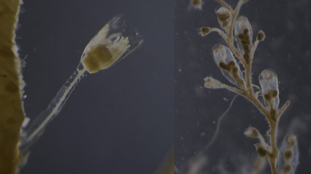
The species Ectopleura larynx is a common species at the Norwegian coast. On the left the released larvae, called actinula. On the right a polyp that usually grows in a large colonies with up to a hundred polyps. Picture Credit: Lara Beckmann

-Lara

You want to learn more about hydrozoans and why it is important to study them? Read more about it in my blog article for Ecology for the Masses: link.

Also, keep up with the activities of NorHydro here in the blog, on the project’s facebook page  and in Twitter with the hashtag #NorHydro.

What’s growing on the shell? – Insights into the Diversity of Hydractiniidae in Norway

This summer I started my master degree project at the University Museum of Bergen and joined the research of Luis Martell and Aino Hosia. I’m a student in the program ‘Biodiversity & Systematics’ at the University of Stockholm and for my 1-year thesis project I wanted to learn more about hydrozoans. Looking for hydrozoan biologists, which there are not so many, I came across the project NorHydro led by Luis and Aino and I decided to go to Bergen to study and learn more about this fascinating group.

My thesis project especially focuses on the diversity of the hydrozoan family Hydractiniidae. Most commonly hydractiniids are encountered on snail- or hermit crab shells where they can build a mat of polyps. Those shells are often inhabited by other animals and the polyps feed on the left-over meals of those and in return defend their host against predators. With their small tentacles, which are equipped with hundreds of cells containing venomous stingers (the nematocysts) they are great predators and catch tiny animals from the plankton.

Schuchertinia allmanii growing on Pagurus pubescens hermit crab. Picture credit: Bernard Picton

Each individual polyp is connected to each other and build a colony of polyps with different functions. Some act as food suppliers, some as reproductive polyps. From those polyps some hydractiniid species grow tiny medusae (jellyfish) which will be released in the water column. The medusa is morphologically comparable to the polyps, equipped with tentacles, a stomach, gonads and nematocysts – but living upside down and not attached to the sea floor. With their ability to swim around they contribute to disperse themselves in the water and can produce a lot of offspring.

Video of a Pagurus bernhardus hermit crab. A hydractiniid polyp colony is growing on the underside of the shell. Video credit: Lara Beckmann

Several species in this family are often used in development biology or immunology research. For example the species Hydractinia echinata led among other organisms to the discovery of stem cells and is still widely used as a so-called model-organism nowadays. This is because this animal group shows a high ability to regenerate and if you cut a hydroid polyp in two pieces, it will just re-grow again to its initial state.

The life-cycle in hydractiniids can include a polyp and medusa stage. Species of the genus Podocoryna release medusae from their colony. Other genera in this family develop only reduced medusae and lack a free-swimming jelly. Illustration credit: Lara Beckmann

The hydromedusa Podocoryna borealis is released from a polyp colony that can grow on various substrates such as snails or worm tubes. Picture credit: Lara Beckmann.

But despite this spotlight to some species, the diversity of this family is still poorly known and there is little attention to hydractiniid species because of their inconspicuousness and difficult identification. To highlight the problem I did some research: how many and what species are commonly recorded in Norway? Which species could potentially occur in this region according to species descriptions? In private observations and ecological surveys only three species were commonly recorded. However, our preliminary results suggest at least 6 species in Norwegian waters with some surprisingly frequent species that are rarely recorded in surveys. This indicates, that there happens a lot of misidentification in the field and we need to get a better estimate of the true diversity of Hydractiniidae in this region in order to improve awareness and to prevent misidentification in the future.

Two polyps of a colony of the species Clava multicornis. You can see reproductive units (so-called sporosacs) growing on the polyp body, which will release eggs or sperm when fully grown. Picture credit: Luis Martell

In order to do that I study this group in much detail. A great advantage of being a taxonomists in these days is that we can use a variety of different sources of information to develop and test for species hypothesis. This so-called integrative approach includes morphological, ecological and genetical information in order to define taxa diversity from several perspectives.

The first step in this process is to gather polyps and medusae of Hydractiniidae from various spots in Norway ranging from Olso, via Bergen and up north to Bodø. I will use already collected samples from previous years and hopefully will also be able to add some more samples from field-trips in the next months.

Getting the plankton-net back on board – and Maryam and me trying to open the jar carefully without spilling the fragile plankton inside. After removing the jar from the net, we directly pick the hydrozoans and put them in a cold environment, so that we can look at them alive when we are back in the lab. Picture credit: Maryam Rezapoor

With fresh material the first step is to take pictures of the living animal. Since hydrozoans tend to shrink and lose a lot of it characteristic traits in the ethanol preservative it is important to take pictures right away when the animal is still alive. Furthermore the samples are identified using a microscope, identification keys and additional literature. With additional collected data such as habitat, location, sample depth and so forth we create a large database for our samples and develop so-called e-vouchers: an electronic data storage for each individual to ensure to gather and store as much information as possible. That makes it possible to go back to the database and look at the specimen data e.g. where did it live? On what substrate was it growing on? How deep was the sampling site? This way I don’t only rely on the preserved material but can easily use the e-voucher material and the additional collected data that can be useful for further analyses.

Besides the morphological and ecological data I also include molecular information: what does the DNA tell us about the specimen? For that I use three different genetic markers (short DNA fragments, or genes) to see how those differ in their sequence of bases between the sampled hydractiniid specimen. In the course of evolution, DNA sequences change due to selection, genetic drift, gene flow and random mutations. That is why we are able to infer evolutionary relationships between biological entities – in this case we are interested in species – and with mathematical models and some simplified assumptions we can delimit species based on their molecular attributes.

Me in the DNA lab waiting for the first results of lab work for this project! Picture credit: Maryam Rezapoor

In order to do so, I will process the sampled material in the lab. First, I extract the DNA from a tiny piece of polyp or medusa tissue and then amplify the specific target region using PCR (Polymerase-Chain-Reaction). This short DNA fragment will further be sequenced which outcome I will check, process and analyze using different computer programs and algorithms. The final results will be summarized and visible in so-called gene trees, revealing the evolutionary history of those three different DNA markers. I will use the gene genealogies to delimit the species and observe how much diversity of hydractiniids we have in Norway.

Using specific genes also facilitate us with species barcodes which are used to create a reference library. This in return enables researchers to identify a specimen without the need to identify the sample using its morphology. This can be the case e.g. for metabarcoding projects (sequencing a mass of DNA from unknown origin) or if samples are unidentifiable. The scientist will then sequence the barcode gene and compare it to the reference library and will eventually be able to tell the species. Those barcodes can be informative in biodiversity studies but by no means substitute taxonomists, which are still needed to describe and identify species when no DNA is available.

A monograph of the Gymnoblastic or Tubularian hydroids by George James Allman, published in 1870. I found this book in the library of the Tjärnö laboratories in Sweden. Allman was a pioneer in hydrozoan research and described many species. Nowadays it is much easier to access original descriptions since many book are available online. You can find a PDF* of this book in the Biodiversity Library. Picture credits: Lara Beckmann

Also, the DNA does not help us when we have no reference we can map the specimen to in order to determine the species. That is why this master project also aims to create a barcode-library for the species in the family Hydractiniidae.

What I really like about this project is that it contributes to our basic understanding of the diversity in our oceans – in this case even in two completely different environments: the water column as well as the seafloor.

Even if hydractiniids are just a little part of the ecosystem, they play their role and influence the ocean in their own way. Also, my work is super diverse and I need to be an allrounder in many respects: trying to find species descriptions I search and read taxonomic literature from the 19th century (coming in Latin, German, English, Swedish or French). Then I work in the laboratory where I combine traditional working methods like drawing, microscopy or photography with modern techniques from DNA sequencing to computational science and bioinformatics.

I love this kind of work, and it is great to contribute just a little bit to reveal the mysteries of our oceans.


– Lara

You want to learn more about hydrozoans and why it is important to study them? Read more about it in my blog article for Ecology for the Masses (link).

Also, keep up with the activities of NorHydro (link to project home page) here in the blog, on the project’s facebook page (link) and in Twitter with the hashtag #NorHydro.

*Link to pdf of A monograph of the Gymnoblastic or Tubularian hydroids by George James Allman