Category Archives: DNA barcode

World Jellyfish Day pt 2: OceanX – gelatinous predators

This summer, researchers from the University Museum of Bergen participated on a research cruise onboard R/V OceanXplorer, the research vessel of OceanX. OceanX is a nonprofit ocean exploration organization founded by Mark and Ray Dalio in 2018. The organization’s mission is to “explore the ocean and bring it back to the world”, and the vessel is designed and constructed by OceanX to conduct ocean exploration, scientific research, and document stories of discovery to share with the public worldwide in a state-or-the art way.

One of the outcomes of the cruise was this amazing short documentary focusing on the work Aino Hosia and the rest of our “Team Jellyfish” (Joan & Luis) were doing onboard.

Today being the World Jellyfish Day, we wanted to share it here – enjoy!


Links:

OceanX home page: https://oceanx.org/

OceanX Youtube channel: https://www.youtube.com/@OceanX

Project ParaZoo: there is a critter inside my jellyfish!

ParaZoo (complete name ‘Metazoan parasites of non-crustacean zooplankton’) is one of the most interaction-focused projects currently running at the Invertebrate Collections of our Museum. This project, funded by the Norwegian Biodiversity Information Centre (Artsdatabanken), aims at studying the different animals that live together inside and on the surface of Norwegian jellyfish. This means that for the next two years we will be looking for tapeworms, flukes, roundworms, and amphipods as ParaZoo tries to answer the question of which of these organisms are associated with gelatinous hosts in Norwegian waters.

ParaZoo is focused on animal parasites and symbionts associated with jellyfish. Thse parasites include roundworms like Hysterothylacium aduncum (left, in Euphysa aurata), amphipods like Hyperia medusarum (middle, on Aequorera forskalea), and flukes like the members of family Didymozooidae (right, in Beroe gracilis). IC: Aino Hosia (left), Katrine Kongshavn (middle), Joan J. Soto-Àngel (right).

Besides jellyfish, arrow worms (Chaetognatha) are also members of non-crustacean zooplankton that host different types of parasites, like this H. aduncum roundworm (Nematoda). IC: Joan J. Soto-Àngel, Luis Martell.

Parasitism and symbiosis are extremely common life styles in the animal kingdom. In fact, some researchers believe that there may be more species of parasites than of free-living animals, given that each free-living species hosts many species of parasites (most of them unique) and those parasites also host their own parasitic tenants. Marine zooplankton is no exception to this trend, and many parasites and symbionts are expected to occur in copepods, krill, and gelatinous zooplankton. Jellyfish and arrow worms, for example, may be important hosts for flatworms and other helminths, yet our knowledge of these animals in Norway is very scarce.

ParaZoo’s logo includes two of the target taxa of the project: roundworms (Nematoda) and hyperiids (Amphipoda). The third main parasitic group covered is flatworms (Platyhelminthes), illustrated by the larvae of Derogenes varicus parasitizing Halopsis ocellata shown in the right side of this figure. IC: Joan J. Soto-Àngel, Luis Martell.

Understanding zooplankton parasites is important because many of them are going to be transmitted to fish, where they may cause serious diseases. To get a better overview of which critters live in non-crustacean zooplankton, ParaZoo will sample, record, and DNA-barcode specimens from all over the country. The collected animals will be included in our museum collections after being identified, documented photographically, and fixed in ethanol. We will then generate an open-access database of information including pictures and DNA sequences that will help with the identification of the parasites. Aquaculture facilities, fishermen, and managers of marine areas will benefit from this database to better plan and counter potential negative impacts caused by the parasites.

Flukes, such as Opechona spp, parasitize gelatinous zooplankton (in this case a sea-gooseberry Pleurobrachia pileus) as larvae called metacercariae. IC: Joan J. Soto-Àngel, Luis Martell.

The larvae of tapeworms (Cestoda) sometimes use jellyfish to reach their definitive hosts: fish. IC: Joan J. Soto-Àngel, Luis Martell.

ParaZoo is committed to present the diversity of jellyfish parasites to all those not familiar with them. In order to do that, we will regularly write entries here on the blog, as well as participate in several academic and not-academic meetings. The official info webpage for the project is available here, so don’t forget to check it out!

Luis

Student visit – Ana González

MSc student Ana González visited the collections last month as part of project NorHydro, where she spent some weeks in the lab working with her samples. Here is an account of her experience:

The challenge of identifying benthic hydrozoans
Hydrozoa is a fascinating but poorly understood group of invertebrates, in part because their identification is not always an easy task. I have been studying benthic hydrozoan communities for over a year now, in particular those living in the shallow waters of Mallorca (Spain), and I have realized that the diversity of forms and structures in the group is higher than I had imagined at the beginning of my studies, and their identification is more difficult than I expected. The assemblages of hydrozoans in the Mediterranean are of course very different from the ones that occur in Norway, but something that both communities have in common is that morphological identification of the animals (i.e. telling which species is present based only on the characteristics we can observe) is challenging, which is why one of the aims of my visit to the University Museum of Bergen last December was to learn a different technique (DNA barcoding) that can help me improve the identification of my samples in cases when the morphology of the specimens is not good enough.

Some of the morphological characters that are used to identify benthic hydrozoans. On the left side a member of Campanulariidae, with a stolonal colony, and on the right side Monotheca obliqua with an erect colony.

DNA barcoding consists in finding a short DNA sequence (the barcode) that is similar for all members of one species but different from all other species. It is a relatively recent tool that –among other things– has helped the scientific community identify specimens that for one reason or the other cannot be identified based on how they look. In some groups, such as many colonial invertebrates, this technique has become a key asset because the colonies are often too young or not reproductive, or the important characters for identifications may be found only in one stage of the life cycle and not in others. For this visit I had the chance to bring all my samples from Mallorca to Bergen and I set to extracting the DNA of selected specimens, amplifying two different barcode genes (COI and 16S), and obtaining clean sequences for them. I discovered that, when it comes to DNA barcoding, every step of the process is important, and being patient and careful is essential.

Me at the DNA lab, running the electrophoresis for my samples.

Getting good results in the DNA lab depends on several factors like not forgetting any step and avoiding contamination as far as possible, but the work does not end there: once you have your sequences they have to be cleaned, quality-checked, and finally compared with others. This means that having a complete and trustworthy database of DNA barcodes is necessary, especially if you want to use the sequence to help you corroborate the identification of a specimen. When done right and with a good database, the DNA barcodes can be useful to detect differences between hydrozoan assemblages growing in different parts of the world or between different substrates and levels of anthropogenic impact, which is what I am doing in my MSc project.

Left: Clytia sp growing on the marine plant Posidonia oceanica. Center: A polyp of Halecium sp, one of the most difficult genera of Hydrozoa to identify based only in morphology, especially when the colony is not reproductive. Right: Eudendrium sp., found in harbours in Mallorca in high abundances.

The analysis of DNA sequences is a powerful tool to compare specimens of distinct populations and in some cases animals that apparently belong to the same species turn out to be completely different (e.g. cryptic species). This is not uncommon for benthic hydrozoans, which have high morphological diversity but also high levels of plasticity, resulting in colonies from different species sometimes being very similar to each other when they grow in similar substrates. As useful as DNA analyses are, however, it is also important to consider their limitations. For example, while the abundance of each species in a given community is important to describe the ecological status of a habitat, estimating abundance is still not always possible from sequence reads in DNA analyses.

Many cryptic species have been discovered in Aglaopheniidae thanks to the combination of DNA barcoding and morphological analysis

The use of DNA barcodes in my work is not limited to my current project, as I hope my identifications and sequences will help a little bit to improve the databases for future studies of hydrozoan communities in the Mediterranean Sea, and maybe even allow other researchers to compare their samples with the species found on other parts of the world. I think that looking closely at each specimen is the best way to truly know variation, so both morphology observations and DNA analyses should be combined to obtain good estimates of the diversity of a taxon in any locality. For example, whenever the DNA analyses reveal differences in two clades that were thought to be the same species, it is time to search for new taxonomic characters that we might have missed before, and for that reason it is also important to have a good knowledge of the morphology of each species. Both morphological and DNA-based identifications have limitations and advantages so, if you have the opportunity to use both, why choose only one?

Ana

Final workshop for hyperbenthic copepods (HYPCOP)

Our first international workshop with from ltr; Anders Hobæk (NIVA), Cessa Rauch & Jon Kongsrud (UMB), Tone Falkenhaug (project leader, IMR), Alexandra Savchenko & Rony Huys (NHM), photo by Alexandra Savchenko

During the last week of September, HYPCOP organized its last and crucial workshop for finishing the project. We invited international collaborators Prof. Dr. Rony Huys and Dr. Alexandra Savchenko from the Natural History Museum in London. Prof. Dr. Huys is a well-known copepod taxonomist and crustacean researcher and published a multitude of species descriptions and books including key identification guides. We were very happy to hear he had time to come and travel to Bergen, paying us a visit while also helping us with species identifications of the many, many copepods we had collected during the two years of our project.


During the two years of the HYPCOP project we collected around 600 specimens from different localities all over Norway, including shallow coastal waters and the deeper parts of the mid-Atlantic Ridge (Loki’s Castle field of active hydrothermal vents). From all those specimens we extracted DNA from the soft tissue of the animal. Therefore, keeping the hard exoskeletons, for morphological identification downstream. This is the most time consuming and challenging part. The species can sometimes only be identified based on minuscule differences in the appearance of its legs. Besides, one needs good taxonomic competence to assign these differences to the thousands of marine benthic copepods species. And this is where the HYPCOP team needed help.

HYPCOP started in May 2020, when a lot of countries, including Norway, were in a lockdown and international travel was difficult or even impossible. Therefore, it was problematic for HYPCOP to invite international researchers for most of the time. Thus, we focused mostly on extracting DNA from our collected specimens and building up a barcode library. But what was missing was the nomenclature of the bulk of the specimens. When finally, our first international researchers could come and have a look at our specimens, it turned out to be an enormous task. With the help of Prof. Dr. Huys and Dr. Savchenko we managed now to have almost 300 assigned names to our DNA library of 500 specimens. Quite a few of those are new species and even new genera.

Kickoff of the workshop, which would take place at Marine Biological Station Espegrend for the duration of a week, photo by Alexandra Savchenko

Rony and Alexandra arrived Sunday evening in Bergen together with project leader Tone Falkenhaug and project technician Cessa. We were stationed at the Espegrend marine biological station in Bergen for the entirety of the week. It was for Tone and Cessa the first time they would finally meet Rony and Alexandra in person, after many months of digital communication. It was a nice relaxing first evening. The next day Anders Hobæk from NIVA and Jon Kongsrud from the UiB joined and we started off the week with a presentation overview of the project.

The overview informed everyone about the program of the week and the state of the art of the project. With the DNA barcode library, we managed to construct a COI phylogenetic tree. Some of the larger clades were already identified down to species level, but many more species names were missing from the smaller clades. It was up to us that week together with Rony and Alexandra to identify these last cases.

Alexandra onboard research vessel Emiliana, photo by Tone Falkenhaug

We also had one day of fieldwork planned, to have us work also with some fresh material. This we did with help of research vessel Emiliana and the Beyer’s sled. Both stationed at Espegrend Marine Biological station. We tried to pick out a nice and dry day for going out with the boat and that happened to be in the mid of the week. We went a little bit outside of the Biological Station, with a depth of around 90 – 120m. The Beyer’s sled is an epibenthic sampler, it is called a sled for its form. We got many fresh samples, but due the net being a little large in its mesh size, we did not get as many small species as we liked.


Therefore, we also tried another sampling method with help of Anders; he had brought with him a light trap. Light traps are very easy to DIY with a bottle and inverted bottle opening, like a funnel, and a small led light on the bottom. You install the trap in the water overnight; the little led light attracts a lot of small hyperbenthic and planktonic (and some bigger) species.

Everyone working hard at the Marine Biological Station Espegrend, assigning species names to specimens, photo by Cessa Rauch

The entirety of the week consisted of many hours working at the microscope, going through literature, dissecting specimens, and assigning species names to the specimens. Eventually with help of Rony and Alexandra, we managed to assign 298 scientific names to 702 specimens in our collection. From those specimens, we extracted DNA from 593 specimens and produced a DNA library, which we uploaded to the BOLDSYSTEMS (Barcode of Life Data System). This library also has all the metadata of our specimens, such as location, depth, size, and pictures of the specimens (either life, fixed and in some cases parts). And it will be publicly available at the end of the HYPCOP project.

The week was demanding but very rewarding and we got many specimens identified, with even a few new species and genera to Norway and possibly new to science; all thanks to the hard work and help of Rony and Alexandra. We therefore also would like to take this opportunity to thank them again for their time and efforts in helping the HYPCOP project move forward! Until next time.

Rony Huys and Alexandra Savchenko helping the HYPCOP project move forward, photo by Tone Falkenhaug

– Cessa

Unraveling copepod secrets one leg at a time

A blog by HYPCOP

Hyperbenthic copepods (HYPCOP) are a very difficult and diverse group to work with, and identification goes painstakingly slow, because some species can only be distinguished from one another based on small details in some of their tiny legs. As of now, we have no specialists in marine benthic copepods in Norway and our greatest resource is our collaborator Anders Hobæk and the detailed drawings of G.O. Sars from the early 1900s .

Working together under guidance of G.O. Sars and Anders Hobæk

Anders is a senior researcher scientist at the Norwegian Institute for Water Research (NIVA) here in Bergen. He is specialized in copepod taxonomy, but his focus was mostly on freshwater copepods, or marine pelagic copepods. Which makes the marine benthic copepods just a little bit more challenging to work with, however, his skills are transferable and so we get together multiple times a year to work on our collection of benthic copepods to dissect them and identify them.

Beginning of June, we had again one of those get togethers in Flødevigen at the Institute of Marine Research (IMR), where Tone Falkenhaug, the project leader of HYPCOP, is situated. For a week we went through the main clades and groups of species that we had DNA barcodes of but not yet a confirmed species name. A lot of the identification was done with help of the rich and detailed illustrations of G.O. Sars1 published work in 1901 – 03 and 1919 – 21, “An account of Crustacea of Norway”

Detailed copepod drawings from G.O. Sars

Sars dedicated a lifetime of identifying and describing a variety of species and he did not neglect the rich and wonderful group of bottom dwelling copepods. Every species he encountered in those early days he described and drew in detail; he did not leave out the smallest details, that as of now, turn out to be of uttermost importance in determining the species. With small copepods you need a good microscope and fine tools. The first thing to look at is the general shape, is it very dorsally flat, like Peltidium purpureum, or more dimensional like Harpacticus flexus?

Sex is also an important feature; females are often characterized by carrying eggs; one egg sack or two egg sacks can already lead you in the right group. Males have often larger antennule made for holding on to females when mating, and other specialized tools that can be species specific. The little claws, called maxilliped, are they large, small, almost invisible? What about the first pair of legs? The second, third and fourth? The fifth pair of legs is often very characteristic for the species and in certain females, like Thalestris longimana, can be a huge in comparison of the rest of its body.

Thalestris longimana, females of this species has relatively large fifth pair of legs

Our work has a continues workflow consisting of, collecting the copepods, extracting their tissue for DNA barcoding, and keeping the exoskeleton. Once the DNA is successfully sequenced, we can take the exoskeleton and dissect the animal leg by leg to finalize the identification. That way the copepod is identified based on its DNA and morphological features, as this is not always mutually exclusive. DNA can be tricky as you need a good reference library to find the correct species, which is as of now, not complete, or even lacking for many species. Besides, there is such things as DNA contamination, cross contamination between species, therefore you always must look at the morphology to exclude that the DNA gives you the wrong species. Together with images of the animals we are building up a valuable reference library of DNA sequences and a museum collection of dissected animals on fixed slides. This way copepod diversity will continue to be valuable for future generations top study.

Working under the eyes of G.O. Sars

-Cessa


1Sars, G. O. 1901-03. An Account of the Crustacea of Norway. Vol. IV. Copepoda Calanoida.- Bergen Museum, Bergen & Christiana. 171 pp. + 109 plates Sars, G. O. 1919-21. An Account of the Crustacea of Norway. Vol. VII. Copepoda. Supplement. – Bergen Museum, Bergen & Christiana. 121 pp. + 74 plates

Fieldwork for two projects

The projects HypCop (bottom-associated copepods) and Hardbunnsfauna (Invertebrate fauna of marine rocky shallow-water habitats) went on a day-trip to three localities last week.

We made the most of the sunny and calm weather to visit a very exposed site on Sotra, where we collected in the tide pools and on the barnacle-encrusted intertidal.

Afterwards, we went to two marinas, Glesvær and Hjellestad, on a quest for some specific species the projects were in need of.

Back in the lab we set to work documenting the colours of the animals by photographing them alive, as the colours tend to face in fixatives.

It was nice day in the field, and it looks like we found the species we were after!

Follow us on Twitter and Instagram as @PlanetCopepod and @Hardbunnsfauna

– Jon, Cessa & Katrine


Bryozoa-workshop at Espegrend

February 14th -18th 2022

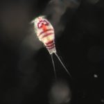
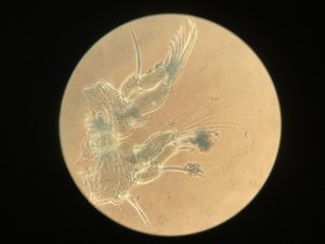
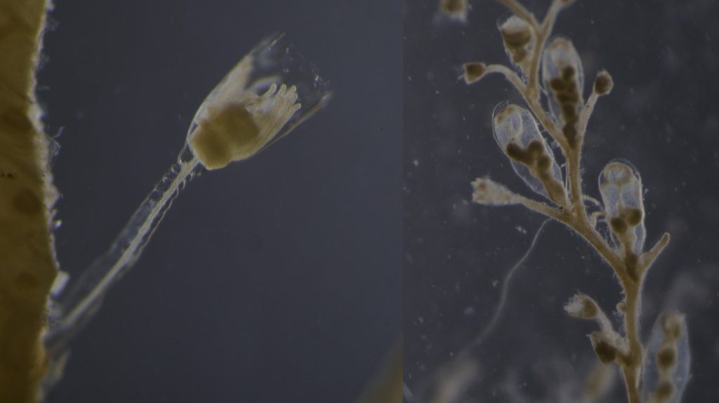
The Bryozoa are maybe not the most famous of animals, so let’s start with a quick rundown: Bryozoa, also known as Polyzoa, Ectoprocta, or moss animals (mosdyr, på norsk) are a phylum of aquatic invertebrates. Bryozoans, together with phoronids and brachiopods, have a special feeding structure called a lophophore, a “crown” of hollow tentacles used for filter feeding, which you can see in action in the video Tine captured:

In Norway we have 292 species registered, of which 281 are marine (Kunnskapsstatus for artsmangfoldet 2020, pdf here). It is estimated that the actual number of species present is higher. Further, several known species are considered “door knocker species” that may establish here within the next 50 years.

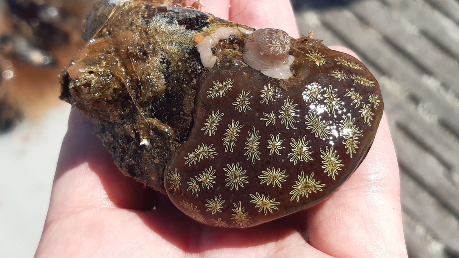
Bryozoa mostly live in colonies made up of tiny individual animals called zooids, which grow in a variety of shapes, and some of them provide structural habitats for other species. They are food for many other animals, namely nudibranchs, fish, sea urchins, pycnogonids, crustaceans, mites and starfish. Marine bryozoans are often responsible for biofouling on ships’ hulls, on docks and marinas, and on offshore structures. They are among the first colonizers of new or recently cleaned structures, and may hitchhike to new places with marine traffic. (Bonus: they have a super interesting fossil record, and this can be used to tell us more about the world in the way back!)

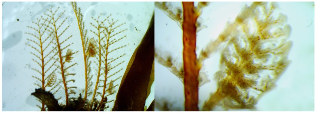
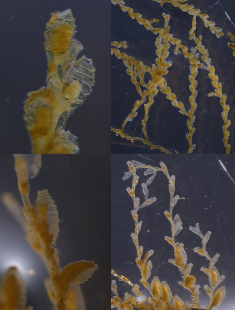
A few of the shapes the colonies can grow in. Pictured are 1: Membraniporella nitida 2: Bugula sp. 3: Flustrella hispida 4: Crisia eburnea

They are one of the focus groups of Hardbunnsfauna: there is still a lot we do not know about them!

Ernst Haeckel – Kunstformen der Natur (1904), plate 23: Bryozoa. Public domain, accessed through Wikipedia

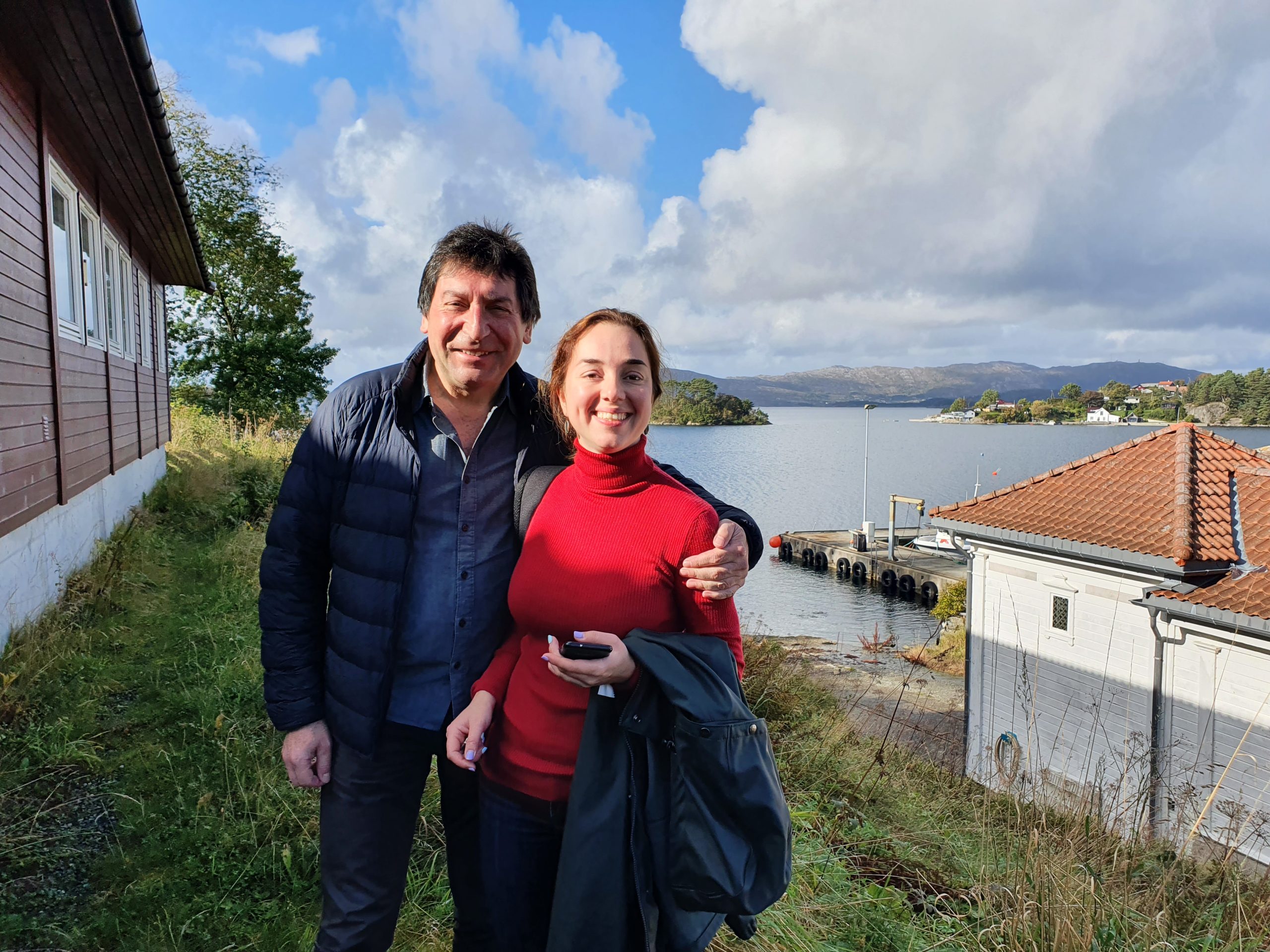
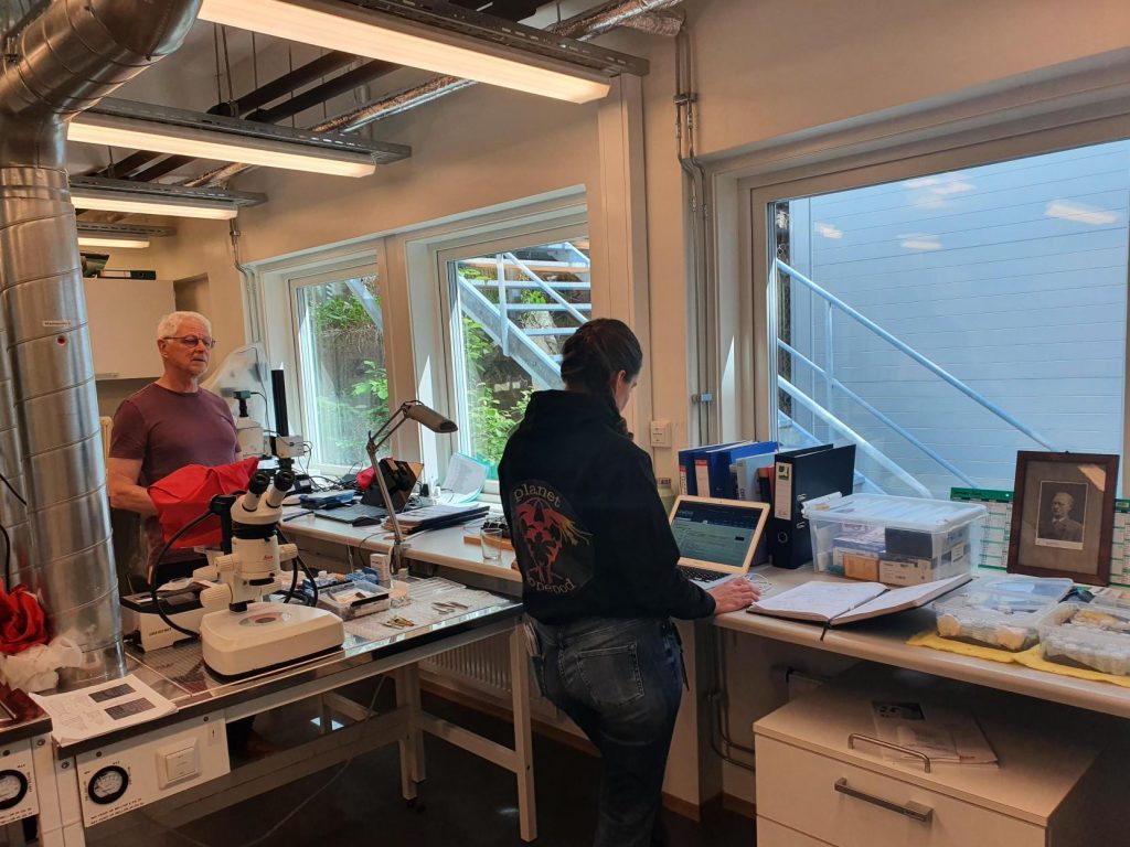
Planning in a pandemic is not easy, and we have had to postpone our plans for this gathering several times. The second week of February we could finally gather our “Team Bryozoa” here in Bergen for a week of in-depth studies of these fascinating animals.

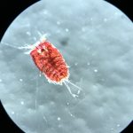
Team Bryozoa (centre), from left Piotr, Mali, Jo and Lee Hsiang, and some of the animals they studied. Group photo by Piotr Kuklinski

In total we were 11 participants;
University Museum of Bergen: Endre, Jon, Tom and Katrine,
Natural History Museum in Oslo: Lee Hsiang and Mali,
NTNU University Museum: Torkild, Tine (MSc. student) and Tiril (MSc. student),
and our two visitors from abroad:
from the Institute of Oceanology, Polish Academy of Sciences came Piotr,
and from the Heriot Watt University (Orkney Campus), Joanne.

The main focus of the workshop was to get as many samples and species as possible identified, work though the DNA barcode vouchers from samples submitted in advance and reach a consensus on which species the dubious ones were, to network with our colleagues, and to include the students in the work and the team. It all went swimmingly, and a we had a very productive and enjoyable week!

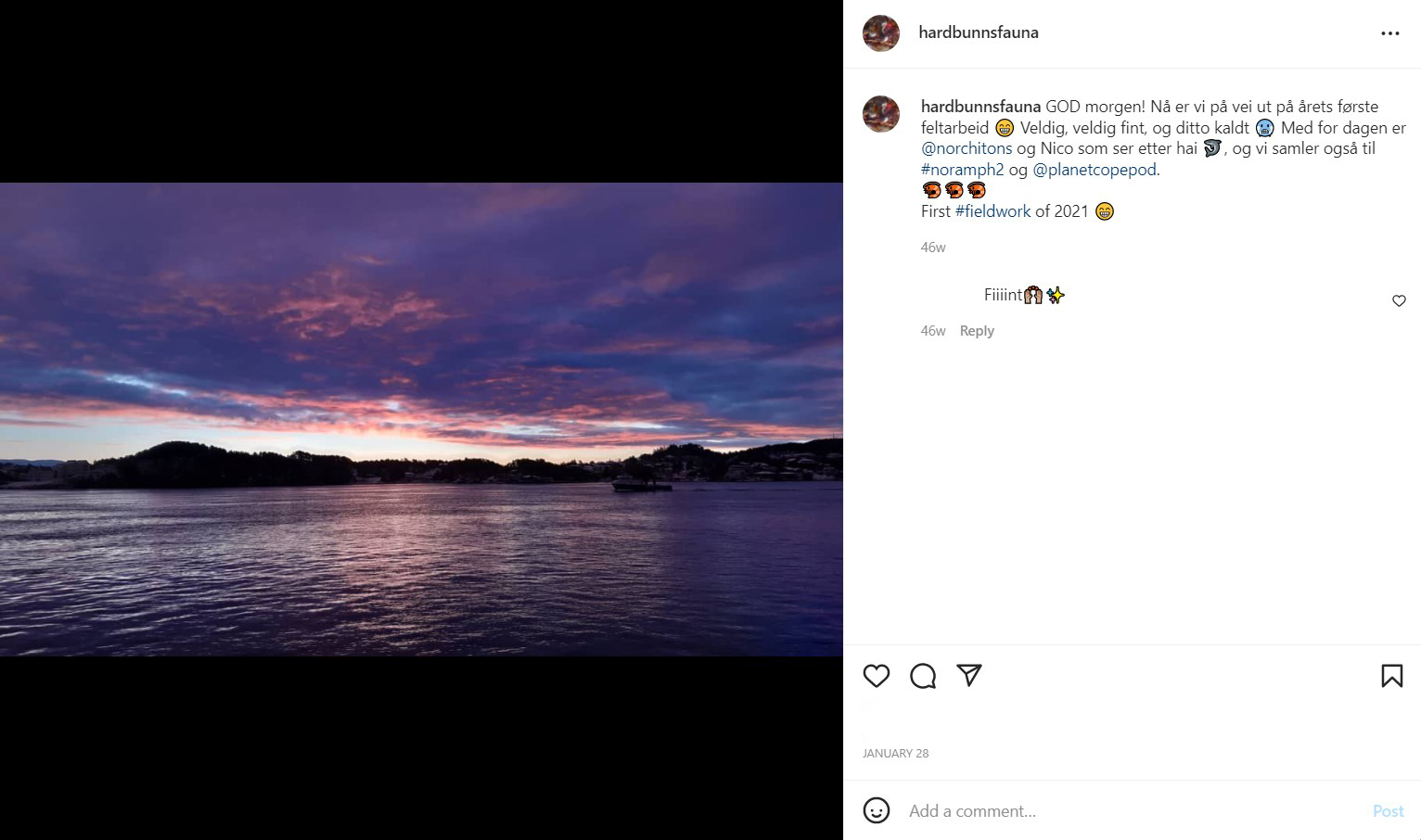
check out @hardbunnsfauna on Instagram for more!


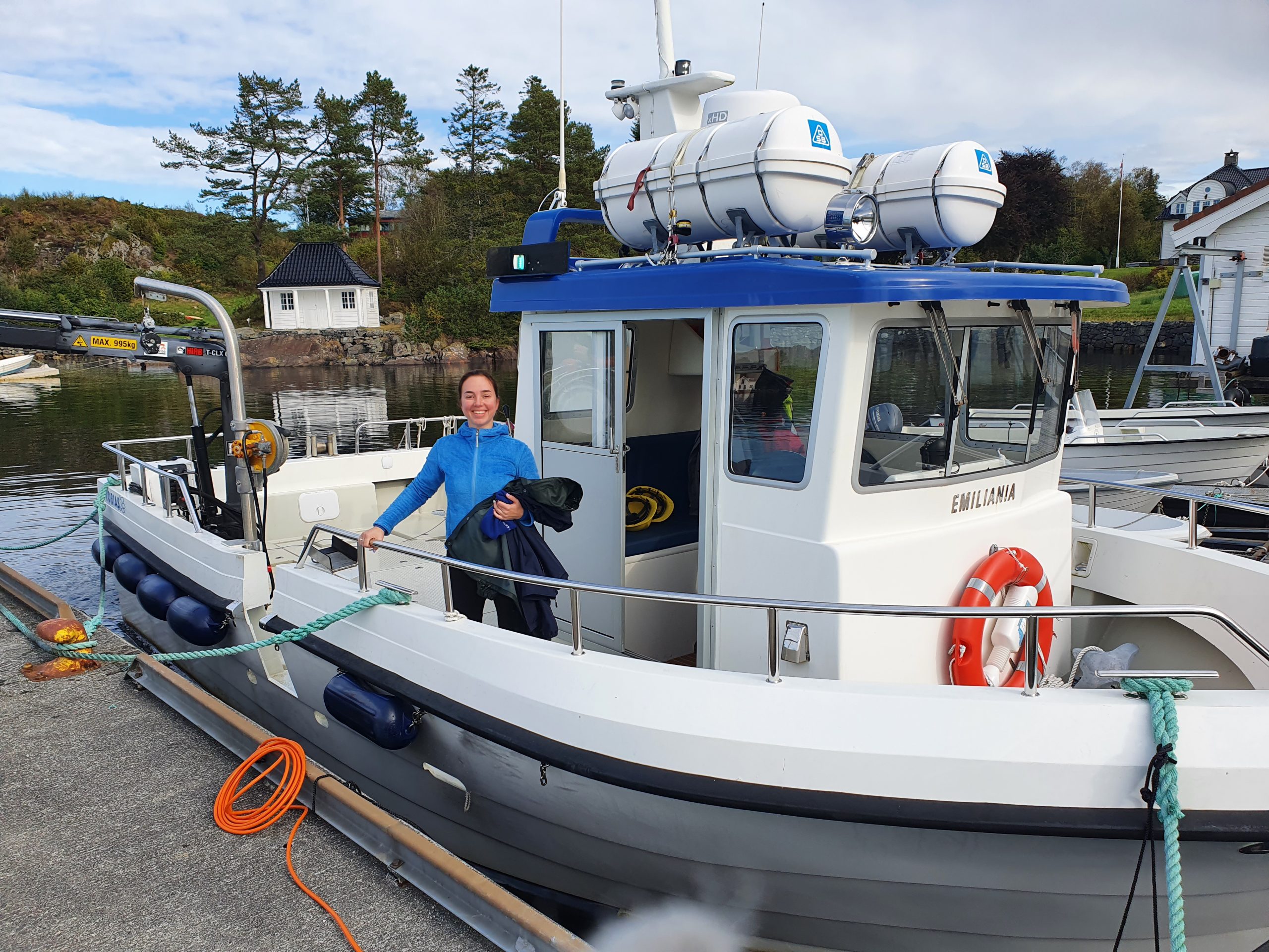
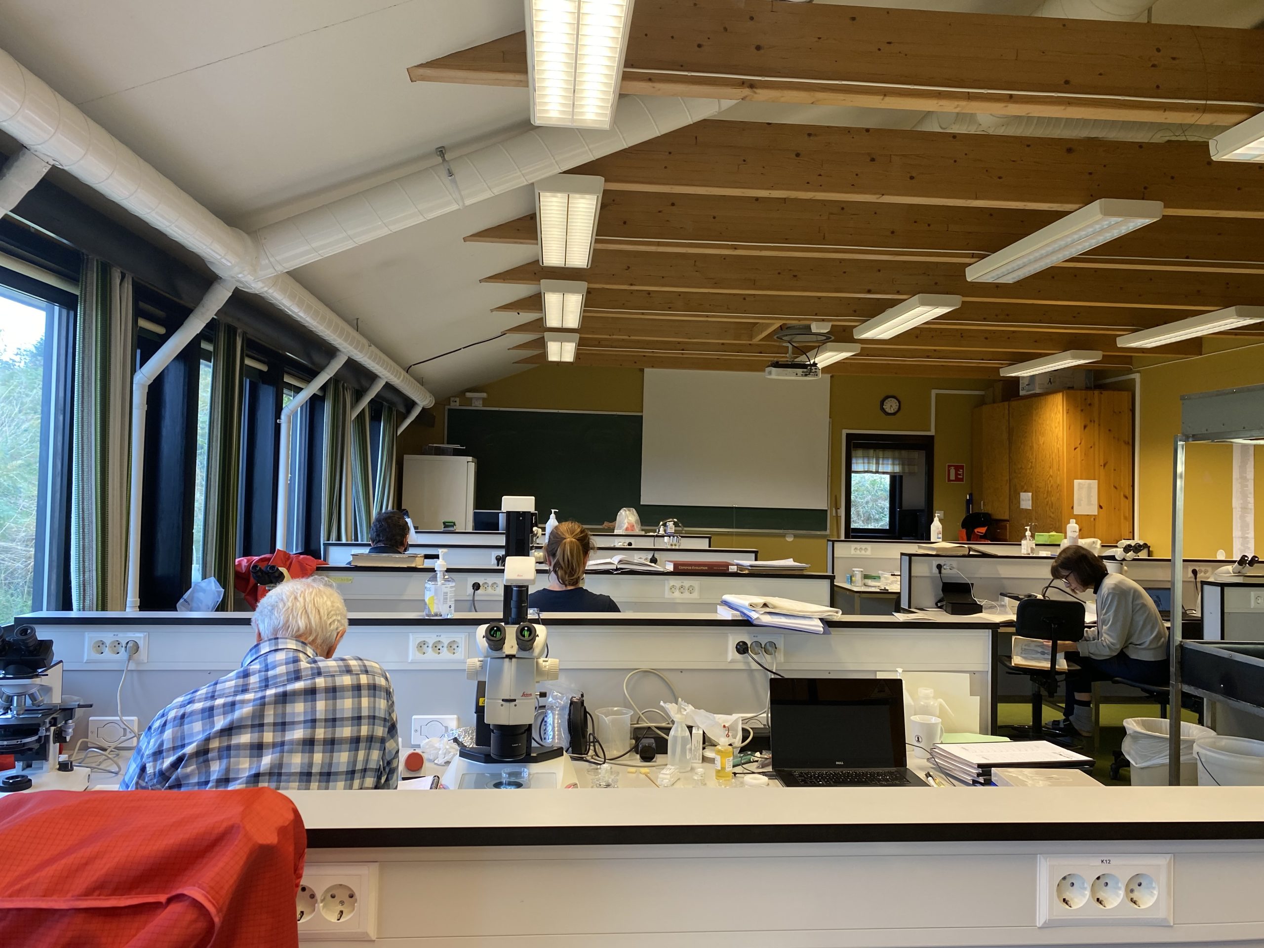
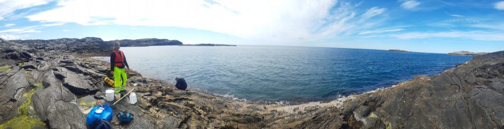
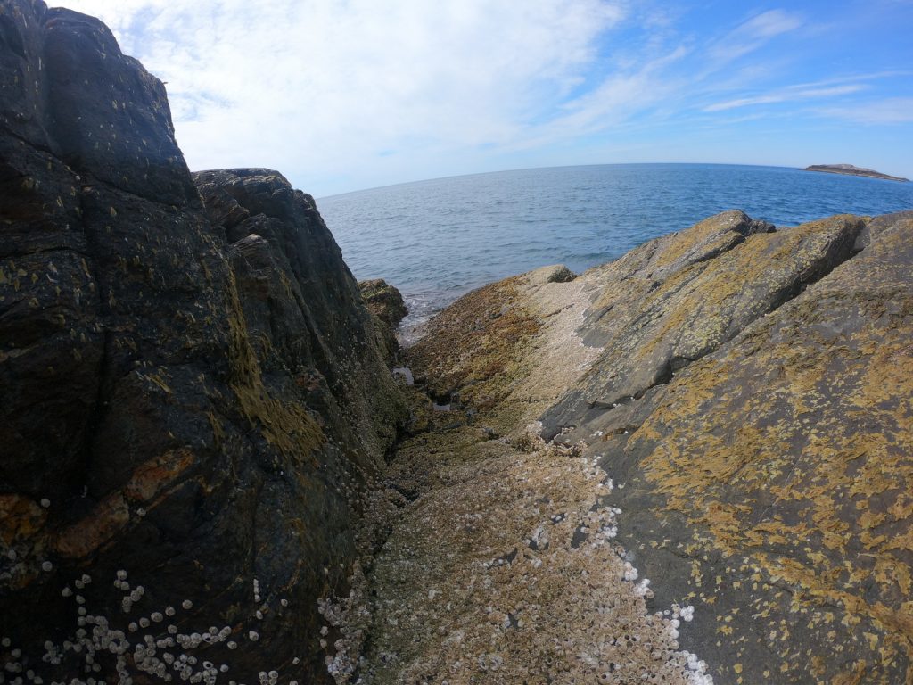
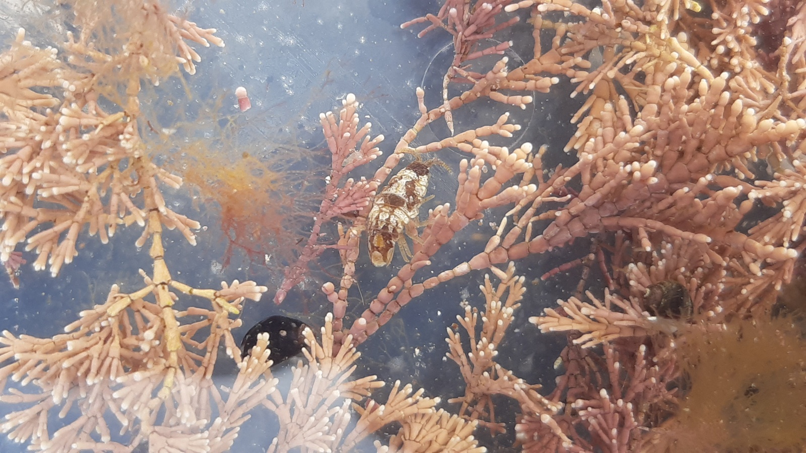
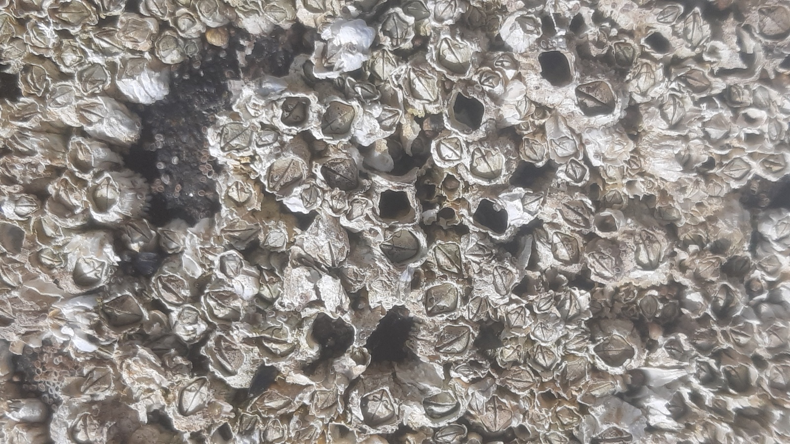
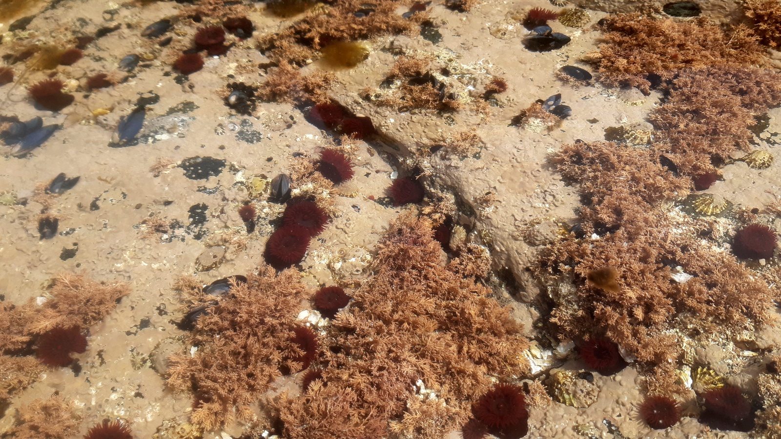
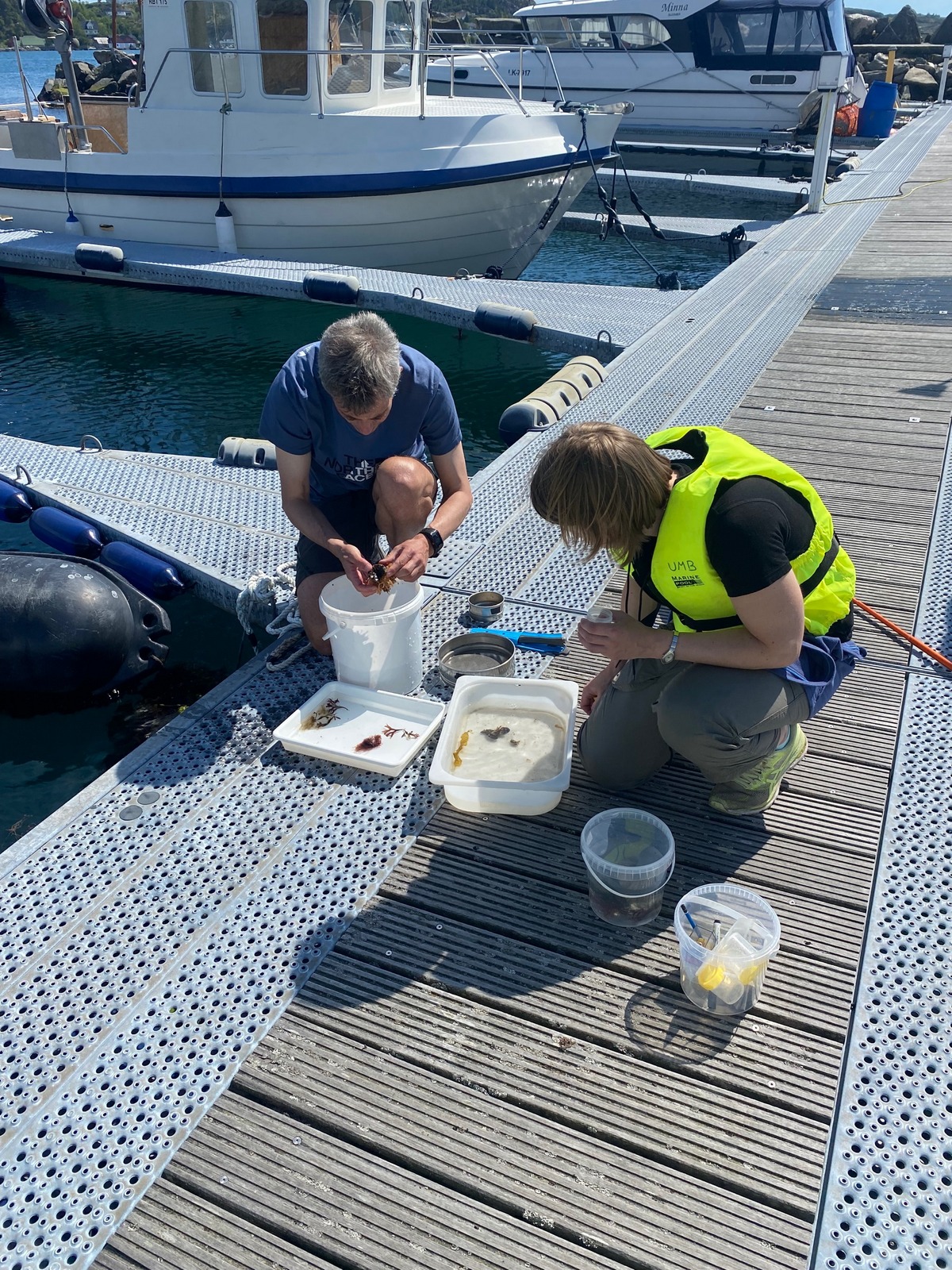
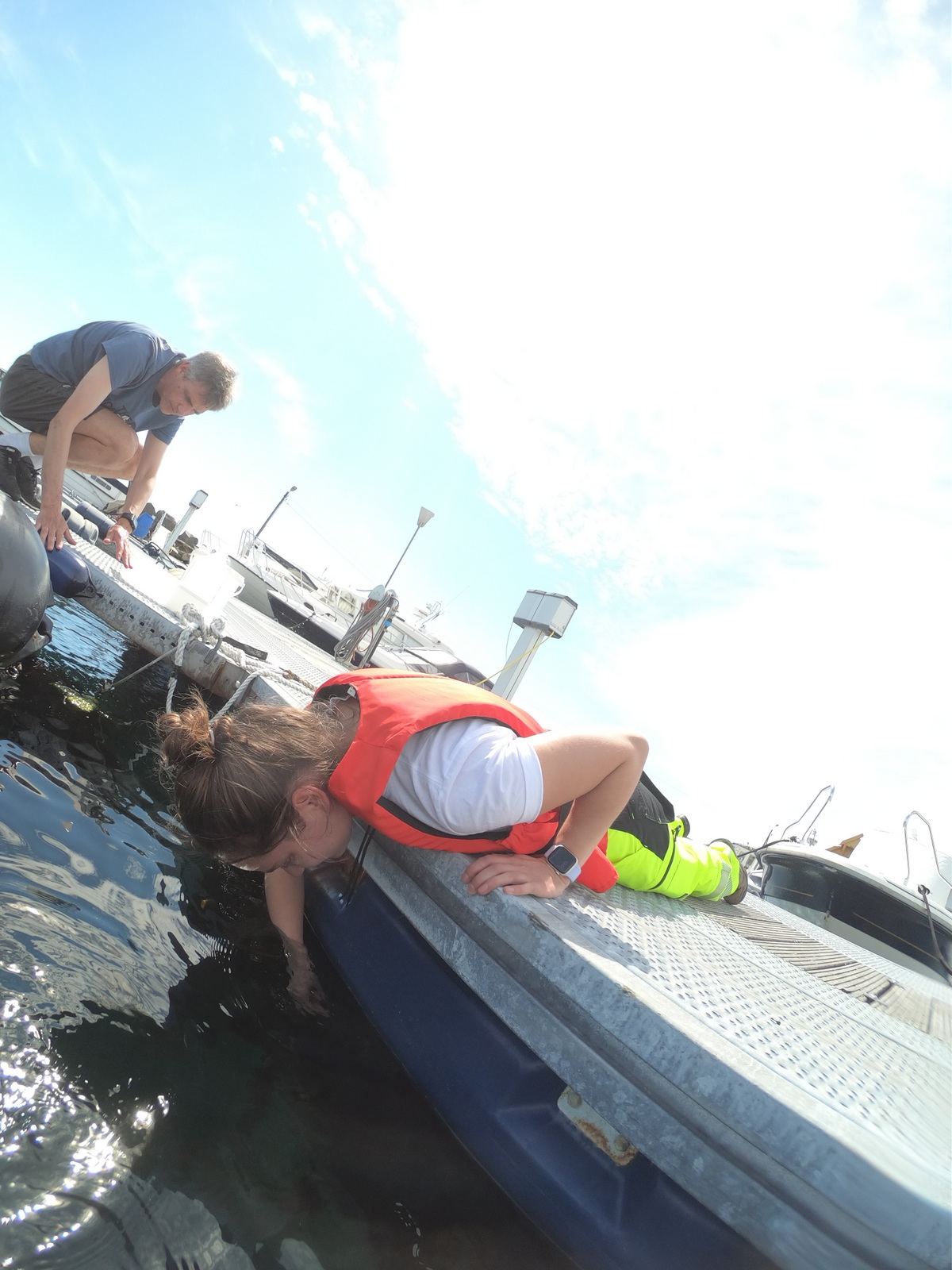
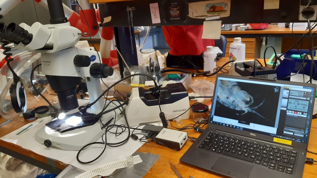
We set up camp on Espegrend Marine Biological station, and combined long days in the lab studying material collected throughout the project with shorter trips out on R/V Hans Brattstrøm.

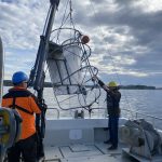
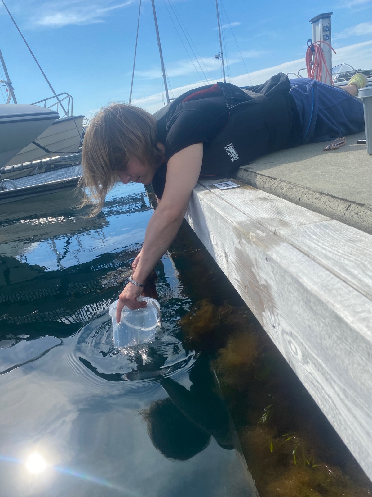
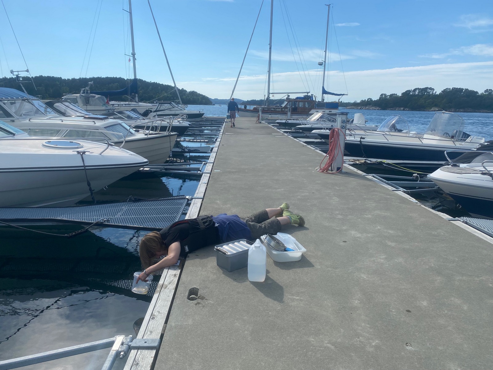
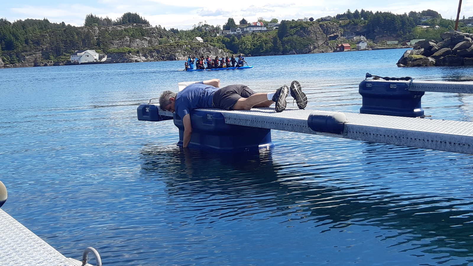
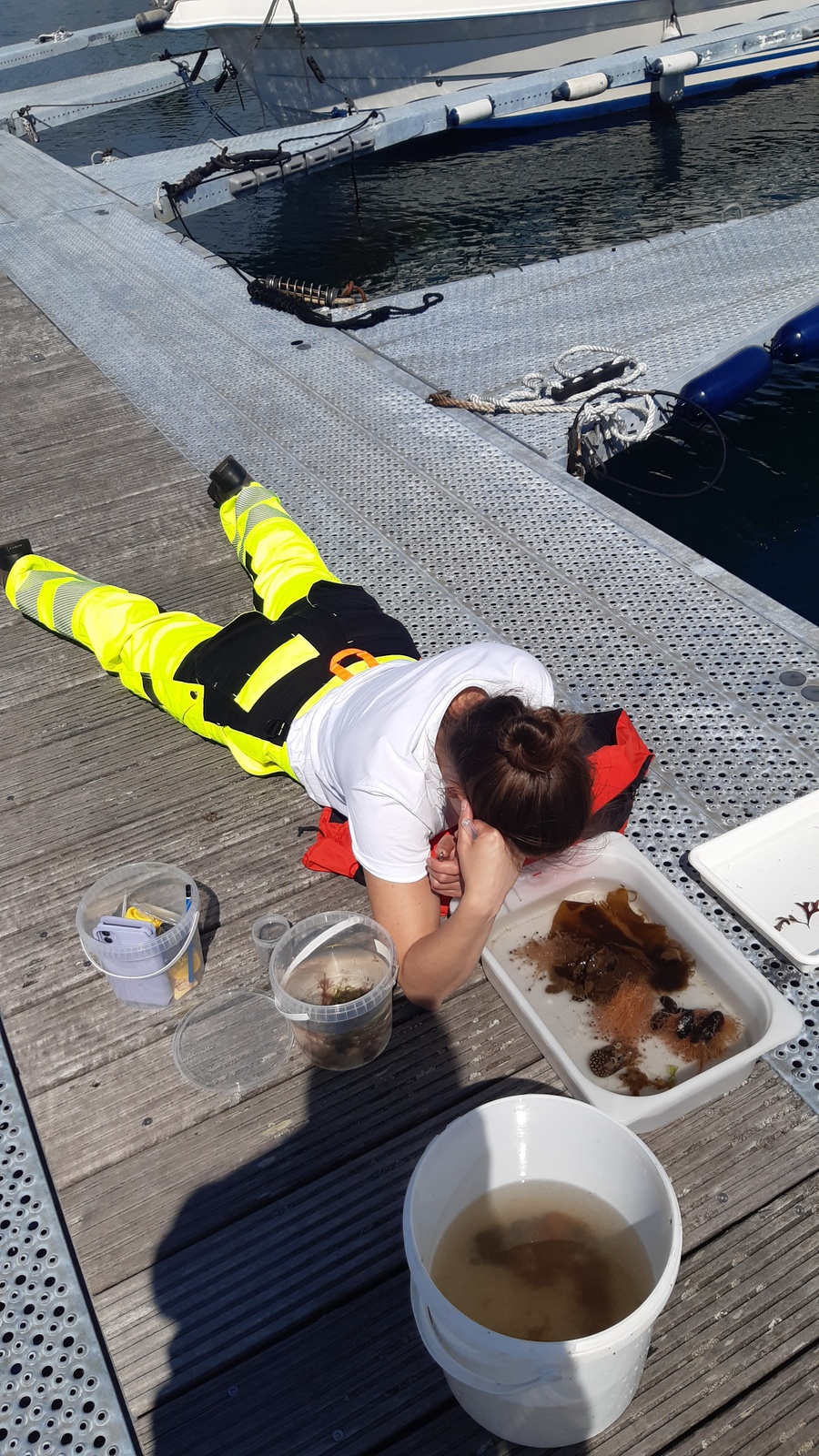
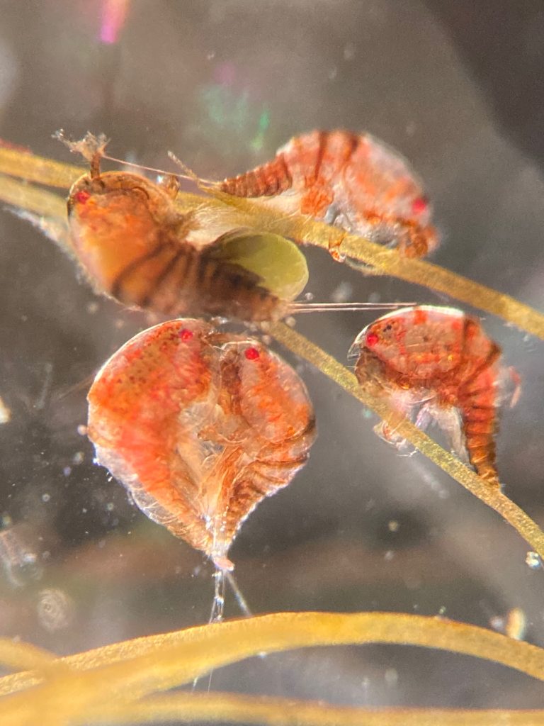
Here we collected live colonies, introduced the students to various collecting methods, and let everyone catch some fresh fjord air.


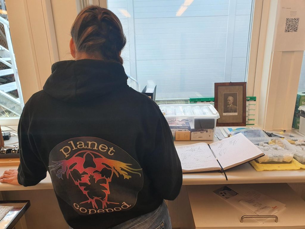
Tine (top left) working together with Mali in the lab and in the field.


Tine is doing her master thesis on the species distribution of Bryozoa in shallow water along the Norwegian coast.

During the workshop she got the chance to have some of the difficult species identifications verified by the experts,  and she prepared a plate of 95 tissue samples that will be DNA barcoded though NorBOL.


Tiril, top left, together with Jon on the ship and working in the lab.


We also had Tiril with us, who is just starting out on what will become a thesis on ascidians (sea squirts), most likely with a focus on species in the genus Botryllus and Botrylloides. 

She worked together with Tom, getting familiar with the literature and the methods used for working on the group. Like Tine, she will be using a combination of traditional morphology based methods and genetic data.


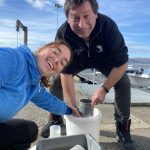
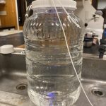
A few impressions from the week

Going forward we’ll first send the plate of tissue samples to CCDB to be sequenced, fingers crossed for good results! During the week, *so many* samples were identified, so we will certainly be preparing more plates during the spring. All the identified samples will be included into the scientific collections of the museum.

Thank you so much to all the participants for their efforts!

-Katrine

Throwback Thursday; HYPCOP workshop at the museum

The project of studying hyperbenthic copepods (HYPCOP) is unique in multiple ways; we study a very unknown group of marine copepod species with very little taxonomic knowledge available here, in Norway. It is challenging as there are more than 700 species described, and possibly more. With pandemic lockdowns, it was hard to have international specialists come and help us, so we had to rely on resources available locally. With so many institutes involved from different corners of Norway, it was not always easy to meet up physically to work on our collection. Hence, when it happens, it is a memorable event, and valuable progress for the project is made.

One of the many species of copepod we have here in the collection at the UiB museum

We have Tone Falkenhaug as the project leader, situated at the Institute of Marine Research in Flødevigen (IMR), than we have our collaborator from Norwegian Institute for Water Research (NIVA), Anders Hobæk and the three technicians at the department of natural history from the University of Bergen. The year before we all got together in Flødevigen, so for 2021 we decided that it would be Bergen to have another workshop.

From ltr; Anders Hobæk, Cessa Rauch, Tone Falkenhaug and Francisca Carvalho making the picture

A year into our project we managed to build up a substantial collection of benthic copepods; currently we have around 460 registered specimens, and 195 off those are barcoded with two different DNA markers, mitochondrial (COI) and ribosomal (16S). What keeps ahead of us is the monster task of working through our specimens to label the DNA barcodes with morphological identifications. It means many hours of very precise work with the finest needles, while sitting at the microscope.

During our workshop in Bergen we got together to work through one of the copepod family trees we generated from their DNA:

Preliminary tree of the COI mitochondrial marker

Anders Hobæk is a taxonomist with many years of experience dissecting copepods, and together we went through the samples one by one. It is very satisfying to be able to identify a specimen and get the to the same species level as the DNA barcode. There are multiple reasons as why we choose to identify species based on morphology.

Not all species are easy to barcode, as copepods, especially the benthic ones, are often so extremely tiny; it is difficult to get good quality DNA extracted from them.

Copepods are tiny; this one with scalebar

The small quantities of copepod DNA goes hand in hand with greater risk of contamination of other surrounding DNA, especially if you work with more general markers. Besides, even if we have the DNA barcode, not all copepod DNA is identified as such, which means that even with the right DNA, when running it through the database, it tells us that we have fly DNA, to give an example. Last but not least, in a lot of cases, we were not able to get good DNA sequences from the copepod extracts, so the only option is identifying them morphologically, by dissecting the animals and with help of literature identify the right genus, or even better, the species.

Species identification with help of literature, here a page from G.O. Sars

Our next workshop shall take place again in Flødevigen, in the meantime we keep you updated about our planet of the copepods.

Follow us for more copepod content @planetcopepod, see you there!


-Cessa

2021 in review for Hardbunnsfauna

Another year of our “Hardbunnsfauna”-project;  Invertebrate fauna of marine rocky shallow-water habitats: species mapping and DNA barcoding (funded by the Norwegian taxonomy Initiative) is coming to an end.

I opted for an easy way to show some of the activities we’ve had on our by selecting a post from each month on our Instagram account to share.

Do give us a follow, if you aren’t already: we are @hardbunnsfauna on both Instagram and Twitter!

Click on the images to expand them

January: Field work on R/V Hans Brattstrøm in gorgeous (but FREEZING) weather

February: our report from field work in Saltstraumen got published

March: Workshop at Espegrend field station together with the projects HypCop and NorChitons

April: results are coming in on some of the DNA barcoding we are doing. Sponges (like the blue one here) are tricky to barcode, but we are getting some interesting results!

May: we have also barcoded a lot of other groups, including a substantial amount of microgastropods (tiny snails)

June: The first master student from the project successfully presented his thesis

July: We played marine invertebrate bingo (did you get a full set..?)

August: Fieldwork in the neighborhood; we sampled invertebrates from the fjord clean-up SUB was doing in Puddefjorden

September: We participated at an event at Os together with Havkollektivet, introducing the invertebrate and vertebrate locals to each other

October: Katrine was on a research cruise with limited internet, but did sample many interesting critters for the project

November: Field work in Haugesund, where Slettaa Dykkerklubb arranged a course on marine biology for divers

December: Pre-end-of-year-hectic-season, but we are enjoying the contributions coming in (physical and electronic) from our wonderful citizen scientists!

Then we wish you all some very
-Katrine

Research internship – Carla García Carrancio

On summer 2021 I had the opportunity to conduct a research visit at the University Museum of Bergen under project NorHydro. Concretely, I was working with the hydrozoan collections, where I got to know first-hand the role played by the curators and the importance of the collections. I examined several specimens and digitalized their associated data creating e-vouchers. Having all the information of specimens in a digital format is very important because it allows other researchers to access the material without having to come to the museum and helps to make the inventory more accessible and organised. I also improved my knowledge of hydrozoan diversity by identifying numerous specimens deposited at the museum. For that, I used some keys for both thecate and athecate hydroids from North—West European waters as well as the guidance of my MSc supervisor Luis Martell.

The main difference between leptothecate and anthoathecate hydroids is the lack of theca in the latter (the theca is a cup-like structure that protects the polyps), but some of them can be very tricky to identify since anthoathecate hydroids may have theca-like structures, and the theca of lepthothecate polyps may be difficult to see at first sight. Also, when you look at a sample, you may found several hydrozoans growing on the same substrate all together, making identification even more difficult. One of the characteristics used to differentiate species is the presence and the shape of the reproductive structures (gonophores), but they are not always present if the polyps are not reproductive.

Sertularella rugosa (top row) is without a doubt one of my favourite hydrozoans. The hydrothecae resemble a bee hive and the colony has a zig-zag appearance. However, it is easy to confuse it with Sertularella tenella (bottom row). Pictures credits: Carla García.

Polyps of the family Campanulariidae. This common family is characterized by the presence of a bell-shaped theca. Pictures credits: Carla García.

During my stay, I also had the chance to go sampling on a research boat, which helped me to understand better the procedures and requirements that are necessary to collect hydrozoans. We used a wide-mouthed plankton net that went up and down at a constant speed to avoid damaging the jellies and other gelatinous organisms from the plankton. After sampling, we took the cod-end to the laboratory. There, the content of the cod-end was poured on a light table. Then, we selected interesting specimens (including hydromedusae belonging to genus Euphysa) with wide mouthed pipettes and transferred them to Petri dishes filled with fresh seawater to observe them better under a microscope.

Towing the plankton-net which went down to 650m to capture some gelatinous organisms. As you can imagine, going up and down such a long distance takes a lot of time, but it is never boring with colleagues like Aino Hosia (right). Picture credits: Carla García.

I was lucky enough to get samples of Euphysa aurata and Euphysa sp., but they did not want to pose for my photo and kept moving around. Picture credits: Carla García.

We took the opportunity to collect some shallow-water benthic hydroids just in front of the Marine Station. Picture credit: Carla García.

Last but not least, I worked at the DNA lab, which allowed me to gain experience in new molecular techniques that I had not used before and to adapt myself to different (and very modern) facilities.

This experience has been simply great for me. I loved the working environment and the fact that everybody was always there to give me a hand. I have learned a lot and I am taking with me many friends that I hope to meet again when I come back to Bergen.

If you want to know more about projects of NorHydro and HYPNO, visit NorHydro’s home page and Facebook page, and check the hashtags #HYPNO and #NorHydro inTwitter.


-Carla García-Carrancio