Category Archives: NorBOL

One Ocean Week 2024

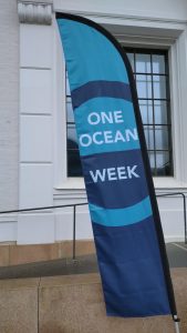
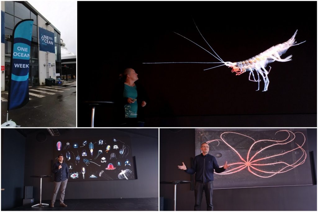
the banner for OOW seen outside the University museum

From April 13th to 19th 2024, the Ocean City of Bergen celebrated the ocean even more than usual(!) in a happening called One Ocean Week (OOW).

One Ocean Week held conferences, meetings, workshops and activities – aiming to pioneer a sustainable use of the ocean.

The marine group at the University Museum was a prolific participant during the event, here are some of our contributions!


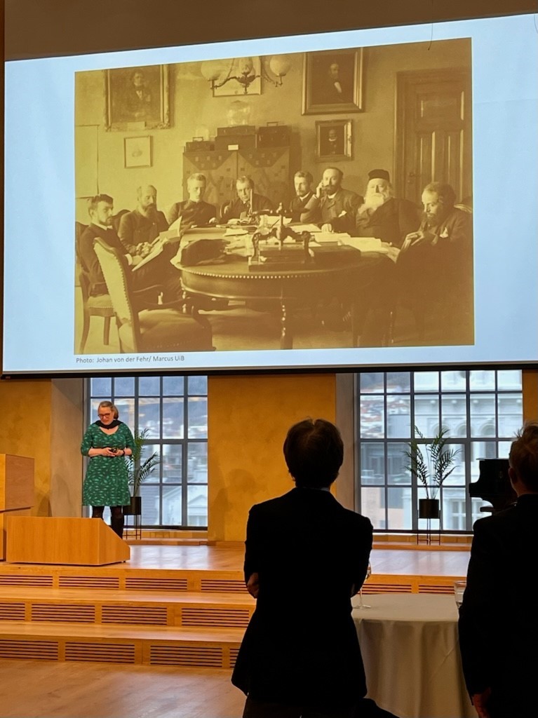
Anne Helene was invited to give the opening talk at the reception in the Aula, placing the Museum and Bergen into the heart of the Norwegian ocean exploration beginning with “The Norwegian North-Atlantic Expedition 1876–78”.

researcher giving a presentation

Anne Helene giving the opening talk during the festivitas in the Aula at the University Museum. Photo: Jenny Smedmark

Screenshot from the documentary. Do follow the link -> to watch if you haven’t already! Photo: OceanX


Aino also made a guest apperance in the aula as the documentary about “Seeing jellyfish as they should be seen” made by the OceanX media team was shown.


Saturday brought a presentation at Skolten about the UiB/OceanX-expedition last August, where Joan and Anne Helene presented some of their exciting new findings together with Henrik and Øyvind from BIO – and even Mattie of OceanX participated, calling in from the OceanXplorer in Singapore!

Presentations of what we found during the OceanX/UiB cruise last summer, where we explored old locatlities in the Norwegian Sea, and visited some of our fjords using the world’s most advanced research vessel. Pictured are Anne Helene, Joan and Henrik – with their animals! Photos: Katrine Kongshavn

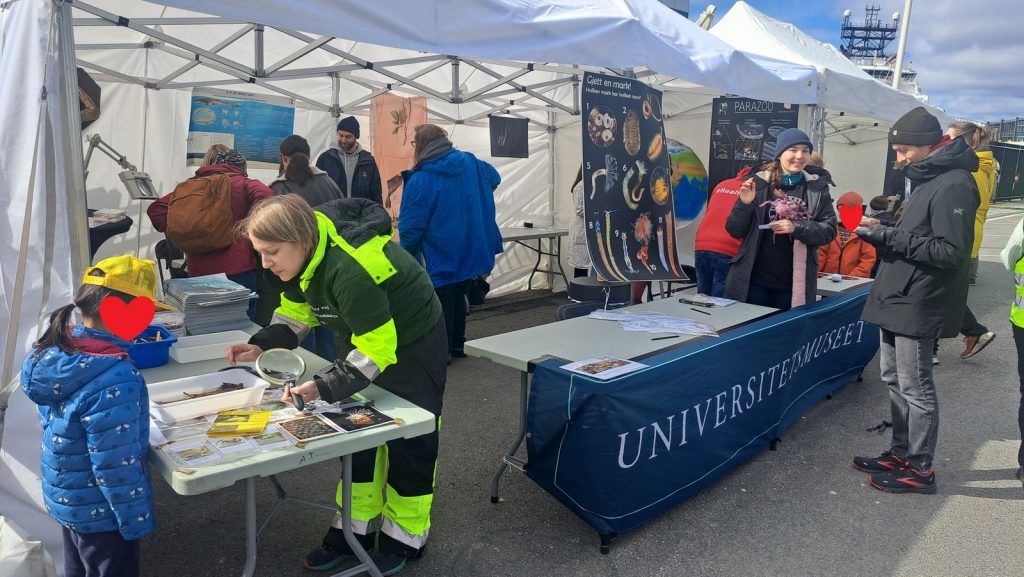
On Sunday the 14th most of us were at Festningskaien with a plethora of activities during the “Family Day”.

Our tent at the Family Day. Photo: Joan J- Soto-Angel

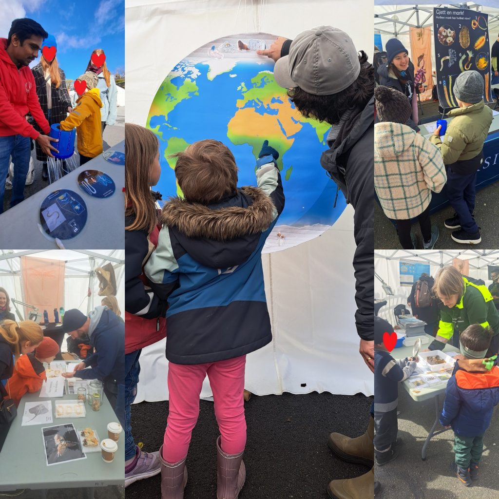
For our “Meet marine biologists from the University Museum” we brought with us five activities focusing on different areas of research that we do.

We had:

  • Sharks (see SharkReferences.com for more), with a shark tooth quiz, info about sharks in Norway, and the chance to see shark skin up close.
  • Bipolar animals (project Pole2Pole) where people could explore the distribution of animals that occur in one- or both- of the poles, and learn more about why that may be the case
  • Polychaetes (project MAnDAriN) with a quiz of trying to match images of colourful bristle worms to their common names
  • A task on Jellyfish parasites(!) (project ParaZoo) where you were to diagnose which parasite our unfortunate jellyfish suffered from, and finally
  • Moss animals (project NorDigBryo) where you could learn more about these animals, see them up close, and pick up a booklet with fun facts and some riddles to solve.

    Our activities. From top left: Jellyfish doctor, bipolar animals, guess the worm, sharks, and moss animals. Images: Joan J. Soto-Angel

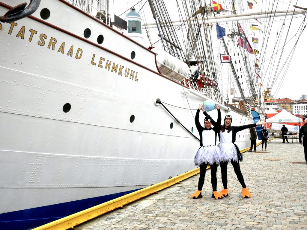
    Two of the museum pedagogues used our tent as the base for their theatre-based activity “The ice is melting! It is for real!” – so we had penguins too!

    The penguins Piia and Pling in action. Photo: Odette Tetlie

    At the Natural History Museum, our researchers offered free talks on topics of their choice; one talk every day (Tues-Fri). The topics covered here were:

    Who eats whom? Marine worms with jaws – delicious and dangerous! (Nataliya)
    Jellyfish in Norway – mostly harmless or murderous monsters?
    (Aino)
    Sharks of Norway
    (Nico)
    Elusive biodiversity: a journey through the less known but most exquisite groups of marine animals (
    Manuel)

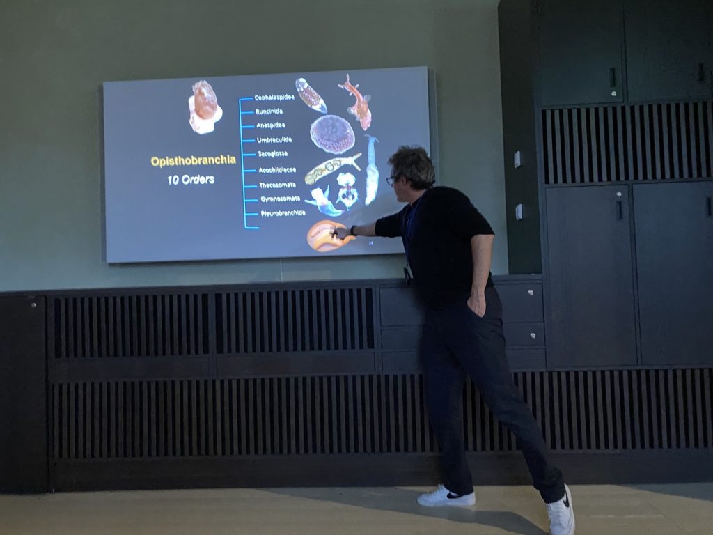
    a person pointing to a phylogenetic tree showing gastropods and how they are related

    Manuel during his presentation at the Natural History Museum. Photo: Cessa Rauch

    Nico also gave a talk at the scientific conference Ocean Outlook: the North Atlantic Ocean climate, deep sea and environment, on the topic “Bioluminescence in deep-sea sharks: evolution and functions.

It was a hectic, but fun week!

-Katrine on behalf of the marine group

Project ParaZoo: there is a critter inside my jellyfish!

ParaZoo (complete name ‘Metazoan parasites of non-crustacean zooplankton’) is one of the most interaction-focused projects currently running at the Invertebrate Collections of our Museum. This project, funded by the Norwegian Biodiversity Information Centre (Artsdatabanken), aims at studying the different animals that live together inside and on the surface of Norwegian jellyfish. This means that for the next two years we will be looking for tapeworms, flukes, roundworms, and amphipods as ParaZoo tries to answer the question of which of these organisms are associated with gelatinous hosts in Norwegian waters.

ParaZoo is focused on animal parasites and symbionts associated with jellyfish. Thse parasites include roundworms like Hysterothylacium aduncum (left, in Euphysa aurata), amphipods like Hyperia medusarum (middle, on Aequorera forskalea), and flukes like the members of family Didymozooidae (right, in Beroe gracilis). IC: Aino Hosia (left), Katrine Kongshavn (middle), Joan J. Soto-Àngel (right).

Besides jellyfish, arrow worms (Chaetognatha) are also members of non-crustacean zooplankton that host different types of parasites, like this H. aduncum roundworm (Nematoda). IC: Joan J. Soto-Àngel, Luis Martell.

Parasitism and symbiosis are extremely common life styles in the animal kingdom. In fact, some researchers believe that there may be more species of parasites than of free-living animals, given that each free-living species hosts many species of parasites (most of them unique) and those parasites also host their own parasitic tenants. Marine zooplankton is no exception to this trend, and many parasites and symbionts are expected to occur in copepods, krill, and gelatinous zooplankton. Jellyfish and arrow worms, for example, may be important hosts for flatworms and other helminths, yet our knowledge of these animals in Norway is very scarce.

ParaZoo’s logo includes two of the target taxa of the project: roundworms (Nematoda) and hyperiids (Amphipoda). The third main parasitic group covered is flatworms (Platyhelminthes), illustrated by the larvae of Derogenes varicus parasitizing Halopsis ocellata shown in the right side of this figure. IC: Joan J. Soto-Àngel, Luis Martell.

Understanding zooplankton parasites is important because many of them are going to be transmitted to fish, where they may cause serious diseases. To get a better overview of which critters live in non-crustacean zooplankton, ParaZoo will sample, record, and DNA-barcode specimens from all over the country. The collected animals will be included in our museum collections after being identified, documented photographically, and fixed in ethanol. We will then generate an open-access database of information including pictures and DNA sequences that will help with the identification of the parasites. Aquaculture facilities, fishermen, and managers of marine areas will benefit from this database to better plan and counter potential negative impacts caused by the parasites.

Flukes, such as Opechona spp, parasitize gelatinous zooplankton (in this case a sea-gooseberry Pleurobrachia pileus) as larvae called metacercariae. IC: Joan J. Soto-Àngel, Luis Martell.

The larvae of tapeworms (Cestoda) sometimes use jellyfish to reach their definitive hosts: fish. IC: Joan J. Soto-Àngel, Luis Martell.

ParaZoo is committed to present the diversity of jellyfish parasites to all those not familiar with them. In order to do that, we will regularly write entries here on the blog, as well as participate in several academic and not-academic meetings. The official info webpage for the project is available here, so don’t forget to check it out!

Luis

A new online resource for identifying ‘fur snail’ hydroids, and a farewell to project NorHydro

Finding the correct name of the hydroids growing on shells of snails and hermit crabs can be notoriously difficult… but fear not! A new online resource is now available and we hope it will help everybody who is interested in these little critters.

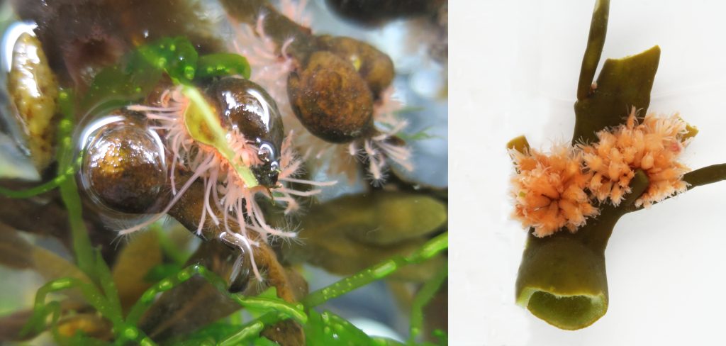
Fig1. Have you seen these pink/orange ‘blobs’ growing on washed-up algae along the coast? They are hydroids of the species Clava multicornis. Read more about this and other Norwegian hydractiniids in the new arter på nett webpages. IC: Luis Martell (left), Katrine Kongshavn (right).

We (the UMB research team of NorHydro: Luis, Aino, Joan, and Lara) have created a series of species identification sheets for all the hydroids and jellyfish belonging to family Hydractiniidae. This family includes some hydroids that can be easily encountered during a walk along the coast or when snorkeling and scuba diving in Norway, and therefore we wanted to provide beach-goers and divers with a tool to find the correct name of these animals.

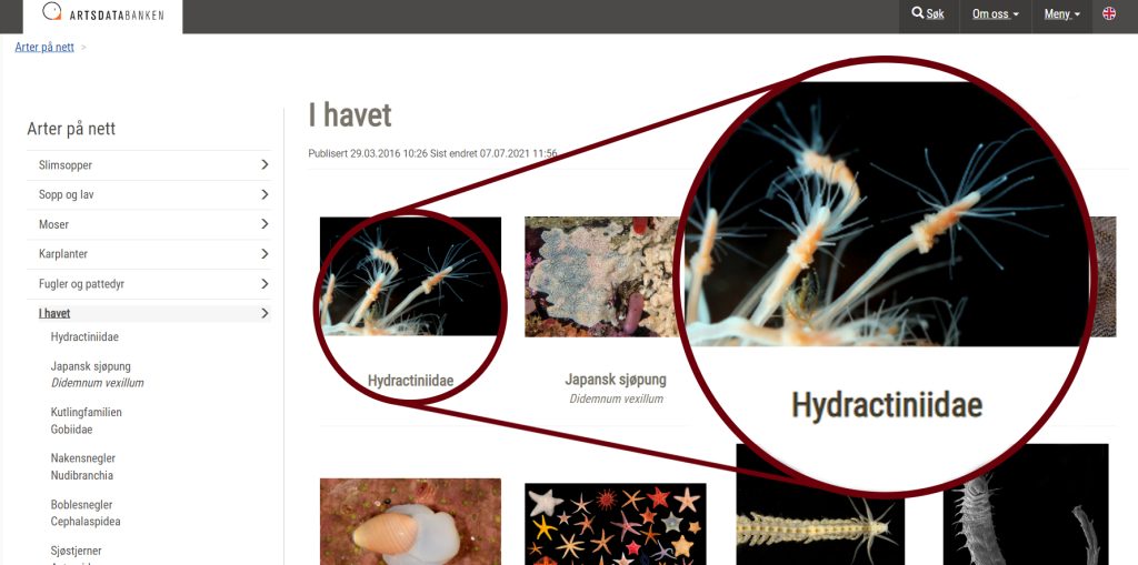
Fig2. The new identification sheets for Hydractiniidae are available inside the section I havet of the Arter på nett platform in Artsdatabanken’s website. IC: Artsdatabanken.

Many hydractiniids are known as ‘snail fur’ because they give a furry appearance to the shells they are growing on, while others grow on rocks or on algae. Identifying the species still requires a little effort, but altogether they are one of the best groups to start with for anyone interested in hydrozoan diversity in Norway. This is what motivated Lara, a MSc student at the time, to created the first version of these sheets (together with some beautiful illustrations) which were then developed further with text from everybody in the team.
The editors and consultants at Artsdatabanken helped us throughout the process, and we are happy to present the final version of this online resource available here:
Arter på nett: Hydractiniidae

Fig3. The anatomy of a polyp colony of Hydractiniidae is best explained with a combination of images at high-magnification and detailed diagrams. IC: Luis Martell (left), Lara Beckmann (right).

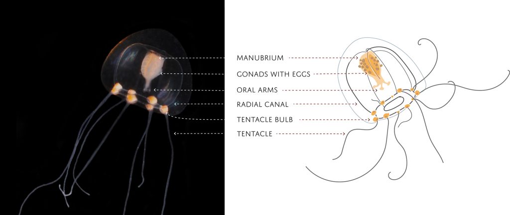
Fig4. Hydractiniid hydromedusae are smaller than 1 cm, but they still have several characters that we use for identification. IC: Joan J. Soto-Angel (left), Lara Beckmann (right).

Fig5. Both the hydromedusa and the hydroid stages of the three species of Podocoryna occurring in Norway are included in the new Hydractiniidae arter på nett. IC: Joan J. Soto-Angel, Bernard Picton, Lara Beckmann

The publication of the Hydractiniidae identification sheets marks the end of our project NorHydro. The project’s main objective was to map the diversity of hydrozoans in Norway, but we also tried to present marine hydroids to all those not familiar with these amazing creatures. During its 3 year’s run, NorHydro produced a detailed inventory of Norwegian hydrozoans through an integrative (morphology + DNA) approach: we collected samples in >90 sites around the country and analyzed existing museum specimens to produce >1200 new records and high-quality, open access DNA barcodes for 160 species. NorHydro’s scientific team identified 12 cases of species complexes, supervised 5 MSc thesis, and participated in >20 outreach activities and presentations in academic events. All in all, NorHydro was an extremely enriching experience and I’m confident its results will help us understand a little better the diversity of marine invertebrates in Norway.

Fig6. Farewell NorHydro!

-Luis

Unraveling copepod secrets one leg at a time

A blog by HYPCOP

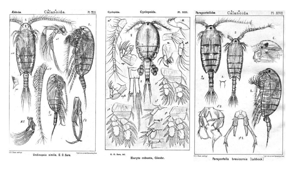
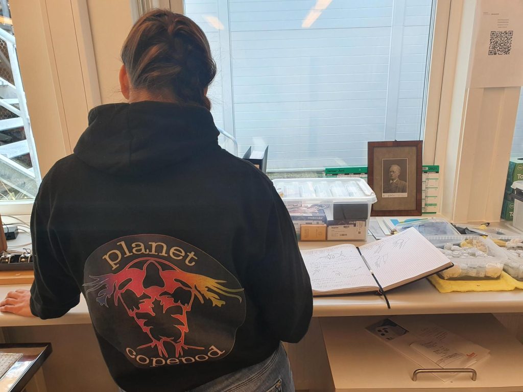
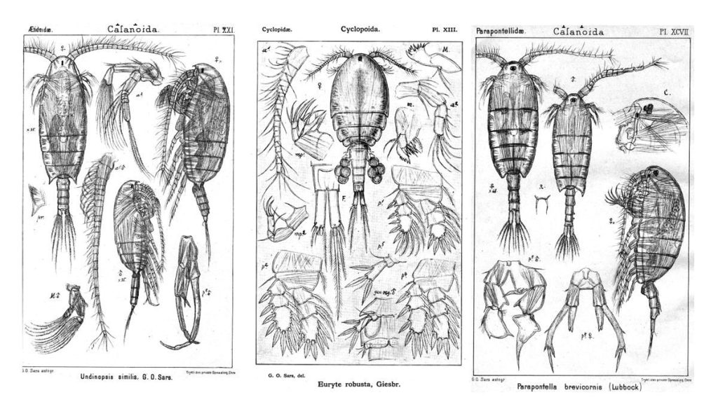
Hyperbenthic copepods (HYPCOP) are a very difficult and diverse group to work with, and identification goes painstakingly slow, because some species can only be distinguished from one another based on small details in some of their tiny legs. As of now, we have no specialists in marine benthic copepods in Norway and our greatest resource is our collaborator Anders Hobæk and the detailed drawings of G.O. Sars from the early 1900s .

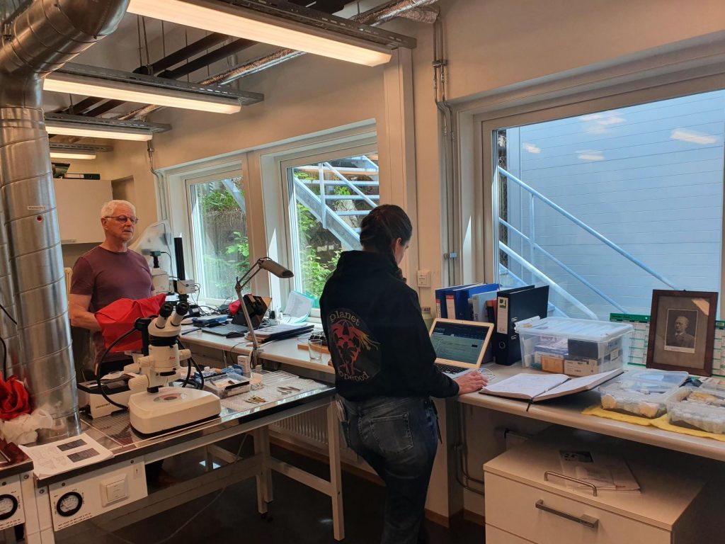
Working together under guidance of G.O. Sars and Anders Hobæk

Anders is a senior researcher scientist at the Norwegian Institute for Water Research (NIVA) here in Bergen. He is specialized in copepod taxonomy, but his focus was mostly on freshwater copepods, or marine pelagic copepods. Which makes the marine benthic copepods just a little bit more challenging to work with, however, his skills are transferable and so we get together multiple times a year to work on our collection of benthic copepods to dissect them and identify them.

Beginning of June, we had again one of those get togethers in Flødevigen at the Institute of Marine Research (IMR), where Tone Falkenhaug, the project leader of HYPCOP, is situated. For a week we went through the main clades and groups of species that we had DNA barcodes of but not yet a confirmed species name. A lot of the identification was done with help of the rich and detailed illustrations of G.O. Sars1 published work in 1901 – 03 and 1919 – 21, “An account of Crustacea of Norway”

Detailed copepod drawings from G.O. Sars

Sars dedicated a lifetime of identifying and describing a variety of species and he did not neglect the rich and wonderful group of bottom dwelling copepods. Every species he encountered in those early days he described and drew in detail; he did not leave out the smallest details, that as of now, turn out to be of uttermost importance in determining the species. With small copepods you need a good microscope and fine tools. The first thing to look at is the general shape, is it very dorsally flat, like Peltidium purpureum, or more dimensional like Harpacticus flexus?

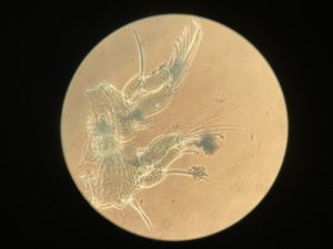
Sex is also an important feature; females are often characterized by carrying eggs; one egg sack or two egg sacks can already lead you in the right group. Males have often larger antennule made for holding on to females when mating, and other specialized tools that can be species specific. The little claws, called maxilliped, are they large, small, almost invisible? What about the first pair of legs? The second, third and fourth? The fifth pair of legs is often very characteristic for the species and in certain females, like Thalestris longimana, can be a huge in comparison of the rest of its body.

Thalestris longimana, females of this species has relatively large fifth pair of legs

Our work has a continues workflow consisting of, collecting the copepods, extracting their tissue for DNA barcoding, and keeping the exoskeleton. Once the DNA is successfully sequenced, we can take the exoskeleton and dissect the animal leg by leg to finalize the identification. That way the copepod is identified based on its DNA and morphological features, as this is not always mutually exclusive. DNA can be tricky as you need a good reference library to find the correct species, which is as of now, not complete, or even lacking for many species. Besides, there is such things as DNA contamination, cross contamination between species, therefore you always must look at the morphology to exclude that the DNA gives you the wrong species. Together with images of the animals we are building up a valuable reference library of DNA sequences and a museum collection of dissected animals on fixed slides. This way copepod diversity will continue to be valuable for future generations top study.

Working under the eyes of G.O. Sars

-Cessa


1Sars, G. O. 1901-03. An Account of the Crustacea of Norway. Vol. IV. Copepoda Calanoida.- Bergen Museum, Bergen & Christiana. 171 pp. + 109 plates Sars, G. O. 1919-21. An Account of the Crustacea of Norway. Vol. VII. Copepoda. Supplement. – Bergen Museum, Bergen & Christiana. 121 pp. + 74 plates

Bryozoa-workshop at Espegrend

February 14th -18th 2022

The Bryozoa are maybe not the most famous of animals, so let’s start with a quick rundown: Bryozoa, also known as Polyzoa, Ectoprocta, or moss animals (mosdyr, på norsk) are a phylum of aquatic invertebrates. Bryozoans, together with phoronids and brachiopods, have a special feeding structure called a lophophore, a “crown” of hollow tentacles used for filter feeding, which you can see in action in the video Tine captured:

In Norway we have 292 species registered, of which 281 are marine (Kunnskapsstatus for artsmangfoldet 2020, pdf here). It is estimated that the actual number of species present is higher. Further, several known species are considered “door knocker species” that may establish here within the next 50 years.

Bryozoa mostly live in colonies made up of tiny individual animals called zooids, which grow in a variety of shapes, and some of them provide structural habitats for other species. They are food for many other animals, namely nudibranchs, fish, sea urchins, pycnogonids, crustaceans, mites and starfish. Marine bryozoans are often responsible for biofouling on ships’ hulls, on docks and marinas, and on offshore structures. They are among the first colonizers of new or recently cleaned structures, and may hitchhike to new places with marine traffic. (Bonus: they have a super interesting fossil record, and this can be used to tell us more about the world in the way back!)

A few of the shapes the colonies can grow in. Pictured are 1: Membraniporella nitida 2: Bugula sp. 3: Flustrella hispida 4: Crisia eburnea

They are one of the focus groups of Hardbunnsfauna: there is still a lot we do not know about them!

Ernst Haeckel – Kunstformen der Natur (1904), plate 23: Bryozoa. Public domain, accessed through Wikipedia

Planning in a pandemic is not easy, and we have had to postpone our plans for this gathering several times. The second week of February we could finally gather our “Team Bryozoa” here in Bergen for a week of in-depth studies of these fascinating animals.

Team Bryozoa (centre), from left Piotr, Mali, Jo and Lee Hsiang, and some of the animals they studied. Group photo by Piotr Kuklinski

In total we were 11 participants;
University Museum of Bergen: Endre, Jon, Tom and Katrine,
Natural History Museum in Oslo: Lee Hsiang and Mali,
NTNU University Museum: Torkild, Tine (MSc. student) and Tiril (MSc. student),
and our two visitors from abroad:
from the Institute of Oceanology, Polish Academy of Sciences came Piotr,
and from the Heriot Watt University (Orkney Campus), Joanne.

The main focus of the workshop was to get as many samples and species as possible identified, work though the DNA barcode vouchers from samples submitted in advance and reach a consensus on which species the dubious ones were, to network with our colleagues, and to include the students in the work and the team. It all went swimmingly, and a we had a very productive and enjoyable week!

check out @hardbunnsfauna on Instagram for more!


We set up camp on Espegrend Marine Biological station, and combined long days in the lab studying material collected throughout the project with shorter trips out on R/V Hans Brattstrøm.

Here we collected live colonies, introduced the students to various collecting methods, and let everyone catch some fresh fjord air.


Tine (top left) working together with Mali in the lab and in the field.


Tine is doing her master thesis on the species distribution of Bryozoa in shallow water along the Norwegian coast.

During the workshop she got the chance to have some of the difficult species identifications verified by the experts,  and she prepared a plate of 95 tissue samples that will be DNA barcoded though NorBOL.


Tiril, top left, together with Jon on the ship and working in the lab.


We also had Tiril with us, who is just starting out on what will become a thesis on ascidians (sea squirts), most likely with a focus on species in the genus Botryllus and Botrylloides. 

She worked together with Tom, getting familiar with the literature and the methods used for working on the group. Like Tine, she will be using a combination of traditional morphology based methods and genetic data.


A few impressions from the week

Going forward we’ll first send the plate of tissue samples to CCDB to be sequenced, fingers crossed for good results! During the week, *so many* samples were identified, so we will certainly be preparing more plates during the spring. All the identified samples will be included into the scientific collections of the museum.

Thank you so much to all the participants for their efforts!

-Katrine

Throwback Thursday; HYPCOP workshop at the museum

The project of studying hyperbenthic copepods (HYPCOP) is unique in multiple ways; we study a very unknown group of marine copepod species with very little taxonomic knowledge available here, in Norway. It is challenging as there are more than 700 species described, and possibly more. With pandemic lockdowns, it was hard to have international specialists come and help us, so we had to rely on resources available locally. With so many institutes involved from different corners of Norway, it was not always easy to meet up physically to work on our collection. Hence, when it happens, it is a memorable event, and valuable progress for the project is made.

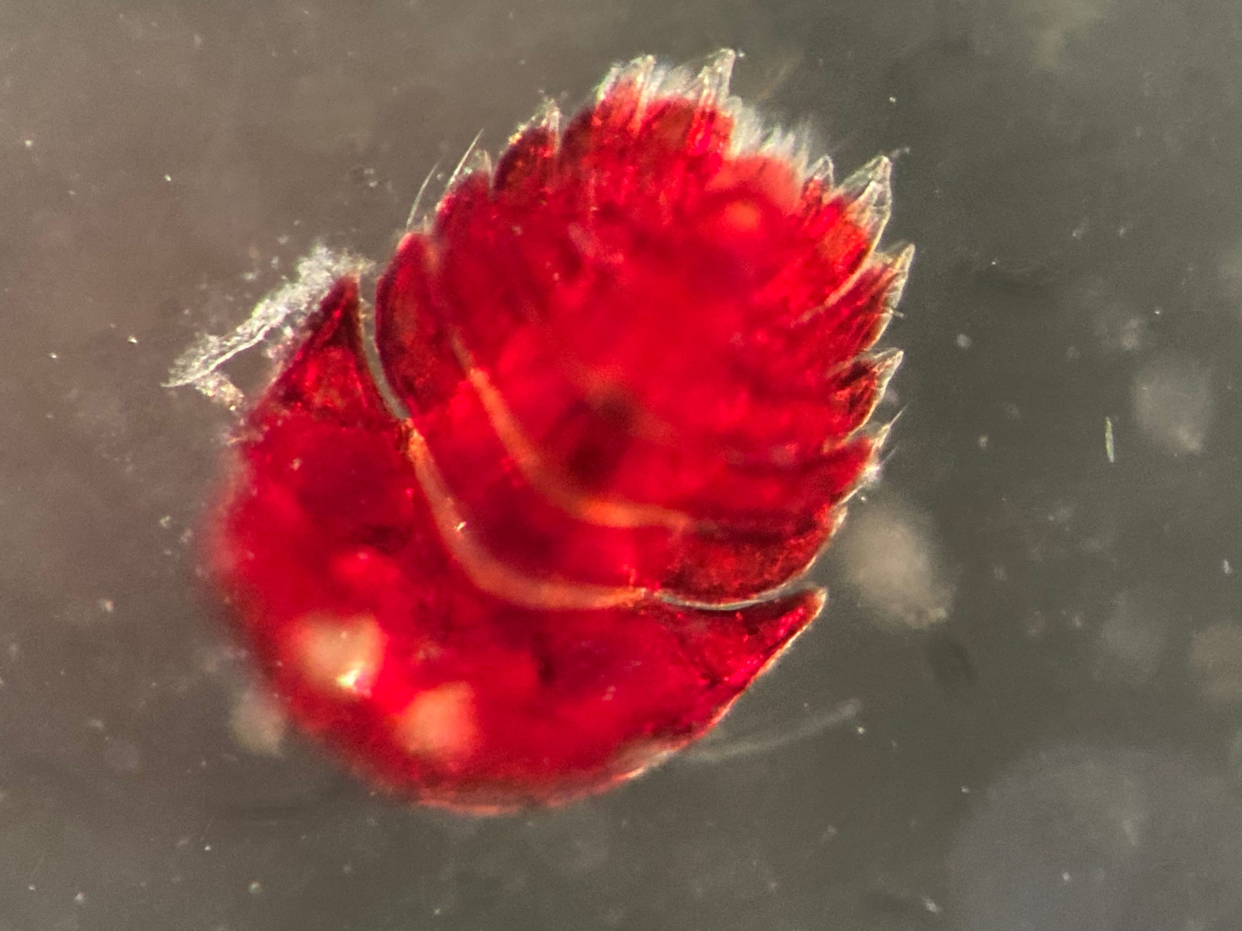
One of the many species of copepod we have here in the collection at the UiB museum

We have Tone Falkenhaug as the project leader, situated at the Institute of Marine Research in Flødevigen (IMR), than we have our collaborator from Norwegian Institute for Water Research (NIVA), Anders Hobæk and the three technicians at the department of natural history from the University of Bergen. The year before we all got together in Flødevigen, so for 2021 we decided that it would be Bergen to have another workshop.

From ltr; Anders Hobæk, Cessa Rauch, Tone Falkenhaug and Francisca Carvalho making the picture

A year into our project we managed to build up a substantial collection of benthic copepods; currently we have around 460 registered specimens, and 195 off those are barcoded with two different DNA markers, mitochondrial (COI) and ribosomal (16S). What keeps ahead of us is the monster task of working through our specimens to label the DNA barcodes with morphological identifications. It means many hours of very precise work with the finest needles, while sitting at the microscope.

During our workshop in Bergen we got together to work through one of the copepod family trees we generated from their DNA:

Preliminary tree of the COI mitochondrial marker

Anders Hobæk is a taxonomist with many years of experience dissecting copepods, and together we went through the samples one by one. It is very satisfying to be able to identify a specimen and get the to the same species level as the DNA barcode. There are multiple reasons as why we choose to identify species based on morphology.

Not all species are easy to barcode, as copepods, especially the benthic ones, are often so extremely tiny; it is difficult to get good quality DNA extracted from them.

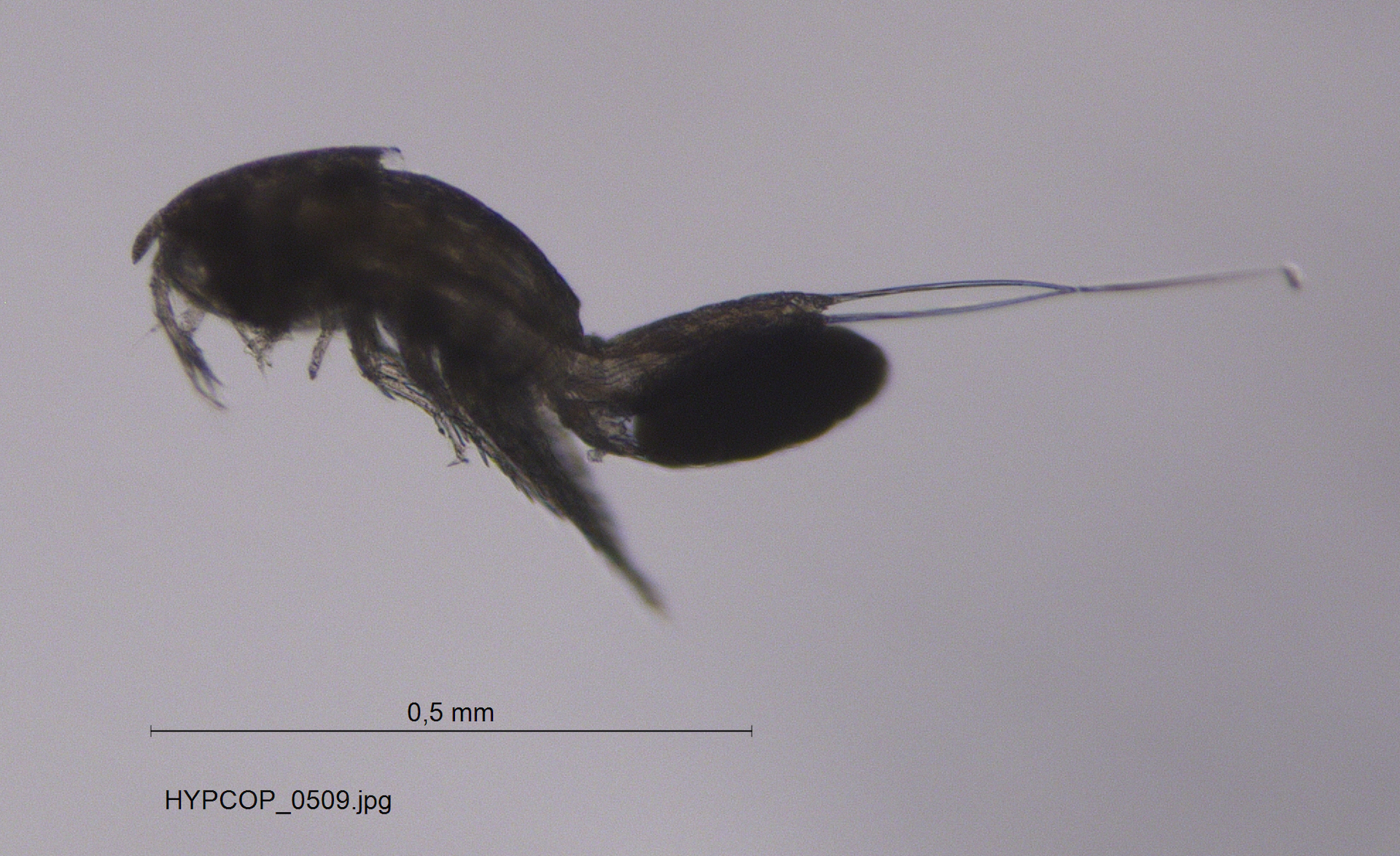
Copepods are tiny; this one with scalebar

The small quantities of copepod DNA goes hand in hand with greater risk of contamination of other surrounding DNA, especially if you work with more general markers. Besides, even if we have the DNA barcode, not all copepod DNA is identified as such, which means that even with the right DNA, when running it through the database, it tells us that we have fly DNA, to give an example. Last but not least, in a lot of cases, we were not able to get good DNA sequences from the copepod extracts, so the only option is identifying them morphologically, by dissecting the animals and with help of literature identify the right genus, or even better, the species.

Species identification with help of literature, here a page from G.O. Sars

Our next workshop shall take place again in Flødevigen, in the meantime we keep you updated about our planet of the copepods.

Follow us for more copepod content @planetcopepod, see you there!


-Cessa

2021 in review for Hardbunnsfauna

Another year of our “Hardbunnsfauna”-project;  Invertebrate fauna of marine rocky shallow-water habitats: species mapping and DNA barcoding (funded by the Norwegian taxonomy Initiative) is coming to an end.

I opted for an easy way to show some of the activities we’ve had on our by selecting a post from each month on our Instagram account to share.

Do give us a follow, if you aren’t already: we are @hardbunnsfauna on both Instagram and Twitter!

Click on the images to expand them

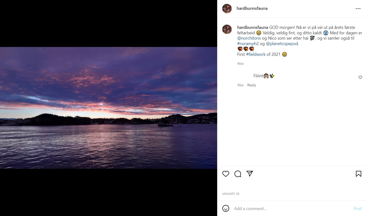
January: Field work on R/V Hans Brattstrøm in gorgeous (but FREEZING) weather

February: our report from field work in Saltstraumen got published

March: Workshop at Espegrend field station together with the projects HypCop and NorChitons

April: results are coming in on some of the DNA barcoding we are doing. Sponges (like the blue one here) are tricky to barcode, but we are getting some interesting results!

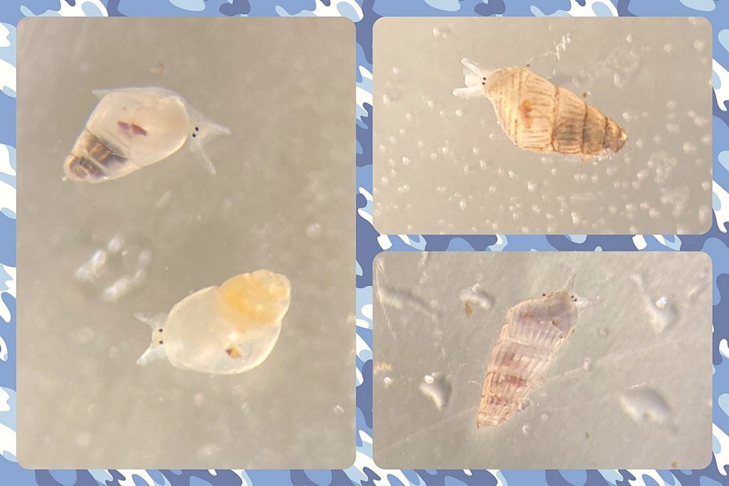
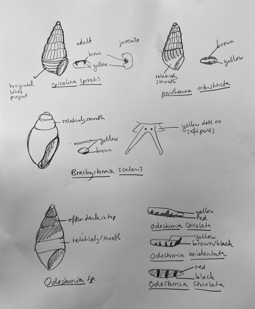
May: we have also barcoded a lot of other groups, including a substantial amount of microgastropods (tiny snails)

June: The first master student from the project successfully presented his thesis

July: We played marine invertebrate bingo (did you get a full set..?)

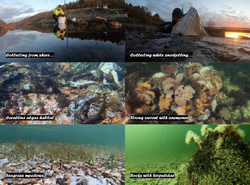
August: Fieldwork in the neighborhood; we sampled invertebrates from the fjord clean-up SUB was doing in Puddefjorden

September: We participated at an event at Os together with Havkollektivet, introducing the invertebrate and vertebrate locals to each other

October: Katrine was on a research cruise with limited internet, but did sample many interesting critters for the project

November: Field work in Haugesund, where Slettaa Dykkerklubb arranged a course on marine biology for divers

December: Pre-end-of-year-hectic-season, but we are enjoying the contributions coming in (physical and electronic) from our wonderful citizen scientists!

Then we wish you all some very
-Katrine

Sampling together in the Sognefjord

From 09 to 13th of May different artsdatabanken projects within the Natural history museum joined efforts during a fieldwork trip to Hjartholm located at the Sognefjord.

The Sognefjord is an interesting fjord for sampling as it is the largest and deepest fjord in Norway and the second largest in the world! This often results in some unique fauna, especially at greater depths. Therefore HYPCOP (Hyper benthic copepods), NORHYDRO (Norwegian Hydrozoa), AnDeepNor (Annelids from the Deep Norwegian Waters) and Hardbunnsfauna (rocky shore invertebrates) travelled toward the small town Hjartholm were we set up laboratory and living space for sampling and processing fresh material.

Hjartholm is located towards the exit of the Sognefjord. From here we would do shallow and deep sampling with help of Research Vessel Hans Brattstrøm

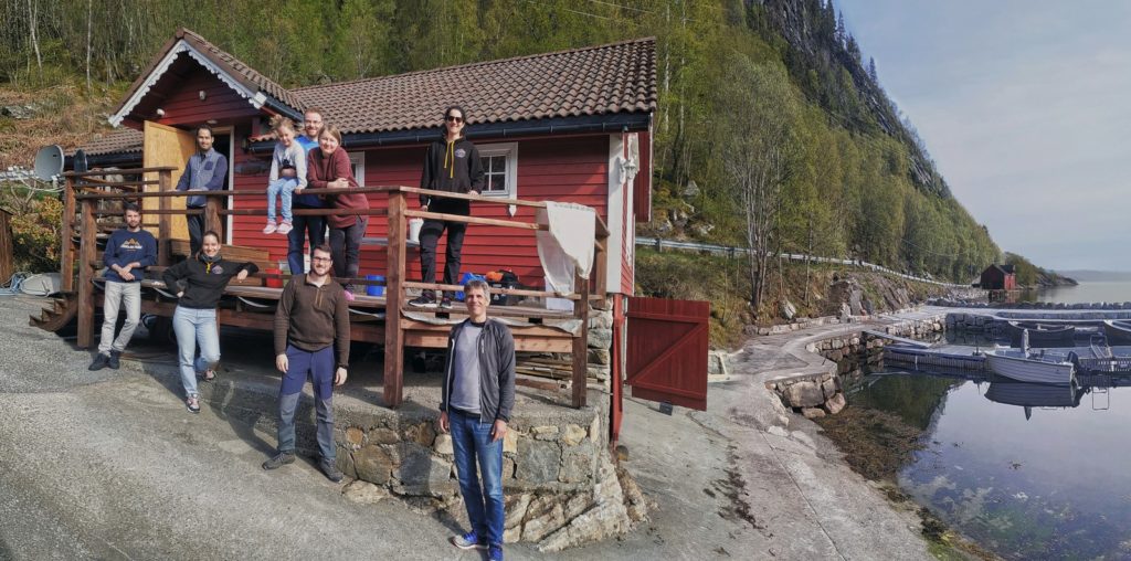
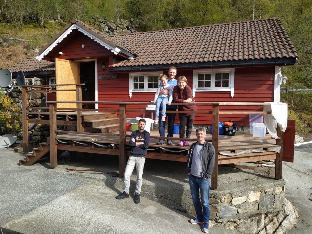
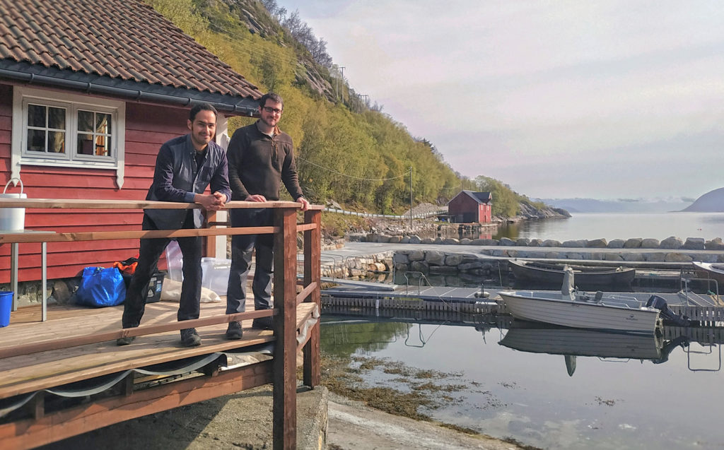
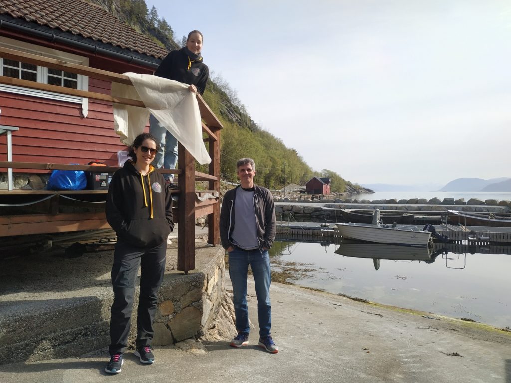
Team members from different projects, Norhydro, HYPCOP, hardbunnsfauna and AndeepNor in front of the boathouse that was transformed into a lab for the occasion

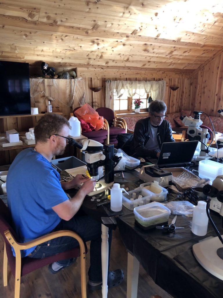
Boathouse communal area turned into a temporary lab

AnDeepNor was on the quest of collecting marine bristle worms (annelida) from the deepest part of the Sognefjord, about 1000m deep.

AnDeepNor researchers from ltr; Miguel Angel Mecca, Tom Alvestad, Nataliya Budaeva, Jon Kongsrud

Jon Kongsrud with the grab

This would be done with the help of research vessel Hans Brattstrøm and a so-called grab. A grab is a device that looks like a clamshell made out of heavy metal. It would be dropped in the water open, and once touching the bottom it would close and grab soft bottom sample.

Unfortunately, on the first day some important machinery for collecting deep samples broke after the third grab. And therefore, AnDeepNor was stuck with only 3 samples for the remaining of the fieldwork days. The good news however is that they did find a great diversity of worms in the only 3 grab samples they found.


Project leader Nataliya with in her hand a plate with clipped tissues from her worms

Once the worms were sorted, preliminary identified and catalogued small tissue was clipped of 96 specimens for barcoding at the University of Bergen DNA laboratory.

All the results of this will be publicly available at the end of the AnDeepNor project in October this year. We are looking forward to their results!


NorHydro has been working hard on collecting hydrozoan samples from different localities in Norway.

NorHydro researchers from ltr Luis Martell and Joan Soto Angel

This time they were more than happy to join the possibility of getting some seriously deep samples from the Sognefjord. With their plankton net they went sampling up to 1200m, which resulted in some beautiful specimens

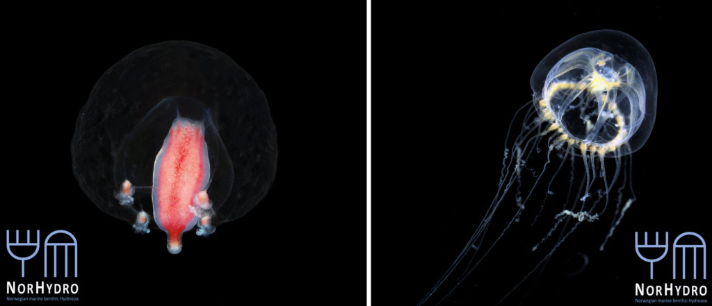
Left: Margelopsis hartlaubii, right: juvenile Melicertum octocostatum

They also took the opportunity to collect some shallow-water benthic hydroids, just in front of the lab where there was a small dock for boats. In the lab they set up a photo-studio to make some beautiful macro images of their collected specimens for everyone to enjoy.

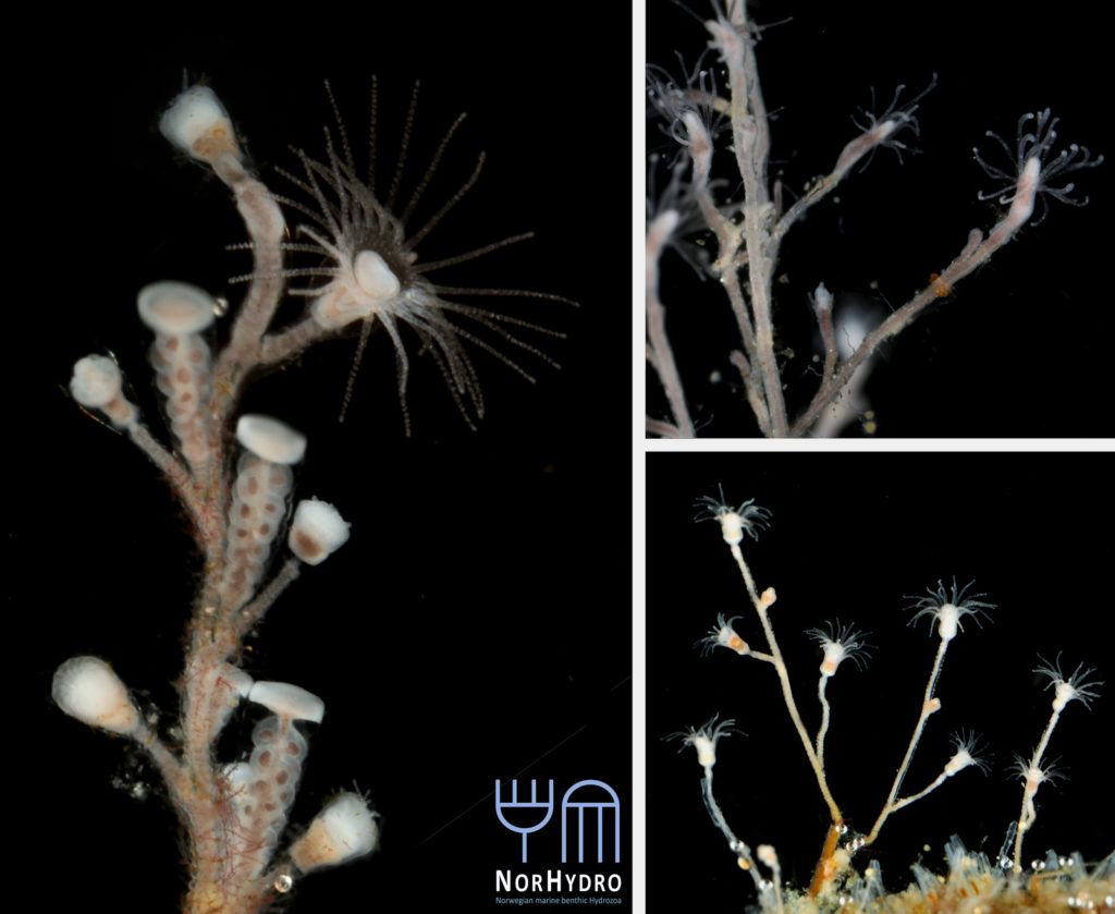
Left: Laomedea flexuosa; top right: Bougainvillia muscus; bottom right: Eudendrium sp.

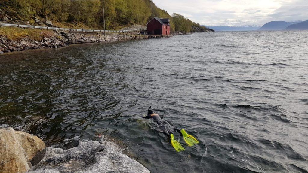
HYPCOP (Picture 9. Team HYPCOP with ltr Francisca Carvahlo, Cessa Rauch and Jon Kongsrud) focus this time was mainly shallow water around the Sognefjord by snorkelling (picture 10. Sampling for Hardbunnsfauna and HYPCOP by means of snorkelling), we sampled from 4 different stations and as you can guess, there were copepods in all of them.

Team HYPCOP with ltr Francisca Carvahlo, Cessa Rauch and Jon Kongsrud

Sampling for Hardbunnsfauna and HYPCOP by means of snorkelling

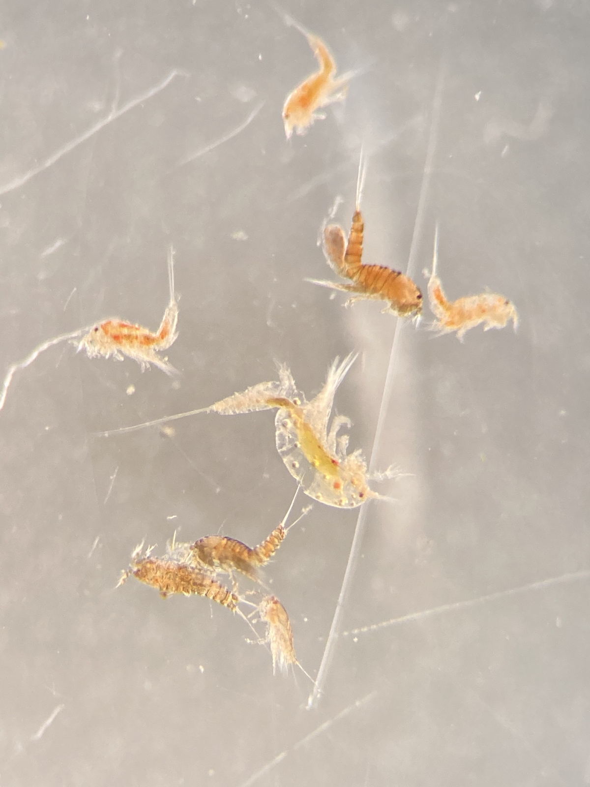
However, some locations had definitively more diversity than others, this mostly had to do with the site being more exposed, or whether there was a lot of freshwater run-off from land that would influence the sites salinity. The fresh collected copepods were photographed and are now ready to be prepared for barcoding in order to determine the species. And although small, they can be very beautiful as well, just not always easy to photograph such active critters.

Even though we had to deal with some gear equipment failure, we still managed to have a very productive week of sampling, in which all the participating projects got their hands-on valuable specimens from the amazing Sognefjord!

Interested to follow up with these projects? You can find us across all social media platforms (Twitter, Instagram and Facebook @hardbunnsfauna, @planetcopepod #NorHydro #AnDeepNor), see you there!

-Cessa, Nataliya & Joan

Sled test for copepods

R.P. sled onboard R/V. H. Brattström

Happy new year to everyone! We managed to start 2021 with a day at sea, testing the R.P. sled for collecting benthic copepods from greater depths . January 27 we went out with research vessel Hans Brattström, crew and research scientist Anne Helene Tandberg who also turns out to be a true sled expert! She would join HYPCOP to teach how to process the samples from the R.P. sled on the boat.


Anne Helene Tandberg (left) joining HYPCOP (Cessa Rauch right) for teaching how to use the sled.

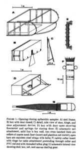
But first, what is an R.P. sled and why is it such an important key in the collection of copepods? The R.P. sled is an epibenthic sampler. That means that it samples the epibenthic animals – the animals that live just at the top of the (soft) seafloor – and a majority of these are often small crustaceans. The “R.P.” in the name stands for Rothlisherg and Pearcy who invented the sled. They needed to collect the juveniles of species of pandalid shrimp that live on the sea bottom floor. These animals are very small so a plankton net was necessary to collect them; a ‘normal’ dredge would not quite cut the job. They needed a plankton net that could be dragged over the bottom without damaging the net or the samples and also would not accidently sample the water column (pelagic); and so, the R.P. sled was born. This sled was able to go deeper than 150m, sample more than 500m3 at the time and open and close on command which was a novelty in comparison to the other sleds that where used in those days (1977). The sled consists of a steel sled like frame that contains a box that has attached to it a plankton net with an opening and closing device. The sled is heavy, ca. 150kg, and therefore limits the vessel sizes that can operate it; the trawl needs to be appropriately equipped including knowledgeable crew. It is pulled behind the vessel at slow speed to make sure the animals are not damaged and to make sure it does not become too full of sediment that is whirled up.


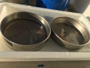
Sieved animals from the decanting process

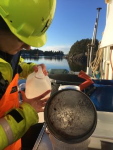
So off we went with r/v Hans Brattström pulling the heavy gear at ca. 700m depth with 1 knot and a bottom time of 10 minutes sampling the Krossfjorden close to Bergen. It was a beautiful day for it with plenty of sun and calm seas. The crew handled most of the sled, leaving sorting the samples up to HYPCOP under the guidance of Anne Helene. Which is not as straight forward as it may sound! The process of filtering the samples after collecting them from the sled is done by decanting, which you can see in this movie from an this blog (in Norwegian) from earlier.

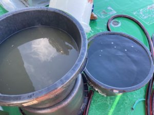
Decanting set-up for R.P. sled samples

Decanting means separating the mixture of the animal soup from the liquid by washing them in a big bucket, throw the liquid through a filter and collect the animals.

Sieved animals from the decanting process

This all needs to be done with care as the animals are often very small and fragile. After collecting, the most time-efficient and best preservation for the samples is to fixate them immediately with ethanol, so they don’t go bad while traveling back to the museum.

Fixating collected animals with technical ethanol

For collecting copepods we use a variety of methods; from snorkeling, to scoping up water and plankton nets, but for greater depths and great quality benthic samples the R.P. sled will be the most important method. We thank Anne Helene for her wisdom and enthusiasm that day for showing HYPCOP how to work with such interesting sampling method


We got some nice samples that will be sequenced very soon so we can label them appropriately. Although this first fieldwork trip off the year was mainly a teaching opportunity, we still managed to sample two stations with plenty of copepods and lots of other nice epibenthic crustacea, and Anne Helene is especially happy with all the amphipods she collected during the day. So for both of the scientists aboard this was a wonderful day – sunshine and lovely samples to bring back to the lab!

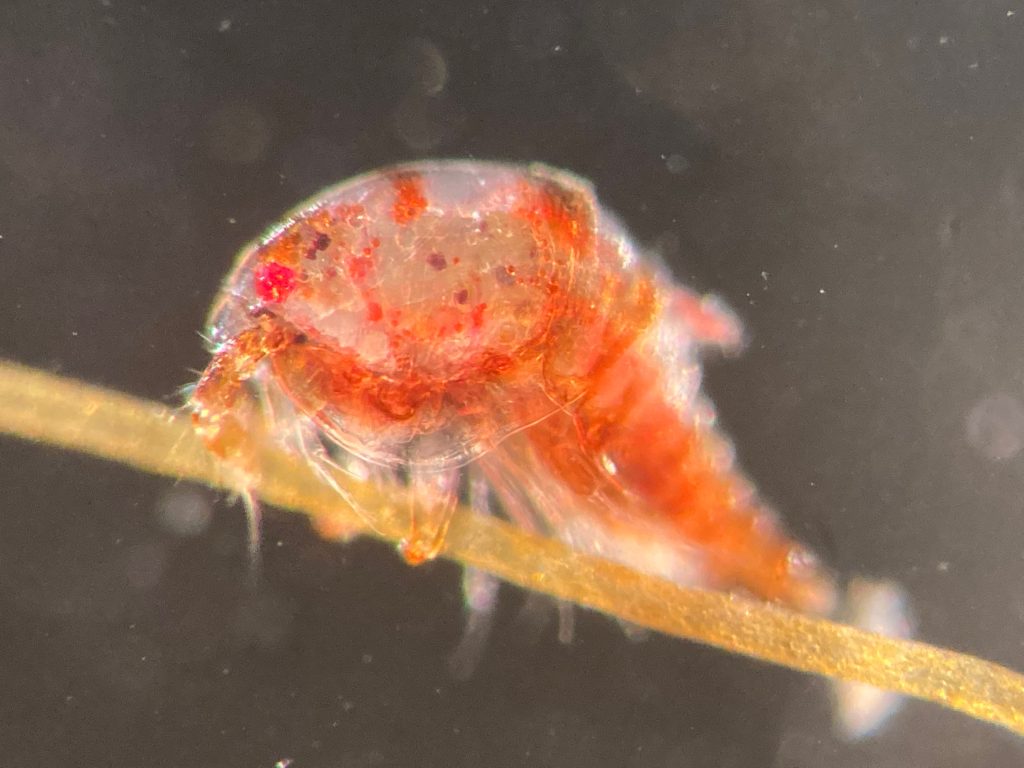
Some fresh copepods caught with the R.P. sled

– Cessa & Anne Helene


Follow HYPCOP @planetcopepod Instagram, for pretty copepod pictures https://www.instagram.com/planetcopepod/

Twitter, for copepod science news https://twitter.com/planetcopepod

Facebook, for copepod discussions https://www.facebook.com/groups/planetcopepod

See you there!

What’s growing on the shell? – Insights into the Diversity of Hydractiniidae in Norway

This summer I started my master degree project at the University Museum of Bergen and joined the research of Luis Martell and Aino Hosia. I’m a student in the program ‘Biodiversity & Systematics’ at the University of Stockholm and for my 1-year thesis project I wanted to learn more about hydrozoans. Looking for hydrozoan biologists, which there are not so many, I came across the project NorHydro led by Luis and Aino and I decided to go to Bergen to study and learn more about this fascinating group.

My thesis project especially focuses on the diversity of the hydrozoan family Hydractiniidae. Most commonly hydractiniids are encountered on snail- or hermit crab shells where they can build a mat of polyps. Those shells are often inhabited by other animals and the polyps feed on the left-over meals of those and in return defend their host against predators. With their small tentacles, which are equipped with hundreds of cells containing venomous stingers (the nematocysts) they are great predators and catch tiny animals from the plankton.

Schuchertinia allmanii growing on Pagurus pubescens hermit crab. Picture credit: Bernard Picton

Each individual polyp is connected to each other and build a colony of polyps with different functions. Some act as food suppliers, some as reproductive polyps. From those polyps some hydractiniid species grow tiny medusae (jellyfish) which will be released in the water column. The medusa is morphologically comparable to the polyps, equipped with tentacles, a stomach, gonads and nematocysts – but living upside down and not attached to the sea floor. With their ability to swim around they contribute to disperse themselves in the water and can produce a lot of offspring.
Video of a Pagurus bernhardus hermit crab. A hydractiniid polyp colony is growing on the underside of the shell. Video credit: Lara Beckmann

Several species in this family are often used in development biology or immunology research. For example the species Hydractinia echinata led among other organisms to the discovery of stem cells and is still widely used as a so-called model-organism nowadays. This is because this animal group shows a high ability to regenerate and if you cut a hydroid polyp in two pieces, it will just re-grow again to its initial state.

The life-cycle in hydractiniids can include a polyp and medusa stage. Species of the genus Podocoryna release medusae from their colony. Other genera in this family develop only reduced medusae and lack a free-swimming jelly. Illustration credit: Lara Beckmann

The hydromedusa Podocoryna borealis is released from a polyp colony that can grow on various substrates such as snails or worm tubes. Picture credit: Lara Beckmann.

But despite this spotlight to some species, the diversity of this family is still poorly known and there is little attention to hydractiniid species because of their inconspicuousness and difficult identification. To highlight the problem I did some research: how many and what species are commonly recorded in Norway? Which species could potentially occur in this region according to species descriptions? In private observations and ecological surveys only three species were commonly recorded. However, our preliminary results suggest at least 6 species in Norwegian waters with some surprisingly frequent species that are rarely recorded in surveys. This indicates, that there happens a lot of misidentification in the field and we need to get a better estimate of the true diversity of Hydractiniidae in this region in order to improve awareness and to prevent misidentification in the future.

Two polyps of a colony of the species Clava multicornis. You can see reproductive units (so-called sporosacs) growing on the polyp body, which will release eggs or sperm when fully grown. Picture credit: Luis Martell

In order to do that I study this group in much detail. A great advantage of being a taxonomists in these days is that we can use a variety of different sources of information to develop and test for species hypothesis. This so-called integrative approach includes morphological, ecological and genetical information in order to define taxa diversity from several perspectives.

The first step in this process is to gather polyps and medusae of Hydractiniidae from various spots in Norway ranging from Olso, via Bergen and up north to Bodø. I will use already collected samples from previous years and hopefully will also be able to add some more samples from field-trips in the next months.

Getting the plankton-net back on board – and Maryam and me trying to open the jar carefully without spilling the fragile plankton inside. After removing the jar from the net, we directly pick the hydrozoans and put them in a cold environment, so that we can look at them alive when we are back in the lab. Picture credit: Maryam Rezapoor

With fresh material the first step is to take pictures of the living animal. Since hydrozoans tend to shrink and lose a lot of it characteristic traits in the ethanol preservative it is important to take pictures right away when the animal is still alive. Furthermore the samples are identified using a microscope, identification keys and additional literature. With additional collected data such as habitat, location, sample depth and so forth we create a large database for our samples and develop so-called e-vouchers: an electronic data storage for each individual to ensure to gather and store as much information as possible. That makes it possible to go back to the database and look at the specimen data e.g. where did it live? On what substrate was it growing on? How deep was the sampling site? This way I don’t only rely on the preserved material but can easily use the e-voucher material and the additional collected data that can be useful for further analyses.

Besides the morphological and ecological data I also include molecular information: what does the DNA tell us about the specimen? For that I use three different genetic markers (short DNA fragments, or genes) to see how those differ in their sequence of bases between the sampled hydractiniid specimen. In the course of evolution, DNA sequences change due to selection, genetic drift, gene flow and random mutations. That is why we are able to infer evolutionary relationships between biological entities – in this case we are interested in species – and with mathematical models and some simplified assumptions we can delimit species based on their molecular attributes.

Me in the DNA lab waiting for the first results of lab work for this project! Picture credit: Maryam Rezapoor

In order to do so, I will process the sampled material in the lab. First, I extract the DNA from a tiny piece of polyp or medusa tissue and then amplify the specific target region using PCR (Polymerase-Chain-Reaction). This short DNA fragment will further be sequenced which outcome I will check, process and analyze using different computer programs and algorithms. The final results will be summarized and visible in so-called gene trees, revealing the evolutionary history of those three different DNA markers. I will use the gene genealogies to delimit the species and observe how much diversity of hydractiniids we have in Norway.

Using specific genes also facilitate us with species barcodes which are used to create a reference library. This in return enables researchers to identify a specimen without the need to identify the sample using its morphology. This can be the case e.g. for metabarcoding projects (sequencing a mass of DNA from unknown origin) or if samples are unidentifiable. The scientist will then sequence the barcode gene and compare it to the reference library and will eventually be able to tell the species. Those barcodes can be informative in biodiversity studies but by no means substitute taxonomists, which are still needed to describe and identify species when no DNA is available.

A monograph of the Gymnoblastic or Tubularian hydroids by George James Allman, published in 1870. I found this book in the library of the Tjärnö laboratories in Sweden. Allman was a pioneer in hydrozoan research and described many species. Nowadays it is much easier to access original descriptions since many book are available online. You can find a PDF* of this book in the Biodiversity Library. Picture credits: Lara Beckmann

Also, the DNA does not help us when we have no reference we can map the specimen to in order to determine the species. That is why this master project also aims to create a barcode-library for the species in the family Hydractiniidae.

What I really like about this project is that it contributes to our basic understanding of the diversity in our oceans – in this case even in two completely different environments: the water column as well as the seafloor.

Even if hydractiniids are just a little part of the ecosystem, they play their role and influence the ocean in their own way. Also, my work is super diverse and I need to be an allrounder in many respects: trying to find species descriptions I search and read taxonomic literature from the 19th century (coming in Latin, German, English, Swedish or French). Then I work in the laboratory where I combine traditional working methods like drawing, microscopy or photography with modern techniques from DNA sequencing to computational science and bioinformatics.

I love this kind of work, and it is great to contribute just a little bit to reveal the mysteries of our oceans.


– Lara

You want to learn more about hydrozoans and why it is important to study them? Read more about it in my blog article for Ecology for the Masses (link).

Also, keep up with the activities of NorHydro (link to project home page) here in the blog, on the project’s facebook page (link) and in Twitter with the hashtag #NorHydro.

*Link to pdf of A monograph of the Gymnoblastic or Tubularian hydroids by George James Allman