Tag Archives: Mollusca

Minute snails in Northern Norway

From 21 till 28th of May, researchers, technicians, and students of the University Museum’s marine section, travelled up North to Torsvåg, close to Tromsø, for joint fieldwork. The participants represented several Artsdatabanken projects that cover marine fungi, hydrozoans, polychaetes, parasites of jellyfish, comb jellies and chaetognaths, bryozoans, marine amphipods and finally the Lower Heterobranchia and Pyramidellidae gastropods. In this blog you can read about the general experience of the fieldwork and more details about the different projects. And here you can read about the fieldwork through the eyes of two master students who joined. And if you want to read more adventures and see more pictures check out this blog post! It was a large group of young and more experienced scientists which created the perfect opportunity for a lot of knowledge transfer.

This was also the first “big” fieldwork trip for the Lower Heterobranchia and Pyramidellidae project, after several sampling events in the Bergen area, which you can read here. Both Lower Heterobranchia and Pyramidellidae include small snails, just a few millimetres in length that are hard to identify (they can resemble other small species of gastropods). The diversity of these tiny sea snails is poorly understood in Norway, and thus, during this project these sea snails will be studied by combining DNA barcoding and shell characters. Sampling will be based on the use of dredges, grabs, and snorkelling.

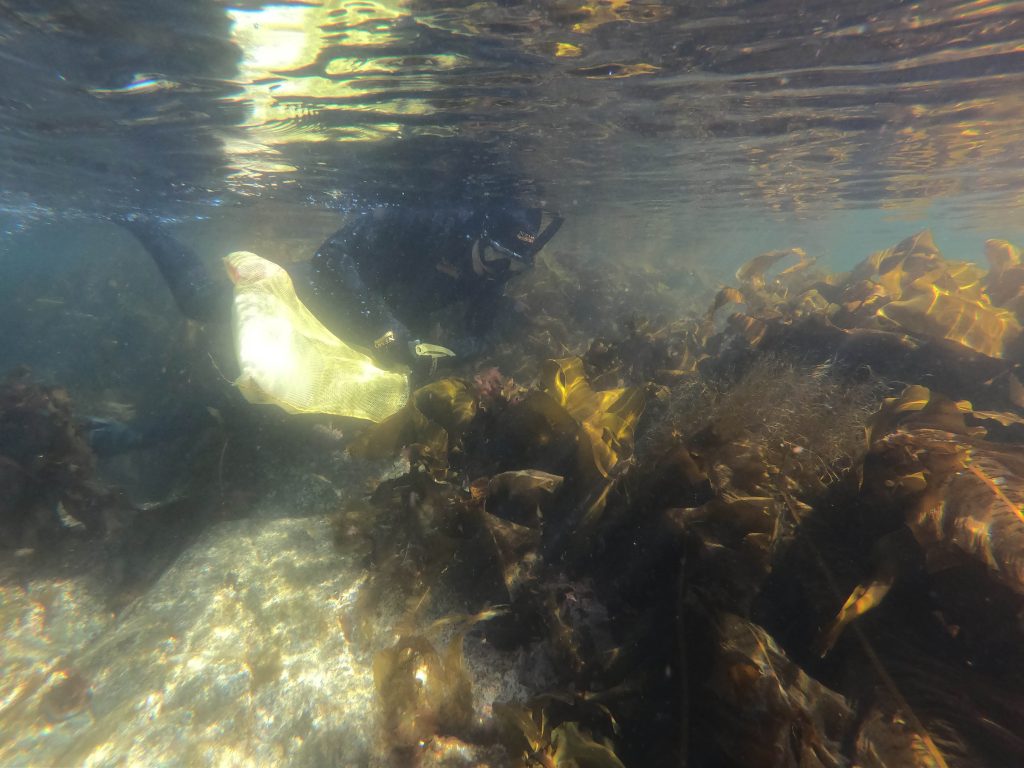
Pyramidellidae are regarded as ectoparasites and are often found living on other molluscs or other marine invertebrates, but also free in soft sediments. The Lower Heterobranchia are often found on algae, for example on the stipes and in between the holdfasts of large kelp. Because of the small size of these snails the best way to collect them is by sampling the substrate they live on. So, this is what we did, we went snorkelling several times in the cold waters of Northern Norway, but thanks to good neoprene layers we were able to keep warm and simultaneously looking like seals!

Underwater shot of a person snorkelling, collecting algae just below the surface

Collecting the right substrates for the Lower Heterobranchs and Pyramidellidae while snorkeling. Photo by Eva Charlotte Samson, UiB

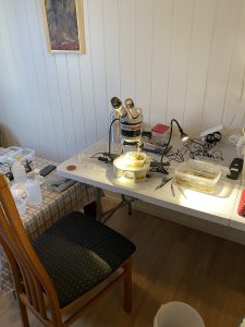
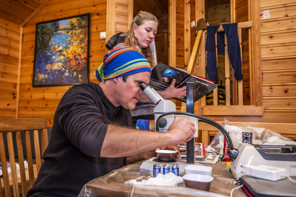
Little lab set-up in the kitchen to sort through the samples.

Little lab set-up in the kitchen to sort through the samples. Photo by Cessa Rauch, UiB.


It was challenging to find our snails; there was plenty of kelp and high diversity of many other taxonomic groups, but the conditions were not exactly right especially for the pyramidellids that seem to prefer areas with strong currents.

So, even though we sampled many different habitats, we often ended up not finding our snails when back in the lab sorting under the microscope.


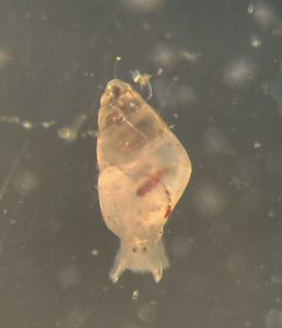
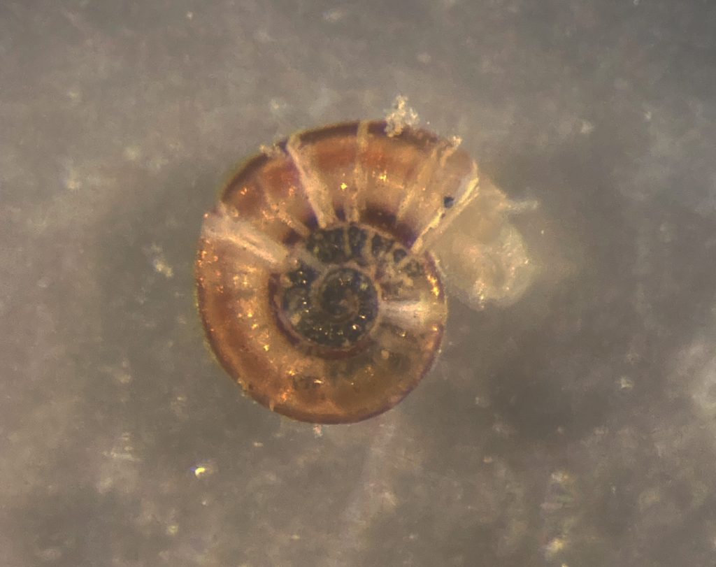
A tiny snail lookin up at the camera

Species of Pyramidellidae; Odostomia turrita. Photo by Cessa Rauch, UiB.


Yet, after collecting a ton of material and spending many hours sorting, we finally found one pyramidellid!

In this case Odostomia turrita.

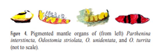
Odostomia are neat little pyramidellids that have glands with distinct colours, which makes somewhat easier the identification of species.

Drawings showing the colour patterns of snails

The different colour patterns distinguish different Odostomia species. Source from Høisæter 2014. (Høisæter, T. (2014). The Pyramidellidae (Gastropoda, Heterobranchia) of Norway and adjacent waters. A taxonomic review).

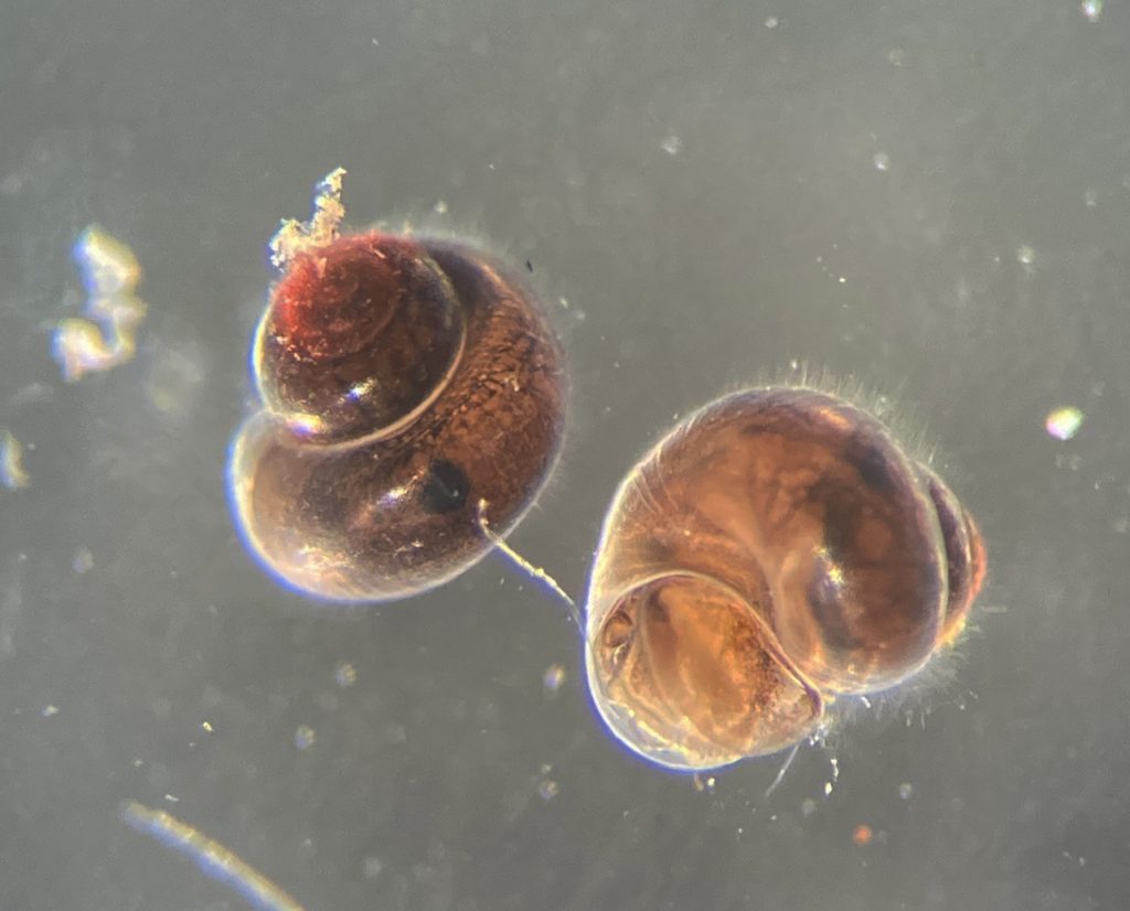
Although minute, the lower heterobranch were “easier” to find… They seem to be less picky with the environment, and on kelp and sand we managed to sample a few different species, amongst others two very similar ones: Ammonicera rota and Omalogyra atomus. In addition, we found in the sand and gravel small snails of the genus RissoellaR. globularis.

Field season has just started, this was a good beginning for a busy Summer with many more blogs to come!

-Cessa & Manuel

CT scan all the things!

Oslo Natural History Museum 02.12.2019 – 06.12.2019

Literature for the week of our CT scanning course


From December 2 till December 6 Justine Siegwald, Manuel Malaquias and I (Cessa) attended a CT scanning course in Oslo at the Natural History Museum.

The course was organized by the Research School in Biosystematics (ForBio) and Transmitting Science.

The group attending the CT scanning course in Oslo at the Natural History Museum

The three of us all work on mollusks and one of the main reasons to deepen ourselves in CT scanning techniques is because of studying the internal anatomy of these often small and delicate specimens.

Species descriptions are not only based on external morphology and/or DNA barcoding, a lot of the species differences are small details within the internal structures. Like the reproductive organs and digestive system (e.g. radular teeth) of the animals. This is very laborious work, and can be challenging especially with the smaller specimens of only a few millimeters long. Besides some species are difficult to collect and can be a rare collection item of the museum. Once you cut these animals to study them, there is no turning back and the specimen is basically destroyed. Being able to see the insides of the animal without touching it would therefore be ideal. CT scanning comes very close to that and with powerful X-ray we can almost see every detail of the insides of the animals without the need to cut them open

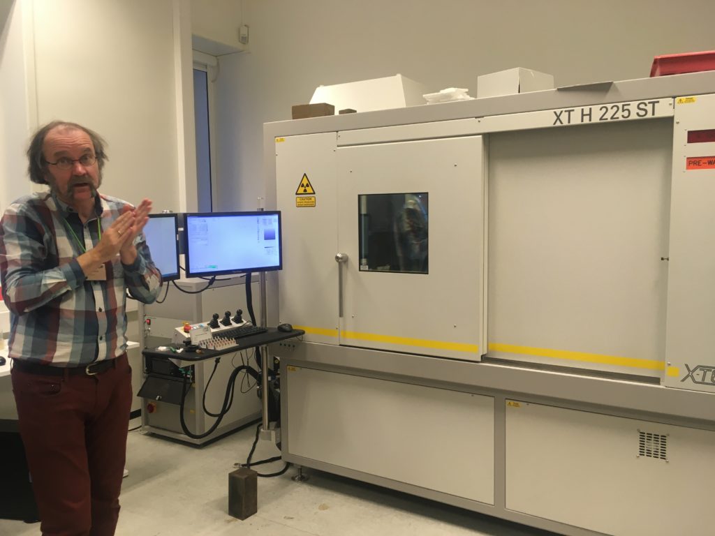
The CT scan in the Natural History Museum of Oslo with one of the teachers in front (Øyvind Hammer)

Lecture about the technology of the CT scan and X-ray

However, how easy as this sounds, CT scanning soft tissue comes with some challenges. Soft tissue means that the x-ray contrast is often very low. Even with modern good x-ray detectors it is still difficult to detect the different internal structures. Therefore, during the course, we were taught how to artificially increase the contrast of the tissue by staining the specimens before mounting them in the CT scan machine.

Coating a specimen with a metal (e.g. gold or platinum) is only useful if you want to see external details of the study object, coating will not help revealing internal structures. For radio contrasting the internal anatomy, you can stain specimens with high density liquids like iodine to enhance the x-ray contrast. So, we went for this option and left our specimens in iodine ethanol solution overnight


After the staining process, we needed the wash out any extra iodine before mounting the specimen into the CT scan. A good scan is not simply pressing the button of the machine; there are a bunch of settings that can be adjusted accordingly (e.g. tube voltage, tube current, filament current, spot size, exposure time, magnification, shading correction, etc.). The scan itself can take hours and file sizes of 24GB for a single object are not uncommon, which means you need a powerful computer with decent software to process this information. Part of the course was also about how to visualize and analyze the files of the CT scan with software like Avizo

Video 1. Software Avizo has a user-friendly interface for analyzing all sorts of CT scanning files.

The week gave us some very promising first results (se video below), and also new insights about how to increase the X-ray contrast of our sea slug samples (lots of hours of staining and very short washing steps).

Video 2. 3D reconstruction of one of Justine’s shell samples

It was a very fruitful week and besides interesting new ideas for scanning our museum specimens, we also met many old and new friends during the week that was as inspiring. ForBio and Transmitting Science did a really great job with setting up this course and I can highly recommend it to anyone who is interested in CT scanning.

Explore the world, read the invertebrate blogs!

-Cessa

Sea slug hunt in Egersund!

I’m always scared to look at the current date, time flies! It was already two months ago that we went on a blitz fieldwork trip to Egersund with a very special group of people. But nevertheless, good times become good memories (and especially good museum specimens) and it definitely does not get too old for a small blog about it.

From January 17 to January 21 a small group of sea slug enthusiasts consisting of a student, citizen scientists, a collaborator and museum members rented a van and drove 7 hours down to our Southern neighbor town Egersund.

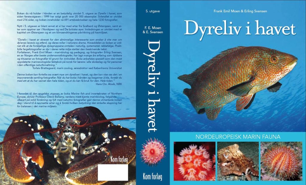
Egersund was not randomly picked as it is the home town to one of Norway’s most productive and dedicated ‘citizen scientist’; Erling Svensen. Author of a number of books and the most well-known and worldwide used ‘Dyreliv I havet – nordeuropeisk marin fauna’ (English Marine fish and invertebrates of Northern Europe), which amateurs and professionals alike use as an extensive research source.

Erling Svensen’s famous book Dyreliv I Havet

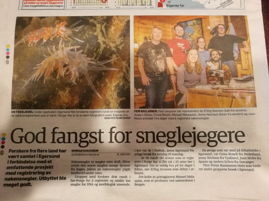
With his almost 5000 dives and counting, Erling knows the critters of the North Sea, big and small, on the back of his hand. Already since the beginning of the sea slug project, Erling was helping providing valuable sea slug species, so it was about time to pay him a visit and bring our team over to make Egersund “biologically unsafe” – enough so to end up in the local news!

We made Egersund unsafe enough to have a small news item about it in the ‘Dalane Tidende’, a local newspaper

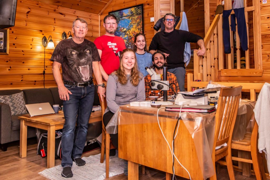
The group consisted of Manuel, Cessa, citizen scientist Anders Schouw, collaborator from Havard University Juan Moles and master student Jenny Neuhaus

From left to right Erling Svensen, Anders Schouw, Jenny Neuhaus, Cessa Rauch, Juan Moles and Manuel Malaquias. Photo by Erling Svensen

Jenny just started her Masters in Marine Biology at the University of Bergen in the fall of 2018, she will be writing her thesis on the diversity of sea slugs from the Hordaland county and on the systematics of the genus Jorunna (Nudibranchia) in Europe. The results of this work will definitely become a blog entry of its own.

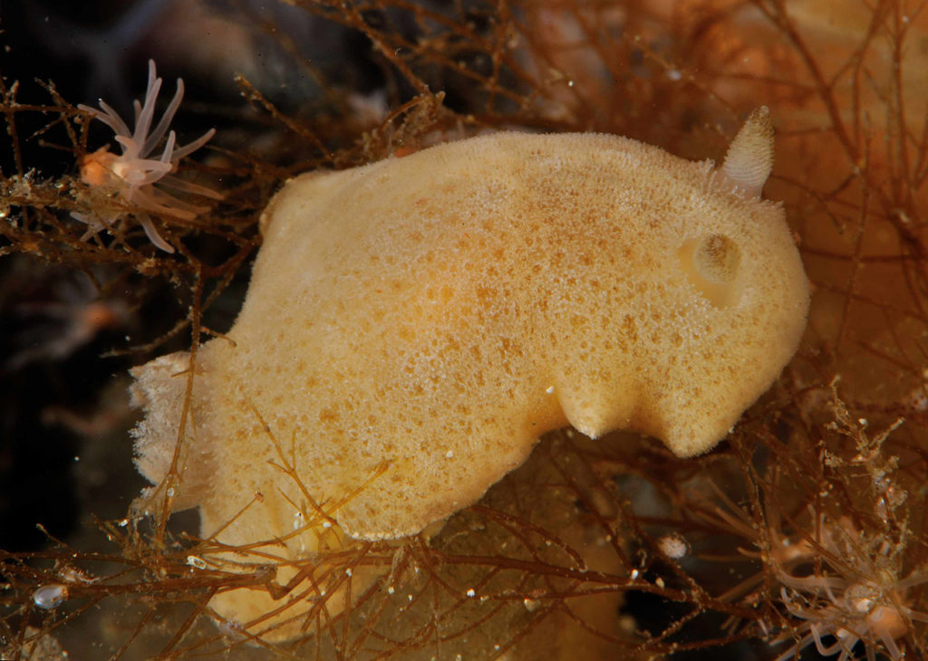
Jorunna tomentosa, Jennies new pet! Photo by Nils Aukan.

Two of the five days of our fieldwork were basically spendt driving up and down from Bergen to Egersund, it left us only with a good 3 days to get an overview of Erling’s backyard sea slug species. Little time as you can imagine. But time was used efficiently, as Anders and Erling are both extremely good sea slug spotters and with help of sea slug specialist Juan and the eager helping hand of Jenny, Manuel and I were able to identify and add 36 species to our museum sea slug database.

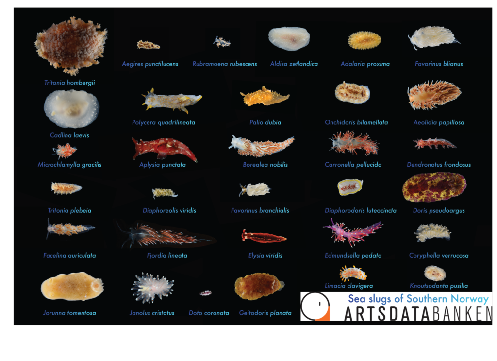
Overview of collected specimens in Egersund

In comparison, we registered 41 one species in Drøbak last year by spending almost a week at the field station! No one thought this would be the outcome (not even Erling himself, as he mentioned that he didn’t find that many sea slug species this time of year on earlier surveys). But we were all very happily surprised, and maybe it was not just luck but also the combination of people we had attending this short field trip. With so many good specialists, either professional or amateur, senior or junior, we were able to work extremely efficient and with a clear communal goal. There was little time spend in reinventing the wheel and explaining the work flow, it was a good valuable exercise that will definitely help us with future brief fieldwork trips and how to make the most from short and tight time schedules. Besides it was a very valuable experience for our student Jenny, as she got first-hand experience with what it’s like to see her study specimens alive, how to handle these fragile individuals, how to sort them from other species and how to document them, which is a good thing to know for her thesis and future career

Jenny learning a lot from the master himself. Photo by Erling Svensen

So yes, all in all our Egersund fieldtrip was short but very sweet!

Furthermore
You want to see more beautiful pictures of sea slugs of Norway! Check out the Sea slugs of Southern Norway Instagram account; and don’t forget to follow us. Become a member of the sea slugs of southern Norway Facebook group, stay updated and join the discussion.

Explore the world, read the invertebrate blogs!

-Cessa & Jenny

Alien species

Collaborative work between the University of Rostock and the Natural History Museum of Bergen

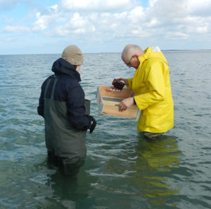
Prof Wolfgang Wranik (yellow coat) sampling in 2015

Prof Wolfgang Wranik (yellow coat) sampling in 2015

Professor Wolfgang Wranik from the University of Rostock in Germany has visited the Natural History Museum during the 13-14th of June to work on a recently detected invasion of an American species of haminoid gastropods observed in southern Scandinavia and the western Baltic Sea.

The species is apparently already reproducing and established in the area, but it is unknown when and how did it make is way across the Atlantic.

A combination of DNA and fine morphological data using scanning electron microscopy is being employed to compare specimens from both side of the Atlantic and confirm the identity of the European specimens.

Animal from Tjärnö, Sweden observed in January 2017.

Animal from Tjärnö, Sweden observed in January 2017.

Global warming is facilitating the spread of southern species into higher latitudes, and the role of shipping and aquaculture activities in re-shaping the distribution of many marine species is well documented. Among haminoids there is fear that a Pacific species established in the Mediterranean Sea since the 1990s (Haminoea japonica) is already displacing the native fauna of molluscs, which raises concerns about the possible impact of the US haminoid in our local environments. 

Manuel Malaquias, Natural History Museum of Bergen, UiB

Happy Easter!

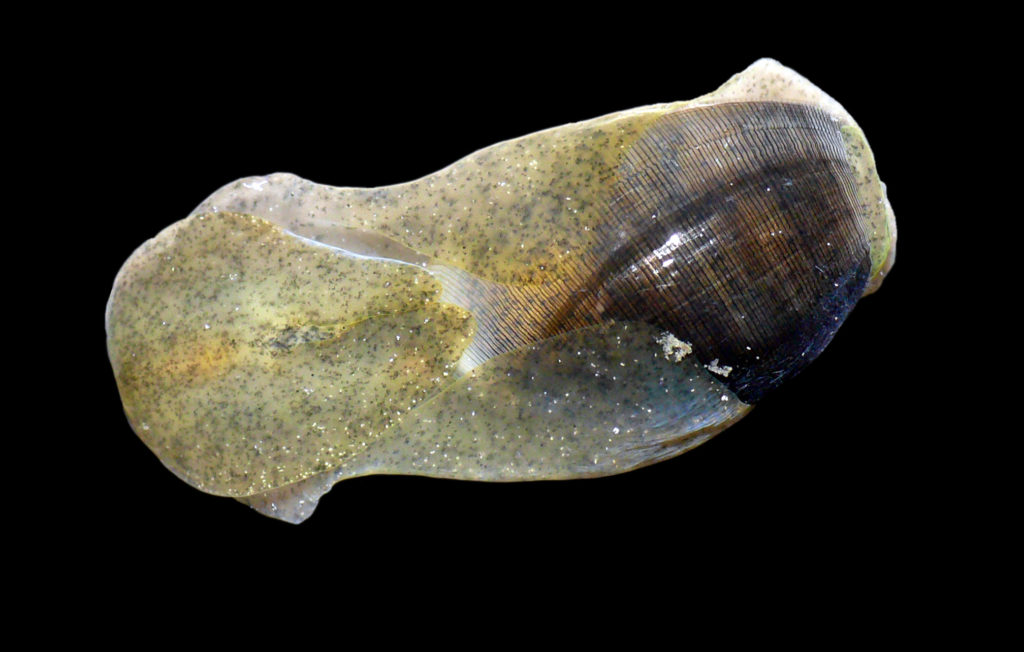
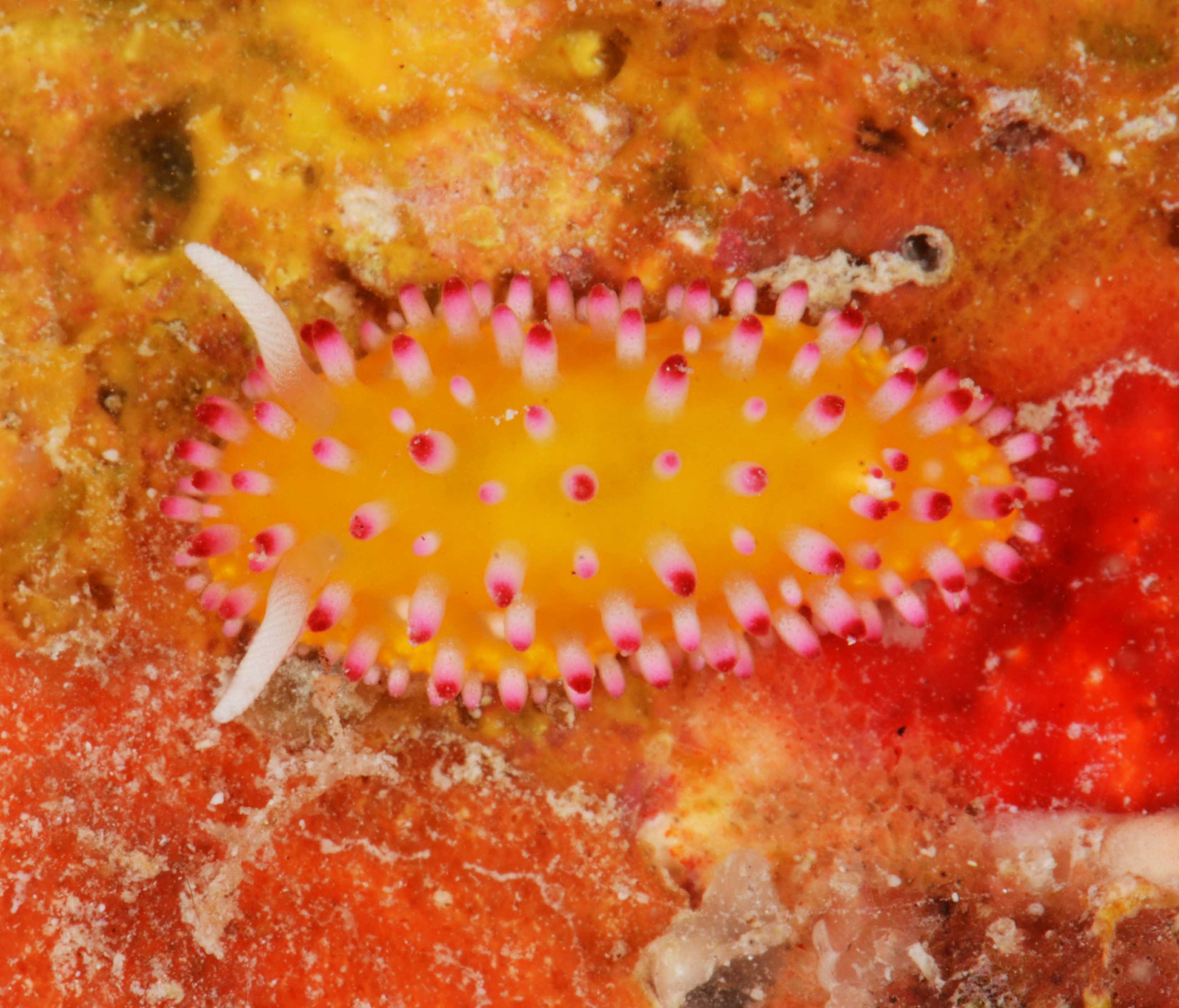
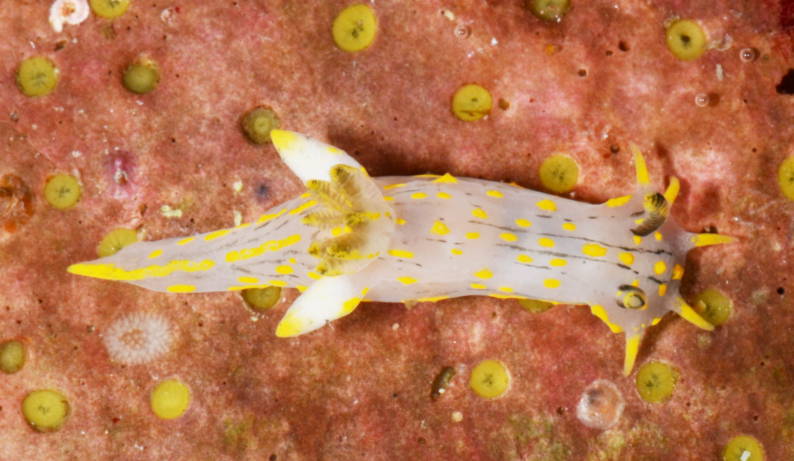
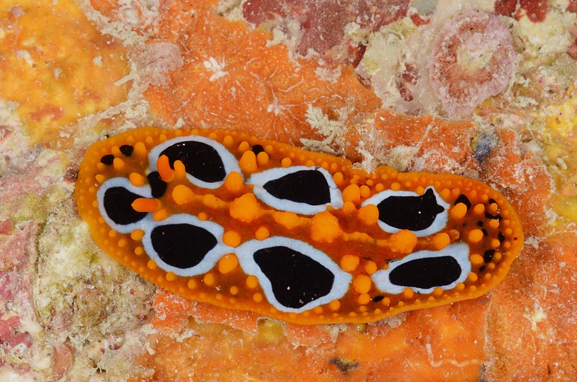
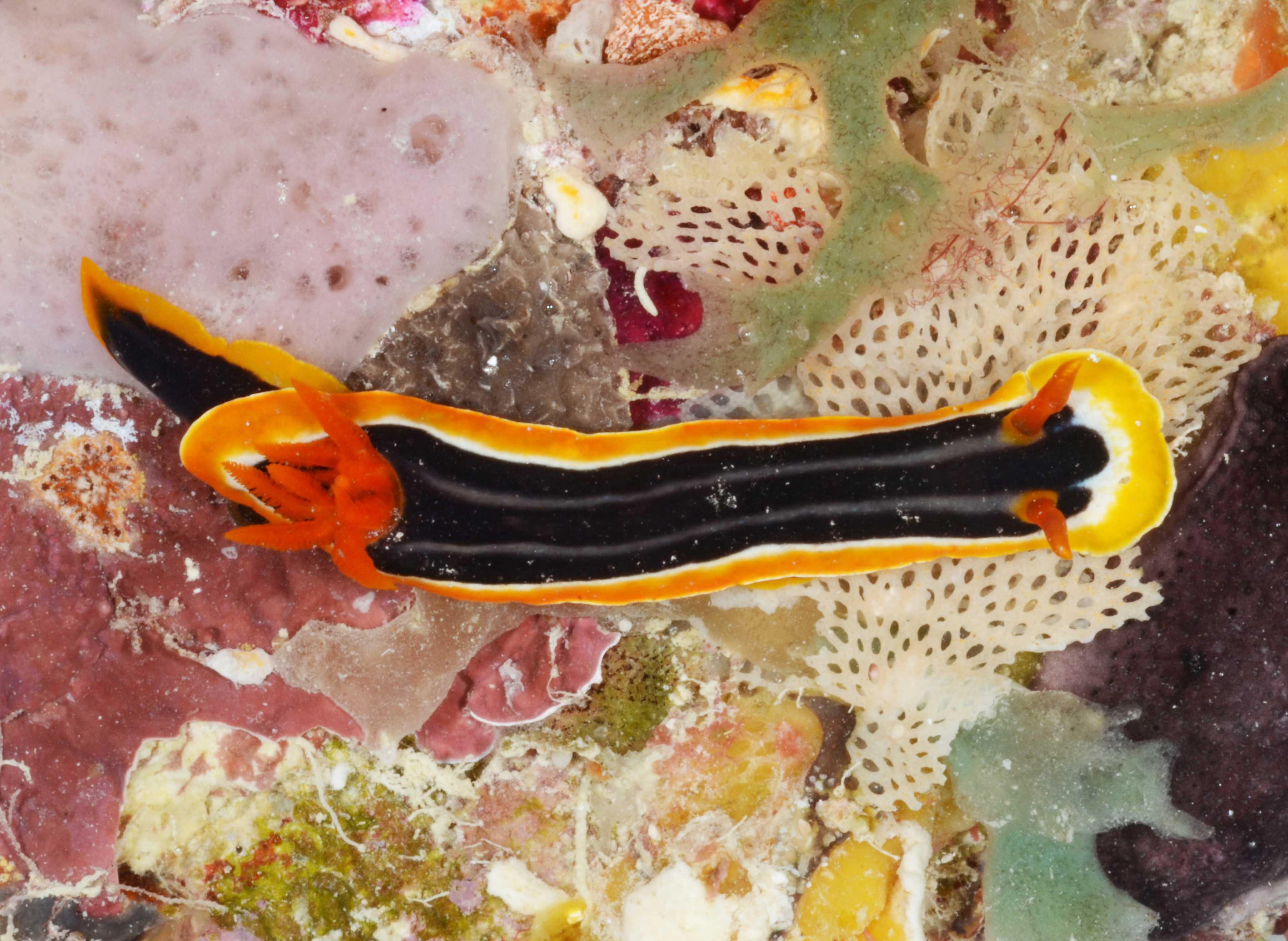
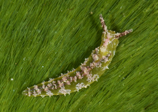
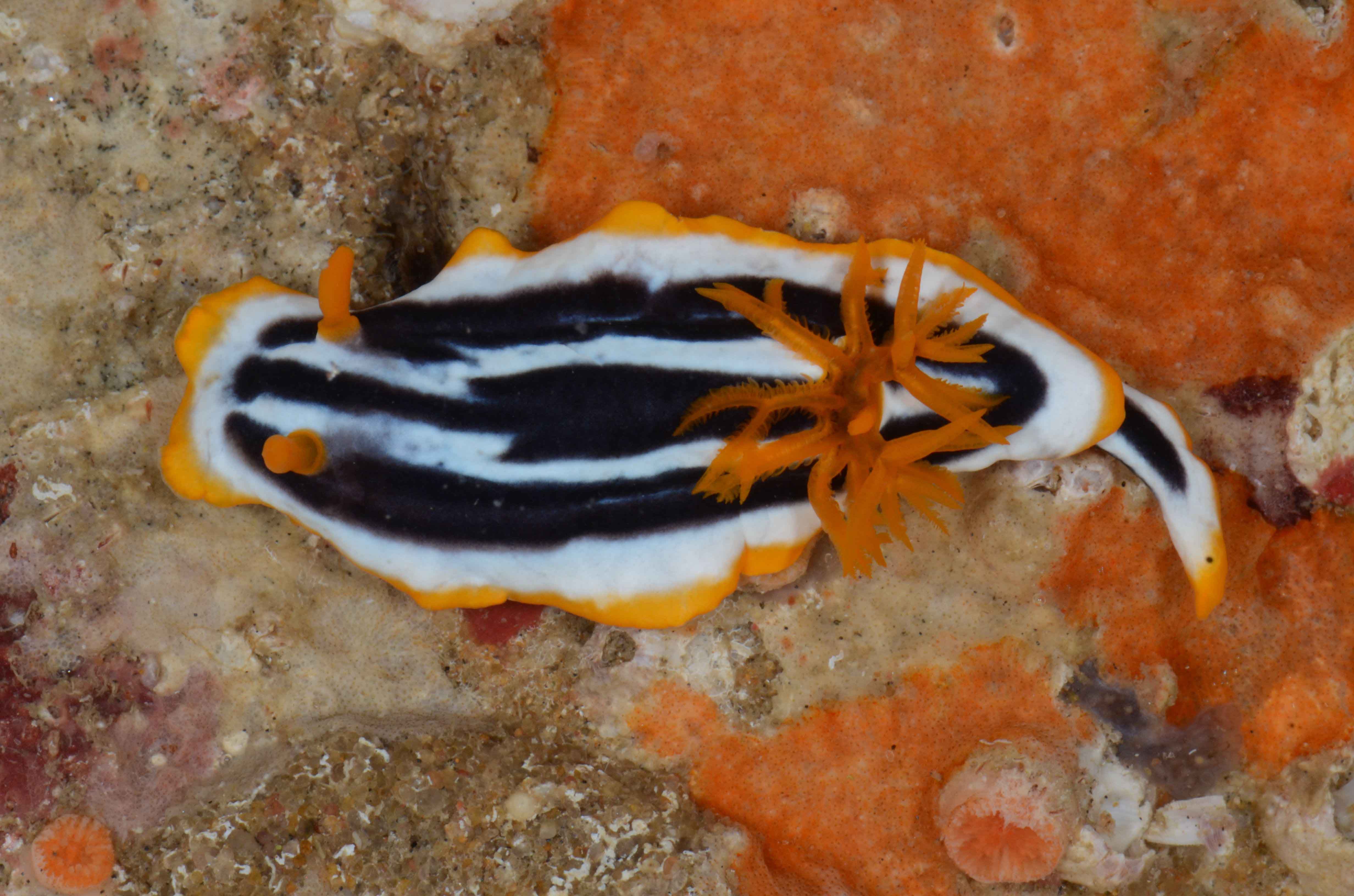
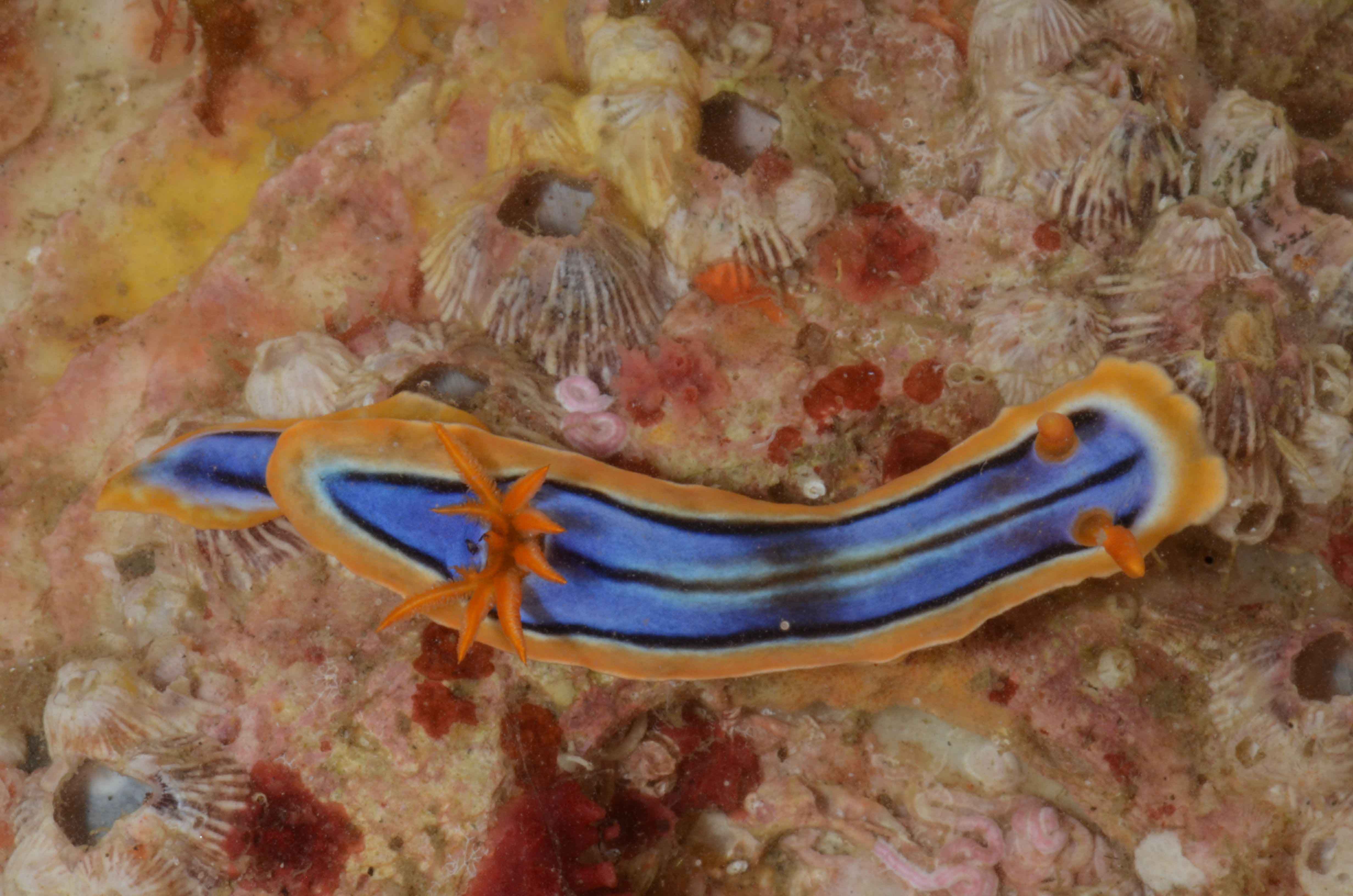
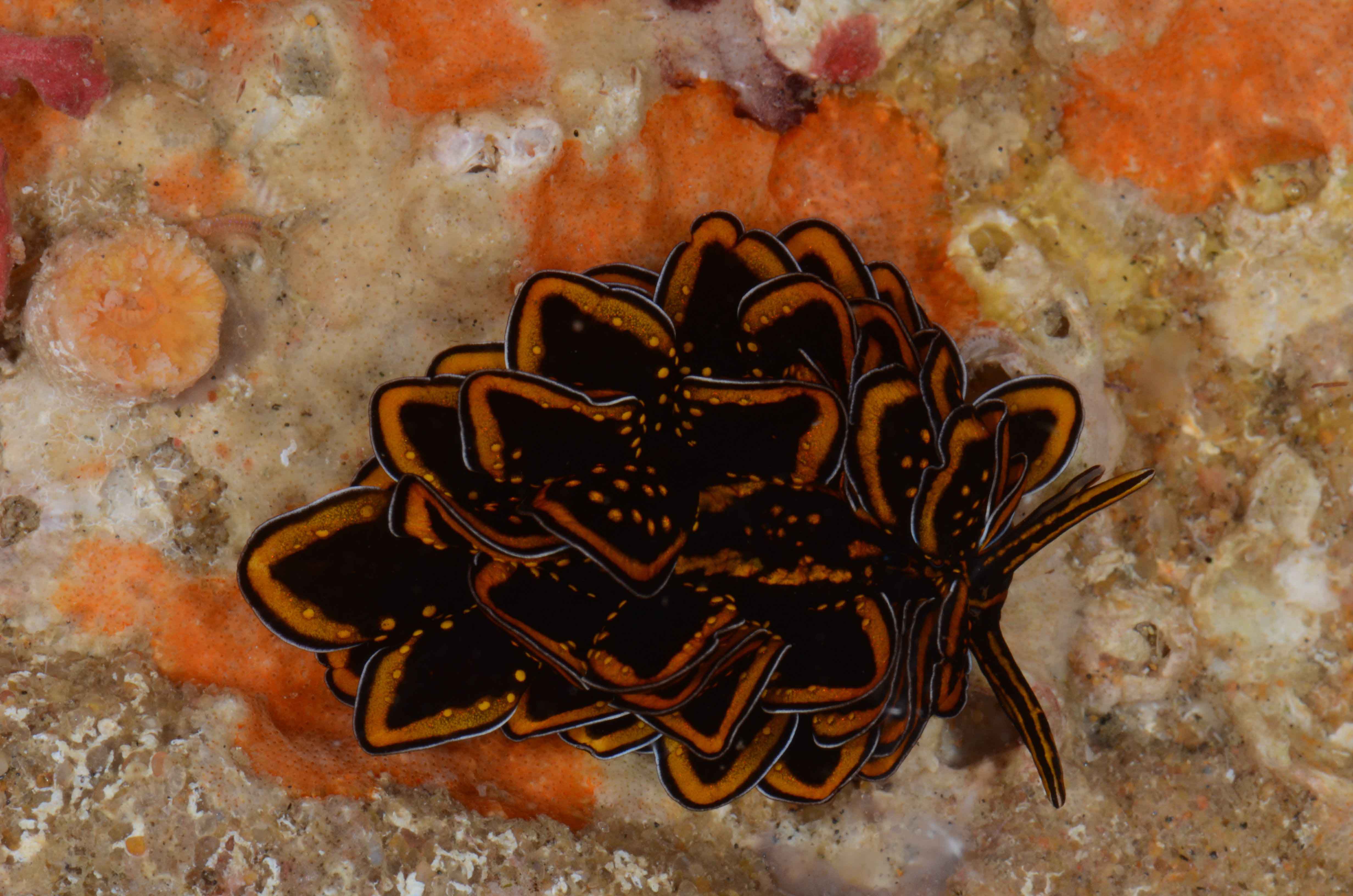
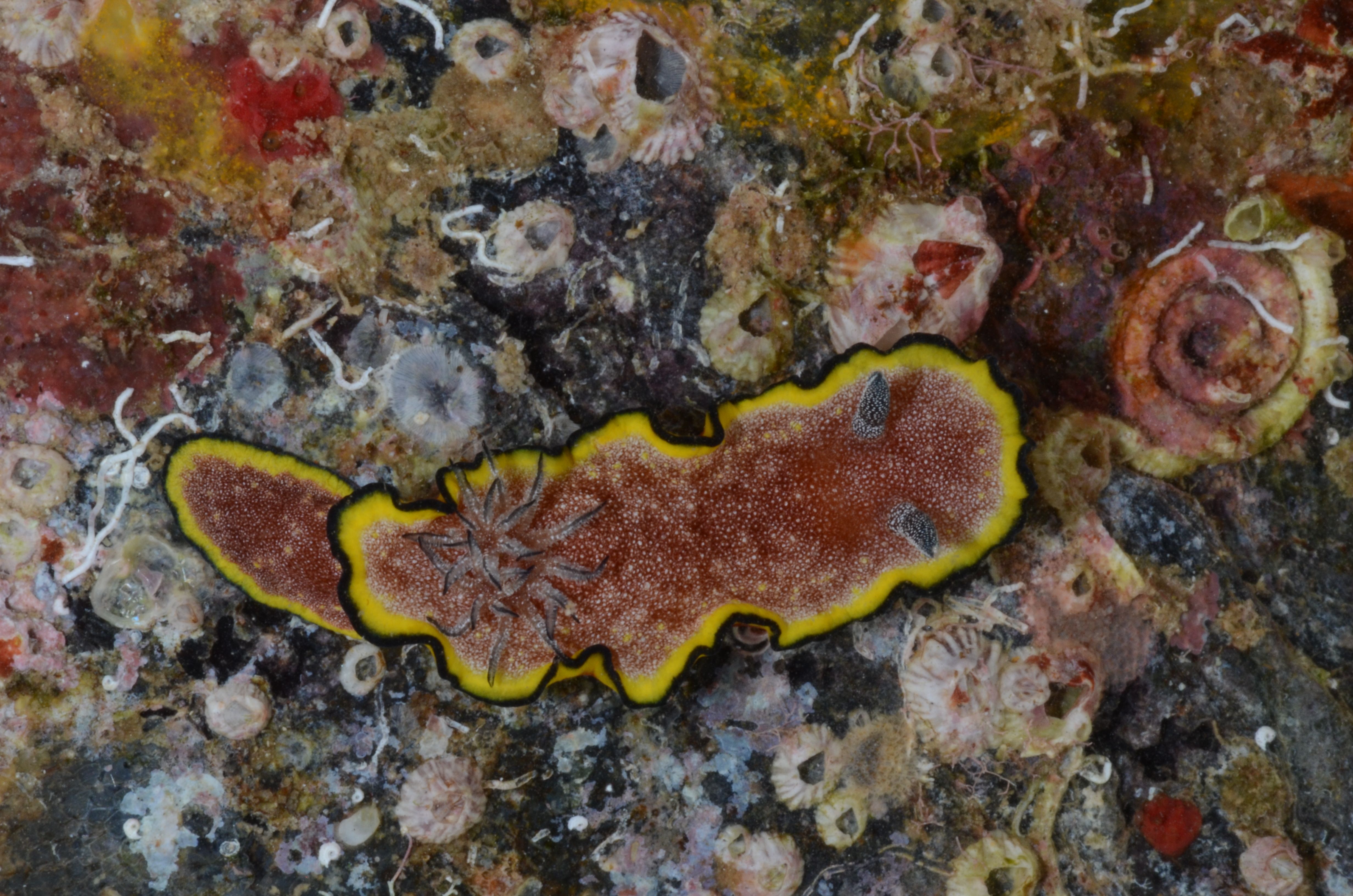
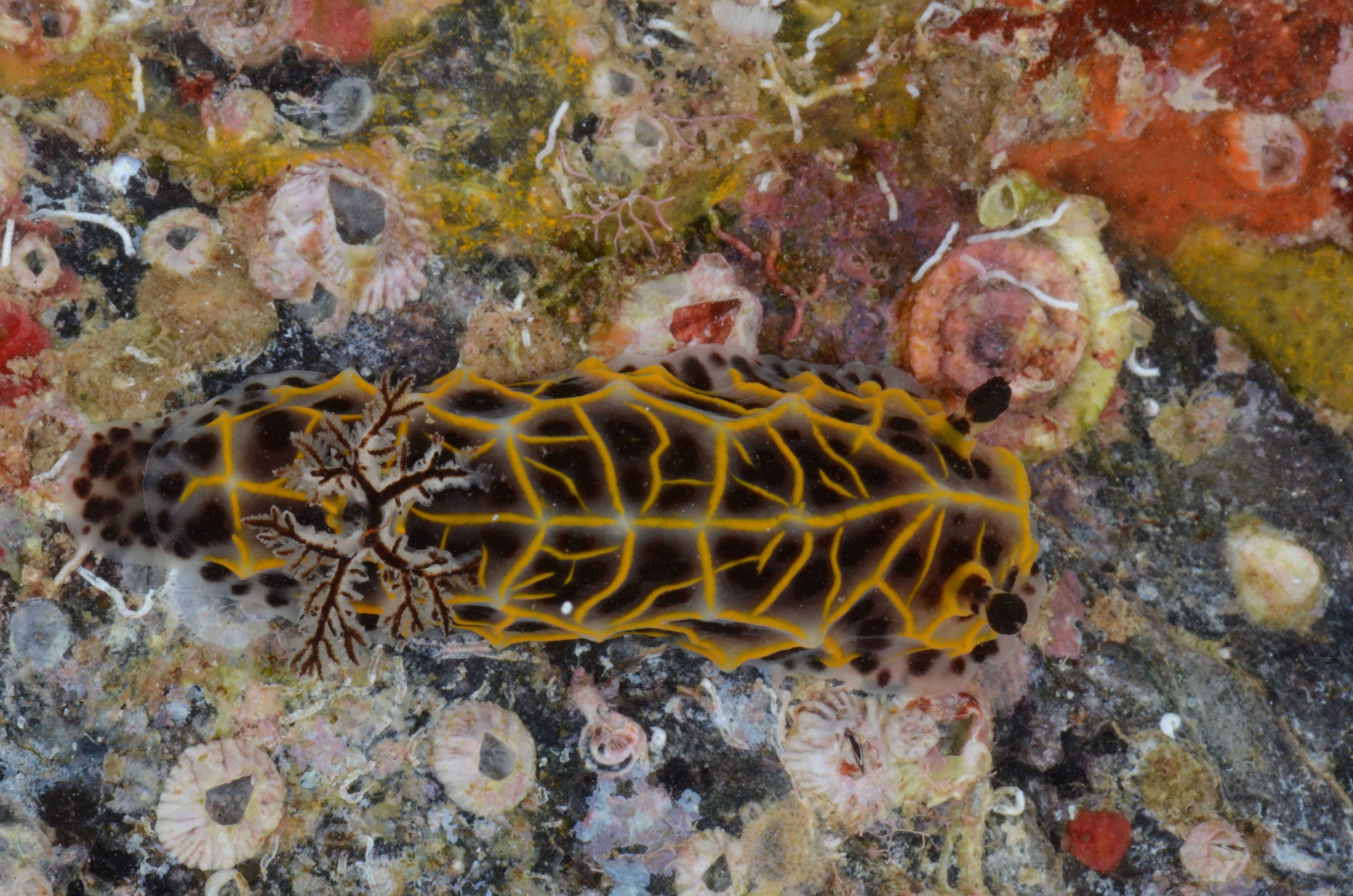
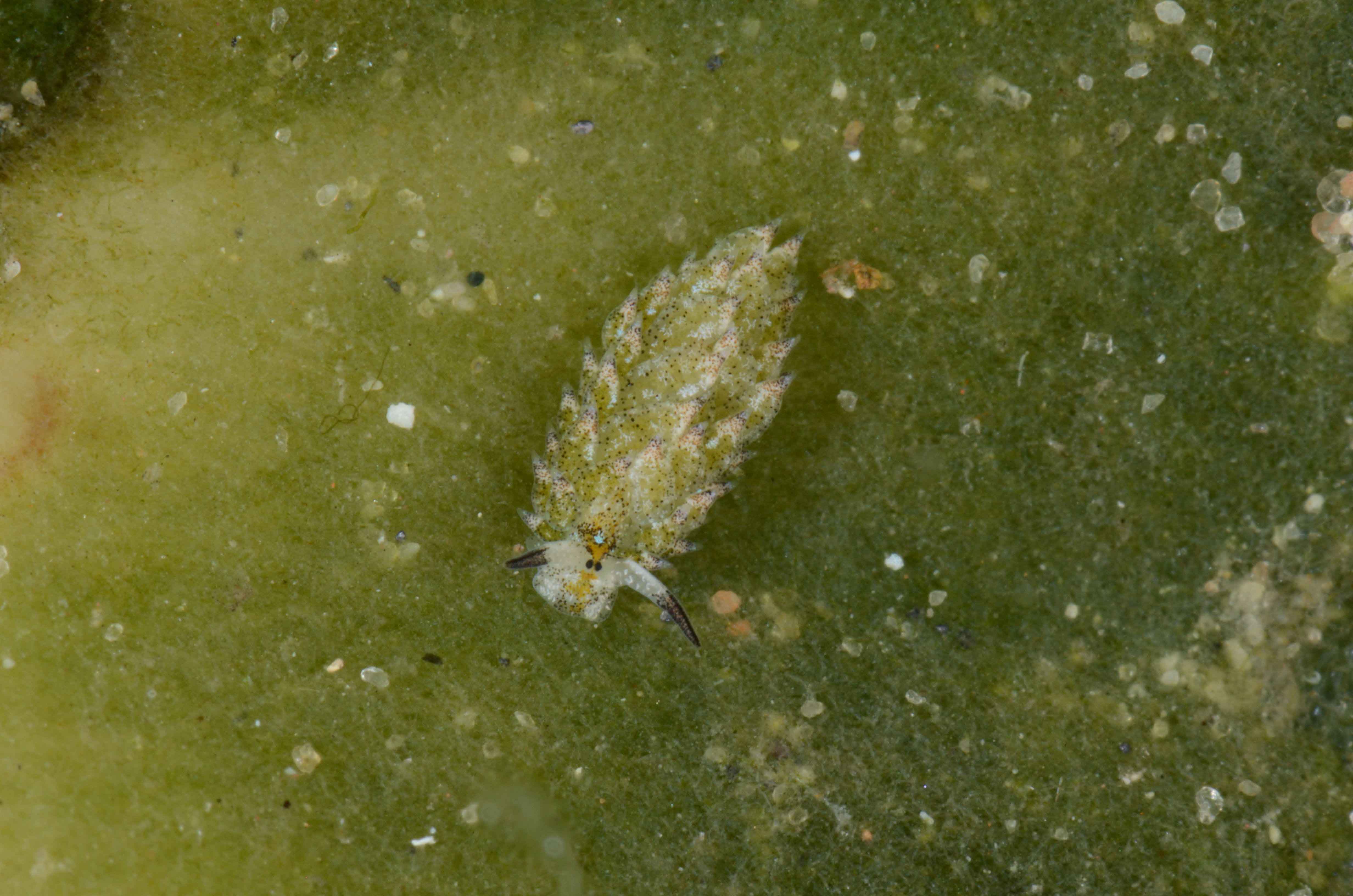
Here’s a collection of some of the “easter bunnies of the sea”, aren’t they amazing?

Photos by Manuel Malaquias, and you can read more about the animals, the field work and the collecting by revisiting the following blog posts:

Sampling for sea slugs in northern Mozambique (East Africa)

Uncovering the origin of species in the Caribbean region – fieldwork in the Florida Keys

Fieldtrip to Mozambique – collecting sea slugs in the most diverse marine biota of the World

More about… Fieldtrip to Mozambique – hunting for seaslugs

Guest researchers: Carlo

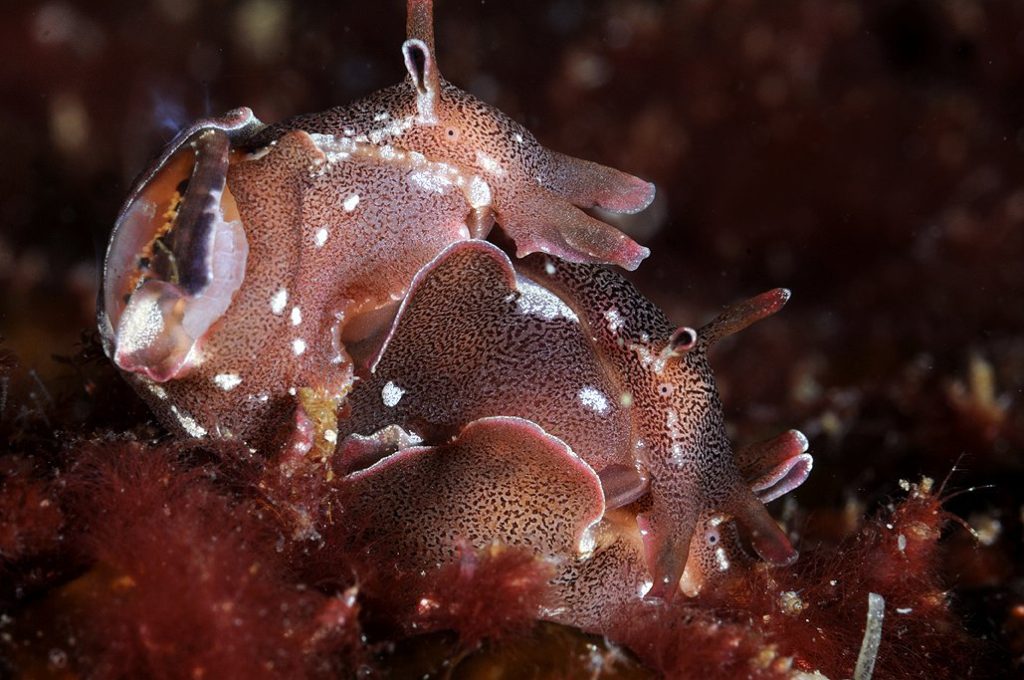
Untangling the diversity and evolution of Sea Hares

Aplysia parvula; Føllingen, Norway; Photo by Nils Aukan

Aplysia parvula; Føllingen, Norway; Photo by Nils Aukan

Sampling and freezing at Askøy

Sampling and freezing at Askøy

Dr Carlo M. Cunha from the Metropolitan University of Santos in Brazil (Universidade Metropolitana de Santos), a world expert in the diversity and systematics of Anaspidea heterobranch gastropods, visited the Natural History Museum of Bergen for a month during January/February 2017 to study our scientific collection of these molluscs. The visit was funded by the University of Bergen´s Strategic Programme for International Research and Education (SPIRE).

The Museum holds a large amount of material from the Scandinavian region, but also from the Mediterranean, Macaronesia islands, Caribbean, and western Indian Ocean.

These marine molluscs commonly known by sea hares comprise around 90 currently known species and have long been of major interest to biologists because of their large and easily accessible nervous system, which form the basis of numerous neurophysiological works.

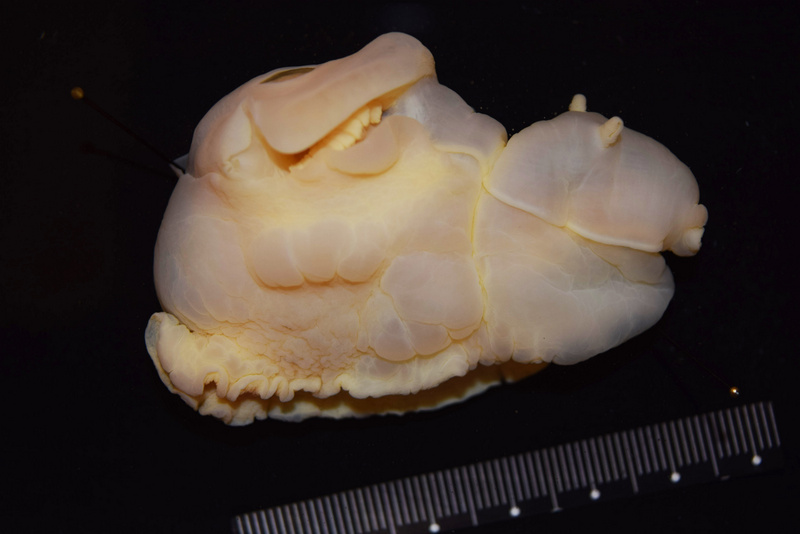
Preserved specimen of Aplysia punctata from norway

Preserved specimen of Aplysia punctata from Norway

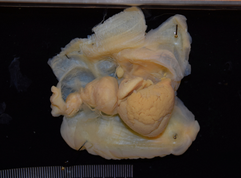
Dissected specimen of Aplysia punctata from Norway

Dissected specimen of Aplysia punctata from Norway

However, the taxonomy of these molluscs and their evolution are still poorly understood. Dr Cunha is using a combination of molecular and morphological tools to learn more about the worldwide diversity of anaspideans and their phylogenetic relationships.

Dr Cunha visit to Bergen has already resulted in the revision and update of the taxonomy of our Anaspidea collection. The Norwegian species of anaspids were revised and redescribed in detail using electron microscopy and DNA barcoding performed in collaboration with Louise Lindblom (University Museum / Biodiversity Labs).

SEM-image of jaws of Phyllaplysia sp from Florida, USA

SEM-image of jaws of Phyllaplysia sp from Florida, USA

Additionally several other species from around the world were studied and will be integrated in ongoing taxonomic revisions. Keep tuned!

-Manuel

We’ve also had Lloyd visiting recently, you’ll find a post about that on the Marine Invertebrates of Western Africa blog: click here

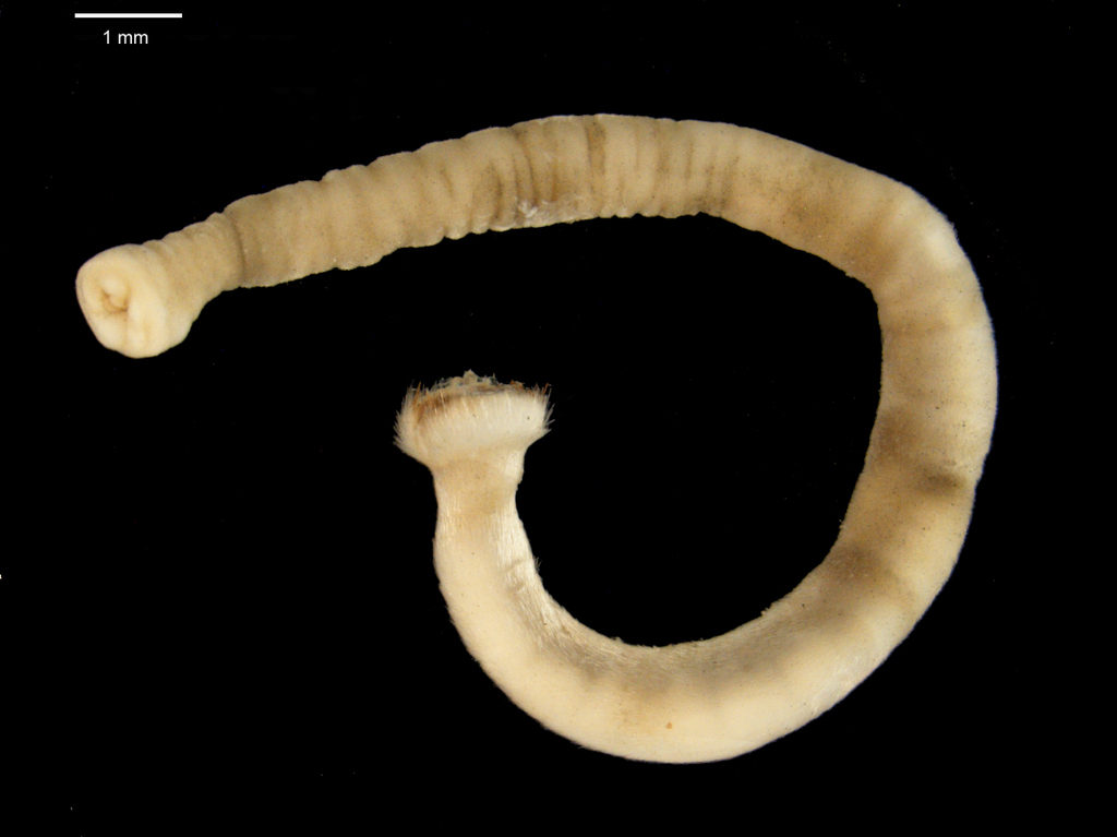
Door #16: Chaetoderma nitidulum- a spiny, shiny mollusc

Molluscs come in a variety of shapes and sizes, but some of the least known are perhaps the Aplacophora, or shell-less molluscs. Instead of a shell, these worm-shaped molluscs have a cuticle covered in calcareous spicules, or sclerites, that give them a beautiful, glistening appearance!

The very first species of aplacophoran mollusc, Chaetoderma nitidulum, was collected from the Swedish west coast and described by the Swedish taxonomist Sven Lovén in 1844. At the time, it was not even known what animal group the new, strange animal belonged to. It had spicules– could it be related to the spiny sea urchins? It had a worm-like body– could it be related to other worm-shaped animals? It would be almost 50 years before it was conclusively recognized as part of Mollusca. Since then, many more species have been discovered, and today close to 500 species of aplacophoran molluscs have been described.

A specimen of Chaetoderma nitidulum from the Norwegian West Coast Photo: N. Mikkelsen

A specimen of Chaetoderma nitidulum from the Norwegian West Coast Photo: N. Mikkelsen

Chaetoderma nitidulum is known today as one of the common aplacophoran molluscs in the East Atlantic, with a distribution from the Svalbard archipelago in the north, to the British Isles in the south. However, taxonomist have been debating the identity of Chaetoderma nitidulum since shortly after it was described. Some researchers have suggested that it could in fact consist of up to six different species. Other researchers have synonymized it with other species, or suggested that it is not a separate species, but only part of a larger species which has a distribution that spans the entire North Atlantic.
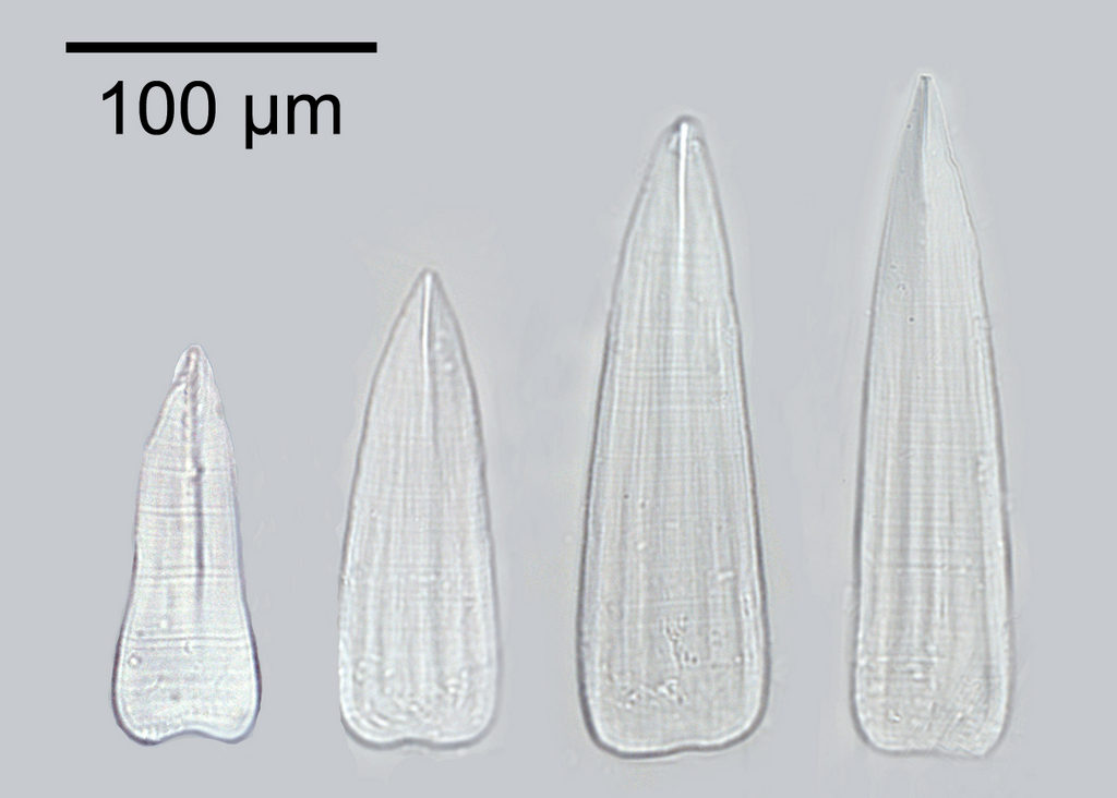
The shape, size and the patterns on the calcareous sclerites covering the body of the aplacophoran molluscs is unique to each species, making it one of the most important characters we have to distinguish between different species.

Calcareous clerites from Chaetoderma nitidulum. Photo: N. Mikkelsen

Calcareous clerites from Chaetoderma nitidulum. Photo: N. Mikkelsen

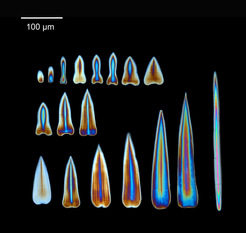
Looking at the sclerites through the microscope equipped with a cross-polarizing filter gives us a shiny, colorful view of the sclerites. The light shines with different colors depending on the thickness of the sclerites, helping us get a good view of the structure of the sclerites.

Sclerites from Chaetoderma nitidulum viewed under cross-polarized light. Photo: N. Mikkelsen

Sclerites from Chaetoderma nitidulum viewed under cross-polarized light. Photo: N. Mikkelsen

We have recently investigated specimens of Chaetoderma nitidulum from different localities from the entire distribution range of the species. Our investigations have revealed a lot of variation between the specimens, both in the calcareous sclerites and in DNA sequences, separating the specimens into at least two different groups. Could it be that Chaetoderma nitidulum actually represents more than one species?

-Nina

Door #9: Research stay of Juan Moles

Juan working at the Museum

Juan working at the Museum

During my stay at the University Museum of Bergen I have been working on the diversity and systematics of Antarctic philine snails. Most of the samples were collected during different cruises on board of the RV Polarstern in the Eastern Weddell Sea, Bouvet Island, and South Shetland Islands (West Antarctica). I photographed all specimens and then clipped them for the DNA analysis (see pictures).


I was able to work at the DNA lab with excellent resources for DNA extraction, amplification, purification, and sequencing.

I am indebted to Louise Lindblom who helped me at the beginning of my crusade there. After a first barcoding of all the material we identified six clades, from which we selected a maximum of three specimens to further sequence the ribosomal genes 16S and 28S and the nuclear gene codifying for the Histone 3.

The first phylogenetic tree with all partitions resulted in the finding of novel clades that now deserve further investigation.

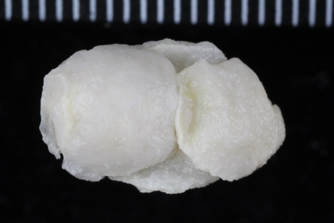
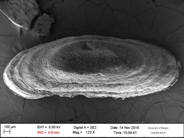
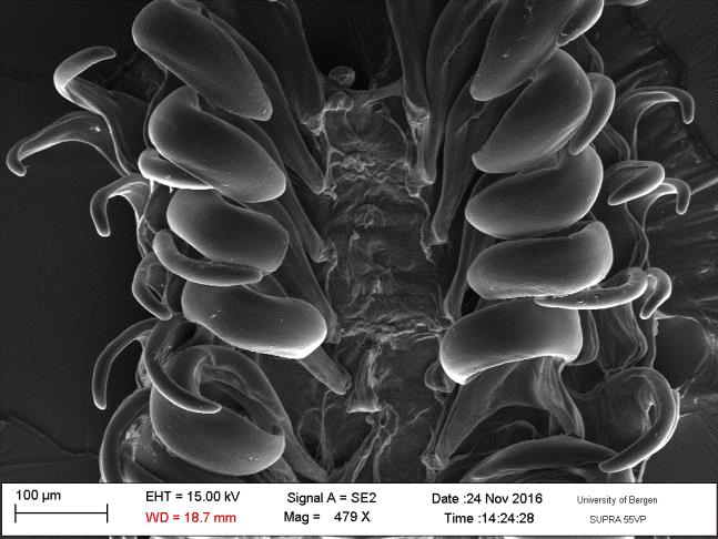
Prof. Manuel António E. Malaquias and his PhD Student Trond Oskars helped me dissecting the material for anatomical analyses. Important taxonomical characters were those related to the male reproductive system, the digestive tract as well, and the shell. After the dissections and drawings of the main parts I prepared the hard structures such as the radula, the shell, and the gizzard plates for Scanning Electron Microscopy (SEM) as well as some soft structures after critical point drying. I could photograph all these material at the same facilities of the museum being helped by Irene and Katrine. After the two months of work, I ended up having huge amount of anatomical and molecular data that deserves further processing. See a picture of the radula and a gizzard plate:

Moreover, I was able to join the student diving club and make several dives to get to know the local flora and fauna. I could even collect some other heterobranch slugs for the barcoding project of the museum. See a couple of pictures of the nudibranch Limacia clavigera and Onchidoris muricata.

Overall, Bergen is a nice city to visit surrounded by nice mountains, good (but not cheap) beers, beautiful fjords, and nice people. I hope I can come back with a postdoctoral position to further enjoy the country and meet more Viking descendants.

-Juan

Travelogue from Jenni’s field-trip to California Academy of Sciences, San Francisco

Sea slugs and San Francisco

Phanerophthalmus sp. from Mozambique. Photo: Manuel A. E. Malaquias

Phanerophthalmus sp. from Mozambique. Photo: Manuel A. E. Malaquias

I am three months into the second year of my masters in marine biology, and was lucky enough to start off this semester with a three week trip to San Francisco in order to collect material for my project.

I am writing my master thesis for the University museum of Bergen on the phylogenetic systematics and evolution of a small marine gastropod.

The title of my project is “Patterns of speciation in the Indo-West Pacific, with a systematic review of the genus Phanerophthalmus (Cephalaspidea, Haminoeidae).

I will be using an integrative taxonomic approach combining fine-scale anatomical dissections and molecular phylogenetics to revise the taxonomy and be able to better understand the relationships of the species. The group is restricted to the shallow waters of the Indo-West Pacific and may therefore be used as a good model to study speciation and the historical biogeography of other organisms in this region.

In order to obtain specimens for this project loans have been made from various museums and academic institutions around the world. In total I have 60 specimens on loan from these various institutions, however they still only represent part of the diversity of the genus with limited geographical coverage. The California Academy of Sciences (CAS) in San Francisco holds the largest collection of sea slugs in the World, including specimens of the genus Phanerophthalmus, with over 100 specimens. So, it was arranged for me to visit this large collection and assess what was important for my project. Travelling to CAS also meant I was able to work alongside Dr. Terry Gosliner, a leading expert in the field of malacology.

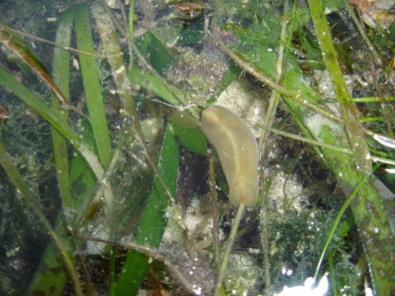
Phanerophthalmus crawling on seagrass

Phanerophthalmus crawling on seagrass

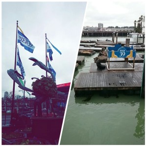
Pier 39 and California sea lions

Pier 39 and California sea lions

So, on January 16th I got on a 10 hour flight to San Francisco. I stayed at a guest house in the Richmond district of San Francisco, about 40 min walk or 30 min bus from CAS.

Waking up on Sunday morning I was a bit jetlagged, but super excited to be in San Francisco. As it was Martin Luther King Jr. day tomorrow (Monday), I had two days to recover from the flight and adjust to the time difference (9 hours behind Bergen!).

I decided to go and explore the city so I took a bus to downtown San Francisco and went to Fisherman’s Wharf, to Pier 39 where the Aquarium of the bay is and also the California sea lions.

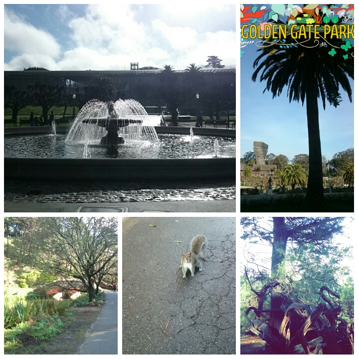
On Monday I went to see where I was going to be spending the next three weeks: at the California Academy of Sciences. Situated in Golden Gate park, the surroundings were beautiful.
Golden Gate park
Golden Gate park

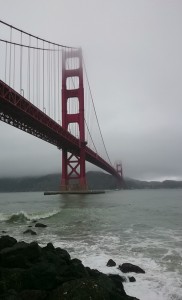
Golden Gate Bridge

Golden Gate Bridge


After visiting the grounds of CAS I wandered over to the Golden Gate Bridge. There was rain in the air and the fog was coming down but the view of the bridge was spectacular.


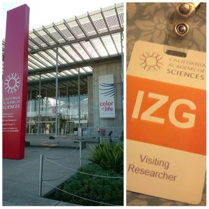
California Academy of Sciences

California Academy of Sciences

Tuesday morning I arrived at CAS eager to dive into the collections. Terry met me at the staff entrance and after a chat and a coffee we got to work. The CAS database contained more than 100 specimens of Phanerophthalmus. The first few days were spent examining labels and matching live photos with specimens. The amount of material was a bit overwhelming and even though I would have liked to look at it all, this would not be possible during my short three week visit. So with guidance from my supervisor, Manuel Malaquias, I was able to focus on the most important specimens. As I am looking at the phylogeny of Phanerophthalmus it is important for me to have specimens which I can extract DNA from. It is also useful to know what these animals looked like live in order to maybe use the external morphology as a character for determining species.

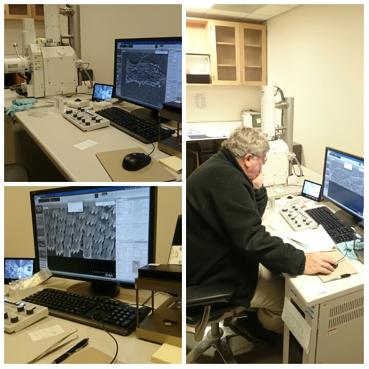
The three weeks flew by so quickly. I spent my days with the collections, dissecting specimens and also got the opportunity to try the academy’s brand new scanning electron microscope. Terry was an amazing host and kept me busy. A huge thank you to him for dedicating so much time towards helping me out. Also, a huge thank you to everyone else at the academy for being so nice and welcoming. After my three weeks at CAS I had a few days to be a tourist in the city. My last weekend in the city happened to be Super Bowl 50 weekend and the city was buzzing with people and events. All in all I had a great visit, and now I have lots of material to carry on working with back in Bergen.

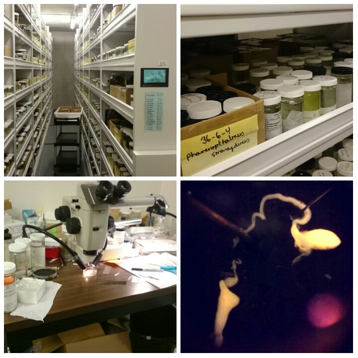
The collections (top), my dissection station (bottom left) and the male reproductive of Phanerophthalmus

The collections (top), my dissection station (bottom left) and the male reproductive of Phanerophthalmus

Scanning electron microscope session with Terry

Scanning electron microscope session with Terry

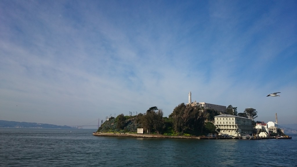
Alcatraz

Alcatraz

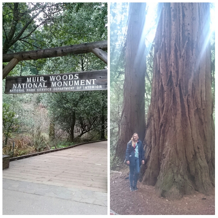
The amazing redwood trees at Muir Woods just outside the city

The amazing redwood trees at Muir Woods just outside the city

Keep calm and focus on sea slugs

Keep calm and focus on sea slugs

-Jenni

Berthella sideralis, a rarity finally documented alive and barcoded!

The Pleurobranchidae sea slug species Berthella sideralis was described by the Swedish malacologist Sven Ludvig Lovén in 1846 based on specimens collect at Bohuslän, in southern Sweden not far from the city of Gothenburg. This species has hardly been mentioned in the literature after its original description, and no images of life species are to our best knowledge available in books, research papers or even web platforms – until now!

A synthesis of the morphological features of B. sideralis can be found in Cervera et al. (2010) who studied in detail two specimens collected during 1930’s in Trondheimfjord as part of a phylogenetic study of the genus Berthella.

Recently, in late November 2015 during a Museum scientific cruise – there is a blog post about this day of field work here – we collected one specimen in Hjeltefjorden (around Bergen) at 220 meters depth using an RP-sledge. This specimen is here documented and was recently genetically barcoded as part of our effort to barcode the Norwegian marine fauna through the NorBOL project.

A live specimen of Berthella sideralis. Ths scale bar i 5 mm. Photo: K. Kongshavn

A live specimen of Berthella sideralis. The scale bar i 5 mm. Photo: K. Kongshavn

Berthella sideralis is only known from Sweden and Norway. In Norway it has been reported between Bergen and Finnmark.

Reference: Cervera, J L., Gosliner, T. M., García-Gómez, J. C., & Ortea, J. A. 2010. A new species of Berthella Blainville, 1824 (Opisthobranchia, Notaspidea) from the Canary Island (Eastern Atlantic Ocean), with a re-examination of the phylogenetic relationships of the Notaspidea. Journal of Molluscan Studies, 66: 301–311.

-Manuel & Katrine