Category Archives: Workshops

10th International Workshop of the Hydrozoan Society

Earlier this year, the Cnidaria and Ctenophora group at UMB organized the 10th International Workshop of the Hydrozoan Society. The event was a success, and we asked visiting researcher Marta Gil to tell us a little more about it.

This is what Marta, who is currently collaborating in our Artsdatabanken projects ParaZoo and NOAH, has to say:

Hello to everyone!

I’m Dr Marta Gil, a researcher at the association Ecoafrik and a member of the Marine Zoology Group at the University of Vigo, Spain. This spring I began a research stay at the University Museum of Bergen to collaborate with Dr Luis Martell on project PARAZOO. I have visited the museum several times, but this visit had a special meaning for me. This year, in May, the 10th Workshop of the Hydrozoan Society was celebrated, organised by the Cnidaria and Ctenophora Group at the Department of Natural History, so I took advantage of my stay in the city and decided to attend.

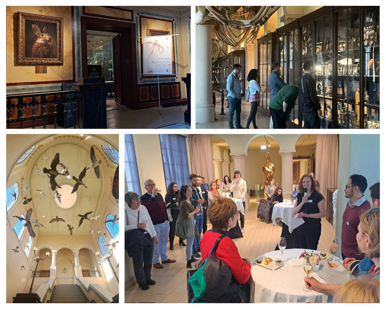
Three images together: one showing the (taxidermied) birds in the staircase of the museum exhibition, one showing participants studying the exhibits in the whale hall - a whale skeleton can be seen over them, and a photo from the reception where the society president is welcoming everyone

Welcome and icebreaker at the University of Bergen Museum of Natural History

This was my first time at this event and I must say it was a very rewarding experience, I don’t think I could have enjoyed it more. We were welcomed by the organisers at the public collections of the Natural History Museum, which was open only for the occasion, creating a fantastic atmosphere to meet the participants and many of the hydrozoan experts.

Over the next three days, we listened to talks from students who are just starting as well as presentations from established researchers who have been working with hydrozoans for years. We have had great speakers such as Dr. Nicole Gravier-Bonnet, Prof. Carina Östman, and Dr.Gill Mapstone; and how to forget the enthusiasm of Prof. Paul Bologna’s group, all the questions and remarks from Dr. Dhugal Lindsay, and the sampling demonstration from our Japanese colleagues Gaku Yamamoto and Aya Adachi? The discussions that took place after each talk and the engaging poster session made this workshop more than a simple scientific meeting, it was a group of friends talking about their favourite animal group: hydrozoans!
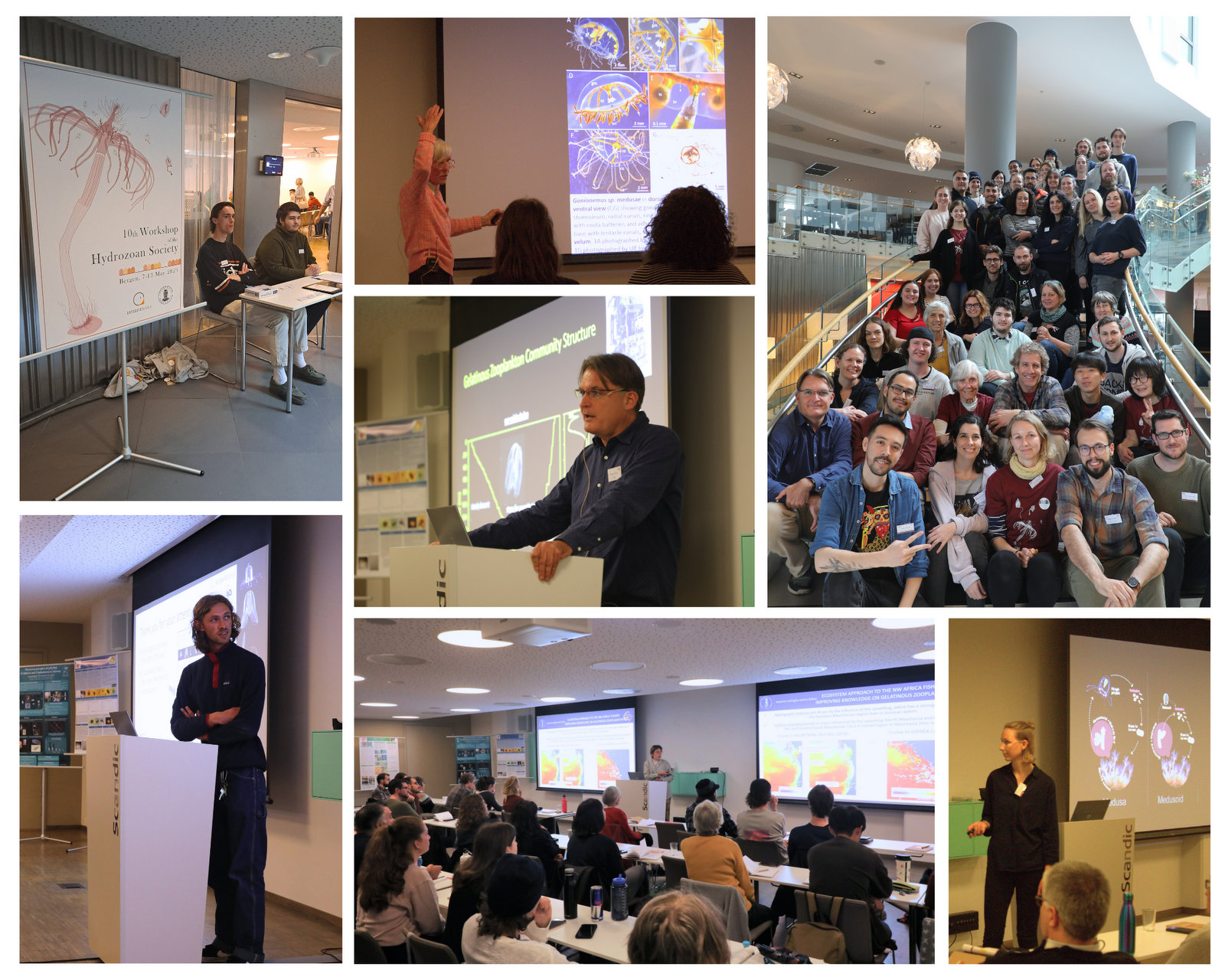
A collage of images showing participants presenting at the venue

The conference part of the Workshop brought many interesting talks from a wide array of topics, together with fruitful and interesting discussions and networking.
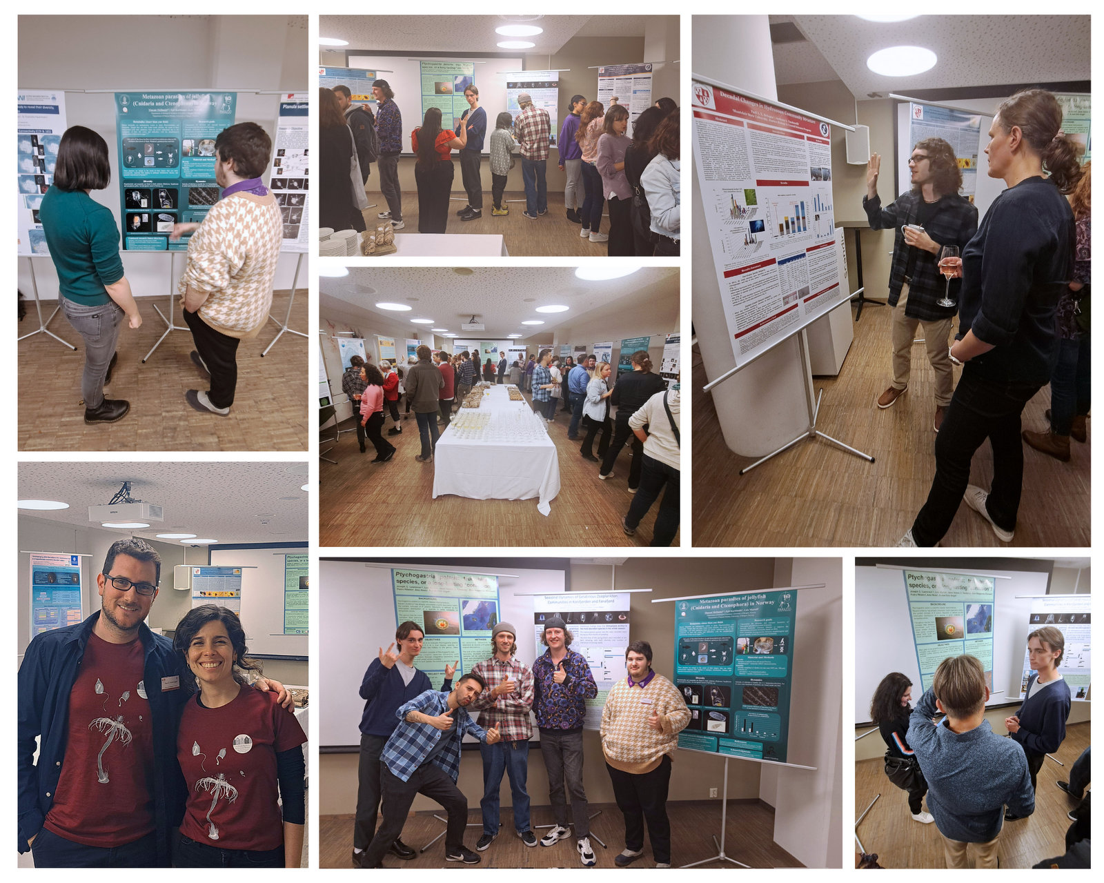
Several images showing people studying large (A0) scientific posters presenting results of their research and projects.

There was a dedicated poster session (and catering) to further discuss about hydrozoans in a more relaxed environment. Many great projects and preliminary results were presented!

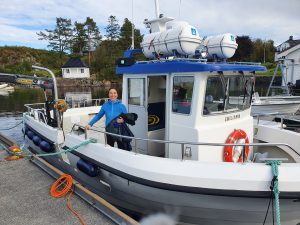
After the talks and presentations were over, the sampling part started, which was the most fun for me! We were able to sample benthic hydroids and pelagic hydromedusae and siphonophores on board the Emiliania and the R/V Prinsesse Ingrid Alexandra, and we later studied them at the Espegrend Marine Biological Station. This part gave us the opportunity to learn and share knowledge, techniques and very useful tips from other experts to study hydrozoans throughout their life cycle.
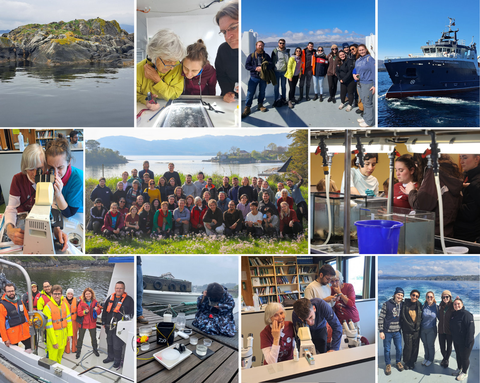
Snapshots from fieldwork, where people are collecting plankton and benthos from ship and shore

Two busy samplings days followed the conference part, where we all exchanged our expertise on sampling, handling and culture techniques. This were a couple of very productive sessions.

To round off an amazing week, on Saturday morning we visited the UNESCO World Heritage Site at Bryggen, where we learned about the history and traditions of Bergen. At sunset, we took the cable car up to Mount Ulriken and, after enjoying the incredible views, we held the award ceremony and the election of the new president of the Hydrozoan Society, who was unanimously elected: Dr. Lucas Leclère!
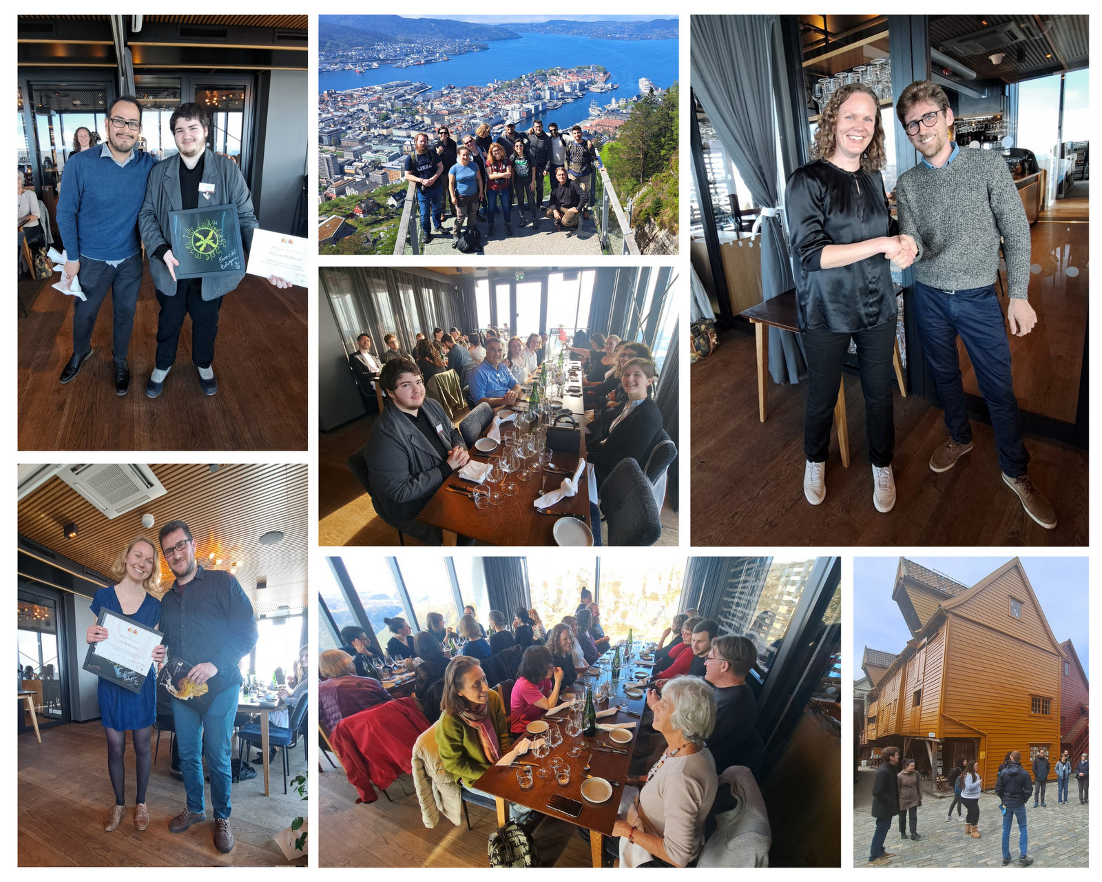
Snapshots from social activities throughout the event; shows people in historical Bryggen, as well as up the city mountains around Bergen

The social activities included a visit to Bryggen, an excursion to Fløyen, and the conference dinner in the top of Mount Ulrikken. The best student presentation awards were given and the new President of the Hydrozoan Society was elected. Exciting times ahead!
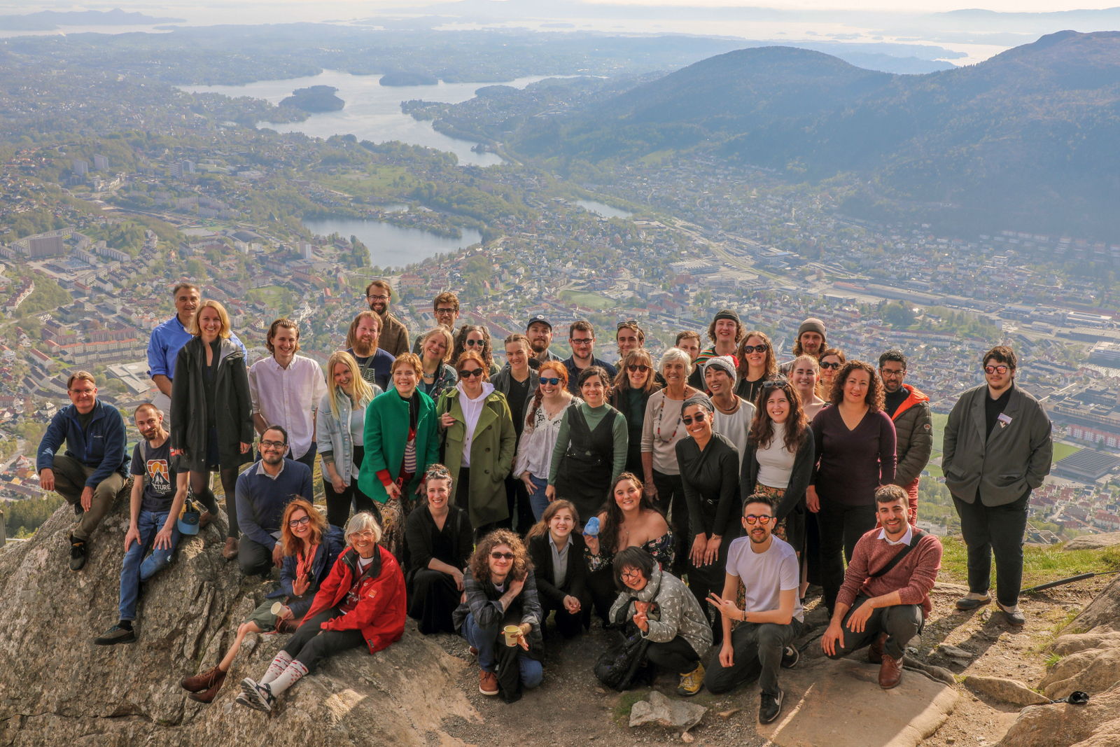
Group photo of the participants up in the mountains, with the city of Bergen in the background

The third group picture we took in just a few days, this time with an incredible view of the city of Bergen as a background.

I would like to close this chronicle by thanking UMB’s Cnidaria and Ctenophora group: Aino Hosia, Luis Martell and Joan Soto, for their incredible work in coordinating the presentations, transfers and field work and for hosting such a rewarding event. I would also like to mention the great work done by all the students of the group, who helped organize the meals on the sampling days and other various activities. Thanks to all of them, the rest of us simply enjoyed the week.

It was definitely an experience worth repeating. I’m already looking forward to the next workshop in France!

-Marta Gil

Final workshop for hyperbenthic copepods (HYPCOP)

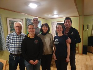
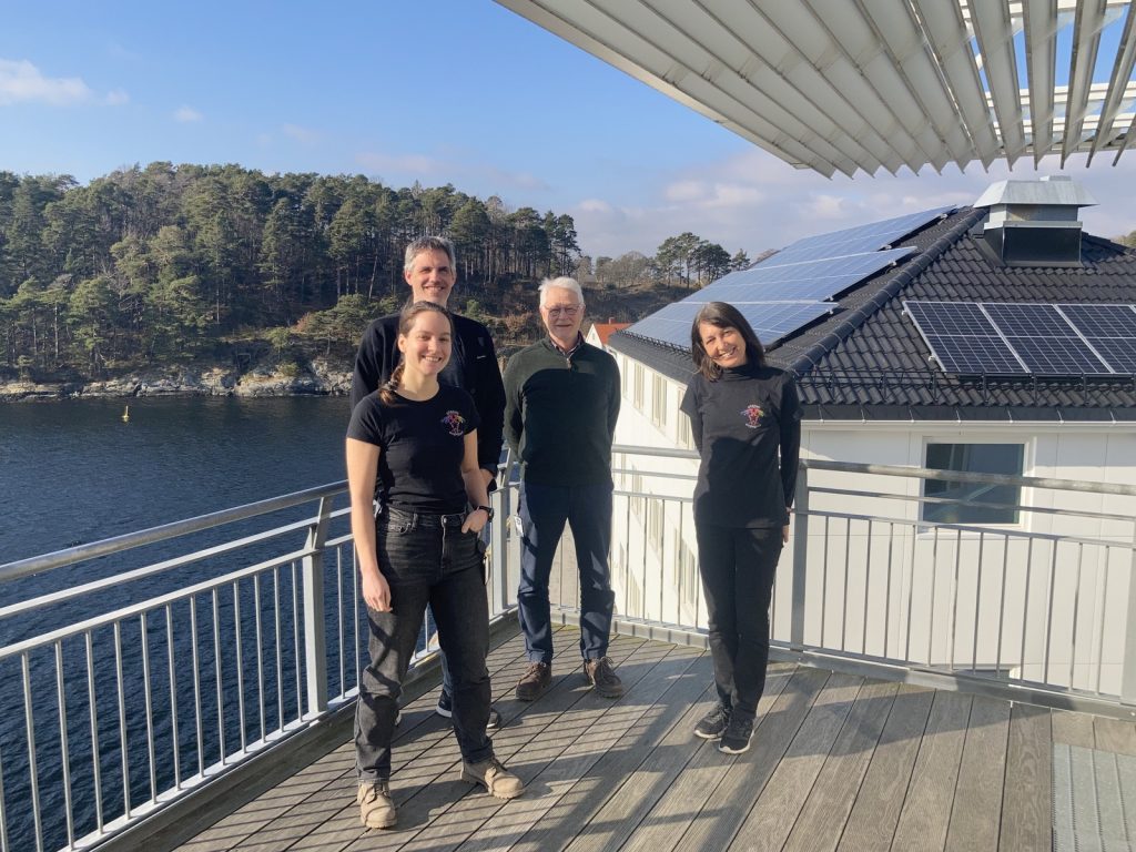
Our first international workshop with from ltr; Anders Hobæk (NIVA), Cessa Rauch & Jon Kongsrud (UMB), Tone Falkenhaug (project leader, IMR), Alexandra Savchenko & Rony Huys (NHM), photo by Alexandra Savchenko

During the last week of September, HYPCOP organized its last and crucial workshop for finishing the project. We invited international collaborators Prof. Dr. Rony Huys and Dr. Alexandra Savchenko from the Natural History Museum in London. Prof. Dr. Huys is a well-known copepod taxonomist and crustacean researcher and published a multitude of species descriptions and books including key identification guides. We were very happy to hear he had time to come and travel to Bergen, paying us a visit while also helping us with species identifications of the many, many copepods we had collected during the two years of our project.


During the two years of the HYPCOP project we collected around 600 specimens from different localities all over Norway, including shallow coastal waters and the deeper parts of the mid-Atlantic Ridge (Loki’s Castle field of active hydrothermal vents). From all those specimens we extracted DNA from the soft tissue of the animal. Therefore, keeping the hard exoskeletons, for morphological identification downstream. This is the most time consuming and challenging part. The species can sometimes only be identified based on minuscule differences in the appearance of its legs. Besides, one needs good taxonomic competence to assign these differences to the thousands of marine benthic copepods species. And this is where the HYPCOP team needed help.

HYPCOP started in May 2020, when a lot of countries, including Norway, were in a lockdown and international travel was difficult or even impossible. Therefore, it was problematic for HYPCOP to invite international researchers for most of the time. Thus, we focused mostly on extracting DNA from our collected specimens and building up a barcode library. But what was missing was the nomenclature of the bulk of the specimens. When finally, our first international researchers could come and have a look at our specimens, it turned out to be an enormous task. With the help of Prof. Dr. Huys and Dr. Savchenko we managed now to have almost 300 assigned names to our DNA library of 500 specimens. Quite a few of those are new species and even new genera.

Kickoff of the workshop, which would take place at Marine Biological Station Espegrend for the duration of a week, photo by Alexandra Savchenko

Rony and Alexandra arrived Sunday evening in Bergen together with project leader Tone Falkenhaug and project technician Cessa. We were stationed at the Espegrend marine biological station in Bergen for the entirety of the week. It was for Tone and Cessa the first time they would finally meet Rony and Alexandra in person, after many months of digital communication. It was a nice relaxing first evening. The next day Anders Hobæk from NIVA and Jon Kongsrud from the UiB joined and we started off the week with a presentation overview of the project.

The overview informed everyone about the program of the week and the state of the art of the project. With the DNA barcode library, we managed to construct a COI phylogenetic tree. Some of the larger clades were already identified down to species level, but many more species names were missing from the smaller clades. It was up to us that week together with Rony and Alexandra to identify these last cases.

Alexandra onboard research vessel Emiliana, photo by Tone Falkenhaug

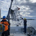
We also had one day of fieldwork planned, to have us work also with some fresh material. This we did with help of research vessel Emiliana and the Beyer’s sled. Both stationed at Espegrend Marine Biological station. We tried to pick out a nice and dry day for going out with the boat and that happened to be in the mid of the week. We went a little bit outside of the Biological Station, with a depth of around 90 – 120m. The Beyer’s sled is an epibenthic sampler, it is called a sled for its form. We got many fresh samples, but due the net being a little large in its mesh size, we did not get as many small species as we liked.


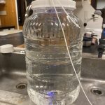
Therefore, we also tried another sampling method with help of Anders; he had brought with him a light trap. Light traps are very easy to DIY with a bottle and inverted bottle opening, like a funnel, and a small led light on the bottom. You install the trap in the water overnight; the little led light attracts a lot of small hyperbenthic and planktonic (and some bigger) species.

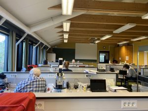
Everyone working hard at the Marine Biological Station Espegrend, assigning species names to specimens, photo by Cessa Rauch

The entirety of the week consisted of many hours working at the microscope, going through literature, dissecting specimens, and assigning species names to the specimens. Eventually with help of Rony and Alexandra, we managed to assign 298 scientific names to 702 specimens in our collection. From those specimens, we extracted DNA from 593 specimens and produced a DNA library, which we uploaded to the BOLDSYSTEMS (Barcode of Life Data System). This library also has all the metadata of our specimens, such as location, depth, size, and pictures of the specimens (either life, fixed and in some cases parts). And it will be publicly available at the end of the HYPCOP project.

The week was demanding but very rewarding and we got many specimens identified, with even a few new species and genera to Norway and possibly new to science; all thanks to the hard work and help of Rony and Alexandra. We therefore also would like to take this opportunity to thank them again for their time and efforts in helping the HYPCOP project move forward! Until next time.
Rony Huys and Alexandra Savchenko helping the HYPCOP project move forward, photo by Tone Falkenhaug

– Cessa

HYPCOP workshop in Flødevigen

From the 7th to the 11th of March the HYPCOP team once more sat together to work on the identification of the species we have in the collection. The strategy was similar as we had in Bergen last year, but this time we looked focus into specific clades. Besides, we met in Flødevigen this time, instead of Bergen, and visited Tone Falkenhaug at her jobsite with the Norwegian Institute of Marine Research (IMR, Havforskningsinstituttet).

The HYPCOP team in Flødevigen, from ltr; Cessa Rauch (UiB), Jon Kongsrud (UiB), Anders Hobæk (NIWA) & project leader Tone Falkenhaug (IMR).

One way for identifying species of hyperbenthic copepods is by looking at their colors. Unfortunately, these get lost as soon as you fixate the samples in technical ethanol. Therefore, we started the workshop with a short sampling trip just out of the bay in front of the research station. We took a small boat from the research station that had a manual operated hinge on the back of the boat, so we could use that for pulling up the grab.

Preparing the small IMR boat with use of the manual hinge and the grab.

Anders Hobæk operating the grab

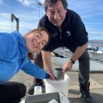
One of the advantages of working with tiny animals is that you sometimes only need small gear to collect them. The grab we used is hand size grab, not much bigger than a 10L basket.

However, as it is made entirely out of metal it is still heavy, which ensures it will be able to “grab” the mud from the bottom when it hits the sea floor.

Once we arrived at a nice location with the boat, we placed the grab over the edge of the boat and let it sink to the bottom which was about 40m deep.

Once the grab would touchdown it would close and engulf softbottom material including the animals that are associated with it. The closed grab would be town back with the manual hinge from the boat. Once onboard, we would empty the grabs content in a bucket and sieve some of the material. This material would go back to the lab for examination.

We carefully examined the sediment, and it was not yet very rich with benthos. We caught a few interesting copepods species, that we documented and fixated for identification.

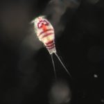
One of the species we caught with the grab

March is not the best season for benthic copepod sampling, the water is still very cold from the winter and most of the small algae needs to grow back. Benthic copepods are much more abundant with rising temperatures and lots of algae growth. Back in the laboratory we started working on our museum collection copepods and assigned clades in our family tree that we would examine first.

Tone Folkenhaug (left) and Anders Hobæk (right) concentrated with dissecting copepods.

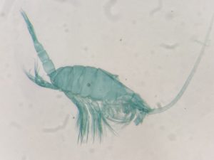
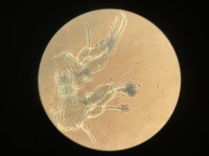
Bigger clades had more priority, and so we took those samples and checked the individual specimens. All the specimens we had in our collection are exoskeleton remnants from the DNA extraction (hence we could have a phylogenetic tree). The exoskeletons are still good for morphology identification but hard to see (due to there translucent nature). Therefore, to help with the identification we would often stain the exoskeletons either with lactophyl blue or lignin pink, which resulted in a visually pleasing collection of prepared slides of different colors.

Slides of Lactophyl and lignin pink stained copepods

Thanks to the workshop we now have manage to identify 145 out the 580 specimens; our efforts for identifying will continue and a new workshop is already planned, we meet again in June and in September, with also this time, help of international researchers!

Stay tuned with @planetcopepod!

-Cessa

Throwback Thursday; HYPCOP workshop at the museum

The project of studying hyperbenthic copepods (HYPCOP) is unique in multiple ways; we study a very unknown group of marine copepod species with very little taxonomic knowledge available here, in Norway. It is challenging as there are more than 700 species described, and possibly more. With pandemic lockdowns, it was hard to have international specialists come and help us, so we had to rely on resources available locally. With so many institutes involved from different corners of Norway, it was not always easy to meet up physically to work on our collection. Hence, when it happens, it is a memorable event, and valuable progress for the project is made.

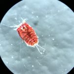
One of the many species of copepod we have here in the collection at the UiB museum

We have Tone Falkenhaug as the project leader, situated at the Institute of Marine Research in Flødevigen (IMR), than we have our collaborator from Norwegian Institute for Water Research (NIVA), Anders Hobæk and the three technicians at the department of natural history from the University of Bergen. The year before we all got together in Flødevigen, so for 2021 we decided that it would be Bergen to have another workshop.

From ltr; Anders Hobæk, Cessa Rauch, Tone Falkenhaug and Francisca Carvalho making the picture

A year into our project we managed to build up a substantial collection of benthic copepods; currently we have around 460 registered specimens, and 195 off those are barcoded with two different DNA markers, mitochondrial (COI) and ribosomal (16S). What keeps ahead of us is the monster task of working through our specimens to label the DNA barcodes with morphological identifications. It means many hours of very precise work with the finest needles, while sitting at the microscope.

During our workshop in Bergen we got together to work through one of the copepod family trees we generated from their DNA:

Preliminary tree of the COI mitochondrial marker

Anders Hobæk is a taxonomist with many years of experience dissecting copepods, and together we went through the samples one by one. It is very satisfying to be able to identify a specimen and get the to the same species level as the DNA barcode. There are multiple reasons as why we choose to identify species based on morphology.

Not all species are easy to barcode, as copepods, especially the benthic ones, are often so extremely tiny; it is difficult to get good quality DNA extracted from them.

Copepods are tiny; this one with scalebar

The small quantities of copepod DNA goes hand in hand with greater risk of contamination of other surrounding DNA, especially if you work with more general markers. Besides, even if we have the DNA barcode, not all copepod DNA is identified as such, which means that even with the right DNA, when running it through the database, it tells us that we have fly DNA, to give an example. Last but not least, in a lot of cases, we were not able to get good DNA sequences from the copepod extracts, so the only option is identifying them morphologically, by dissecting the animals and with help of literature identify the right genus, or even better, the species.

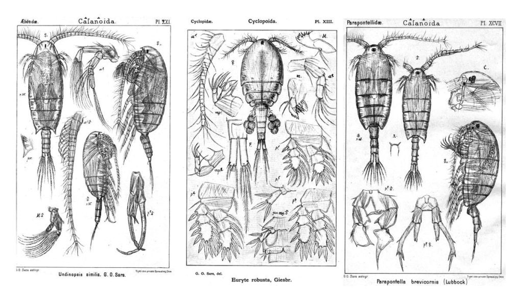
Species identification with help of literature, here a page from G.O. Sars

Our next workshop shall take place again in Flødevigen, in the meantime we keep you updated about our planet of the copepods.

Follow us for more copepod content @planetcopepod, see you there!


-Cessa

CT scan all the things!

Oslo Natural History Museum 02.12.2019 – 06.12.2019

Literature for the week of our CT scanning course


From December 2 till December 6 Justine Siegwald, Manuel Malaquias and I (Cessa) attended a CT scanning course in Oslo at the Natural History Museum.

The course was organized by the Research School in Biosystematics (ForBio) and Transmitting Science.

The group attending the CT scanning course in Oslo at the Natural History Museum

The three of us all work on mollusks and one of the main reasons to deepen ourselves in CT scanning techniques is because of studying the internal anatomy of these often small and delicate specimens.

Species descriptions are not only based on external morphology and/or DNA barcoding, a lot of the species differences are small details within the internal structures. Like the reproductive organs and digestive system (e.g. radular teeth) of the animals. This is very laborious work, and can be challenging especially with the smaller specimens of only a few millimeters long. Besides some species are difficult to collect and can be a rare collection item of the museum. Once you cut these animals to study them, there is no turning back and the specimen is basically destroyed. Being able to see the insides of the animal without touching it would therefore be ideal. CT scanning comes very close to that and with powerful X-ray we can almost see every detail of the insides of the animals without the need to cut them open

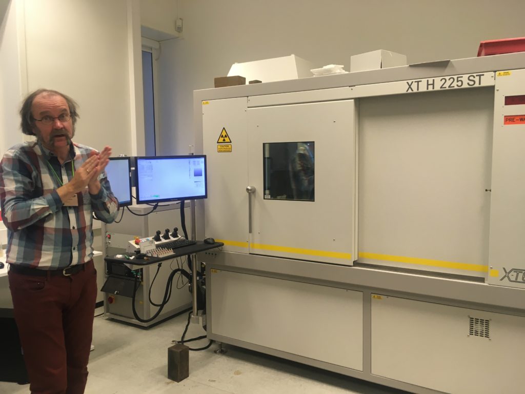
The CT scan in the Natural History Museum of Oslo with one of the teachers in front (Øyvind Hammer)

Lecture about the technology of the CT scan and X-ray

However, how easy as this sounds, CT scanning soft tissue comes with some challenges. Soft tissue means that the x-ray contrast is often very low. Even with modern good x-ray detectors it is still difficult to detect the different internal structures. Therefore, during the course, we were taught how to artificially increase the contrast of the tissue by staining the specimens before mounting them in the CT scan machine.

Coating a specimen with a metal (e.g. gold or platinum) is only useful if you want to see external details of the study object, coating will not help revealing internal structures. For radio contrasting the internal anatomy, you can stain specimens with high density liquids like iodine to enhance the x-ray contrast. So, we went for this option and left our specimens in iodine ethanol solution overnight


After the staining process, we needed the wash out any extra iodine before mounting the specimen into the CT scan. A good scan is not simply pressing the button of the machine; there are a bunch of settings that can be adjusted accordingly (e.g. tube voltage, tube current, filament current, spot size, exposure time, magnification, shading correction, etc.). The scan itself can take hours and file sizes of 24GB for a single object are not uncommon, which means you need a powerful computer with decent software to process this information. Part of the course was also about how to visualize and analyze the files of the CT scan with software like Avizo

Video 1. Software Avizo has a user-friendly interface for analyzing all sorts of CT scanning files.

The week gave us some very promising first results (se video below), and also new insights about how to increase the X-ray contrast of our sea slug samples (lots of hours of staining and very short washing steps).

Video 2. 3D reconstruction of one of Justine’s shell samples

It was a very fruitful week and besides interesting new ideas for scanning our museum specimens, we also met many old and new friends during the week that was as inspiring. ForBio and Transmitting Science did a really great job with setting up this course and I can highly recommend it to anyone who is interested in CT scanning.

Explore the world, read the invertebrate blogs!

-Cessa

Spring time is hydrozoan time

Spring means warmer temperatures, increasing daylight, flowers blooming everywhere… and a lot of outdoor and outreach activities for our projects at the Invertebrate Collections of the museum! In particular, project NorHydro started last month with a kick-off workshop (read more about it here) and we have not stopped collecting specimens, attending conferences, teaching courses and joining sampling trips since then. But why is it that spring is such an important time for hydrozoans and NorHydro?

Well, for starters, spring is the reproductive season for many species of hydrozoans in Norwegian waters. Most Norwegian hydrozoans are seasonal animals and for many of them the increasing temperatures and daylight of March and April trigger a shift in the life cycle towards the production of reproductive stages.

Tiny hydromedusae were already being produced in many of the hydroids we collected, like these colonies of Sarsia tubulosa (left) and Obelia dichotoma (right). Pictures: Luis Martell

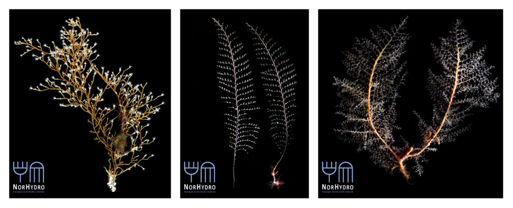
Colonies of Eudendrium vaginatum (left), Kirchenpaueria pinnata (middle), and Halecium halecinum (right). Photos: Luis Martell and Joan J. Soto Àngel

The reproductive stages are often necessary to correctly identify a hydroid colony to species level, so we took advantage of the season to collect the very first samples of the project during the kick-off workshop. More than 50 samples of hydrozoans were collected thanks to the efforts of all the participants, and special thanks are due to NorHydro’s team of hydrozoan experts for identifying the specimens and making for a great start of the project.

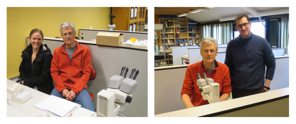
The hydrozoan team: Aino (left), Peter (in both pictures), and Joan (right), hanging around in the lab of the Marine Biological Station during the kick-off workshop. Pictures: Luis Martell

Peter Schuchert from the Natural History Museum of Geneva was a very special participant in this meeting, where he kindly shared his extensive expertise with us. Peter’s knowledge of hydrozoans is impressive, and it was a real pleasure to learn from him on everything from collecting methods to preservation techniques and species identities. Completing our team of experts were our very own Aino Hosia from the University Museum, who stopped by to share some of her experiences on hydrozoan diversity in the region, and Joan J. Soto Àngel from the Sars International Centre for Marine Molecular Biology, who collected many specimens and participated in several interesting discussions about the expected outcomes and potential subprojects of NorHydro.

Our samples were full of hydroids, most of which were documented and preserved for future DNA work. Pictures: Luis Martell and Joan J. Soto Àngel

Of course one of the great advantages for NorHydro during the workshop was the possibility to learn from the activities and results of our sister projects Sea Slugs of Southern Norway and Hardbunnfauna, and to share experiences with them and with the members of the university’s student dive club (SUB-BSI). With such amazing partners and professional collaborators, it was impossible for the kick-off workshop not to be a big success, so thank you all for giving NorHydro the best possible start!

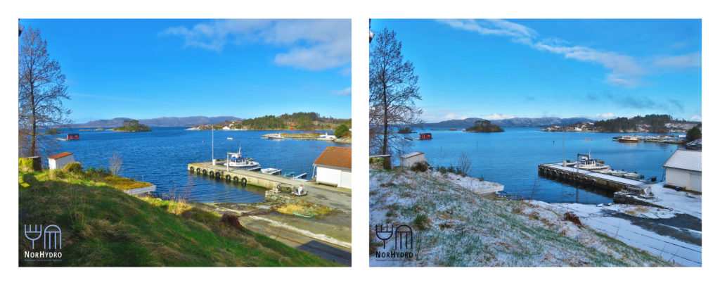
We had all types of weather during the workshop, going from almost-summer to snowy-winter in the same day! Pictures: Luis Martell

Keep up with NorHydro here on the blog, as well as in the Facebook page of Hydrozoan Science  and in Twitter with the hashtag #NorHydro.

Luis

JESS! It’s World Jellyfish Day!

November 3 is World Jellyfish Day, and it is the perfect opportunity to celebrate the gelatinous creatures of the world by sharing experiences and plans with friends and colleagues. We at the Invertebrate Collections take every chance to share our love for jellies, which is why Aino and I were particularly happy to participate in the Jellyfish Research in Svalbard (JESS)-Workshop held last week in Tromsø (22-23 October).

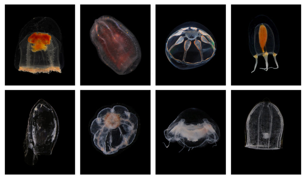
Some examples of Arctic jellies. From left to right, in the top row: Catablema multicirratum, Beroe abyssicola, Botrynema ellinorae, Euphysa flammea; bottom row: Dimophyes arctica, Sminthea arctica, Bathykorus bouilloni, Aglantha digitale. Photos: HYPNO

Loved or hated, jellyfish are the kind of animals that either mesmerize beachgoers or make them go straight out of the water. Fortunately, all participants at the JESS Workshop belonged to the first category of people, and we had a very nice and productive meeting discussing methods, state-of-the-art, challenges, and opportunities of working with jellyfish in Arctic waters.

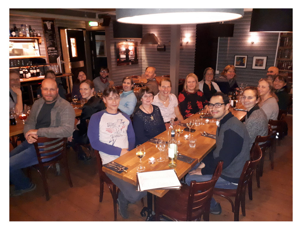
Happy jellyfish hunters in Tromsø

The workshop covered sessions on sampling, data management, ecology, and diversity, including an interesting discussion on how to obtain more (and better quality) jellyfish data from current plankton monitoring protocols. It was an international meeting (20 participants from more than 10 different countries) neatly organized by the University of Tromsø, but it still felt a lot like a bunch of friends getting together to talk about one fascinating subject, which is something I really enjoyed.

Sampling protocols and data curation were some of the most discussed topics during the JESS Workshop. Photos: Joan J. Soto Àngel

All that talking about feeding and predation made us hungry!

While the JESS Workshop was not exactly held on November 3rd, the spirit of commemoration of our gelatinous neighbors was present during the entire event. Celebrating World Jellyfish Day may be a rather recent activity (I could not find any reference of the first time this date was observed, but most likely it only started a couple of years ago), but being fascinated by the movement, color and shapes of jellyfish is certainly not a new thing. There has always been a lot of mystery surrounding the gelatinous inhabitants of the sea, so in a way it was only natural for the origin of the date dedicated to jellyfish to be as much of an enigma as the animals themselves. Mysterious or not, don’t miss the chance to celebrate your local jellies today!

Acknowledgements
Aino Hosia and Sanna Majaneva did a superb job organizing the JESS Workshop and making us feel at home in Tromsø: thank you so much for that! Many thanks as well to all the participants and speakers for the motivating talks and discussions.

Further reading
What could be better than adding some jelly-related reading to the celebration of World Jellyfish Day? I personally love the classics, so I would always recommend Sir Arthur Conan Doyle’s “The Adventure of the Lion’s Mane” for a case of a peculiar encounter with the beautiful Cyanea capillata. Enjoy!

– Luis

Teaching DNA barcoding in Siberia

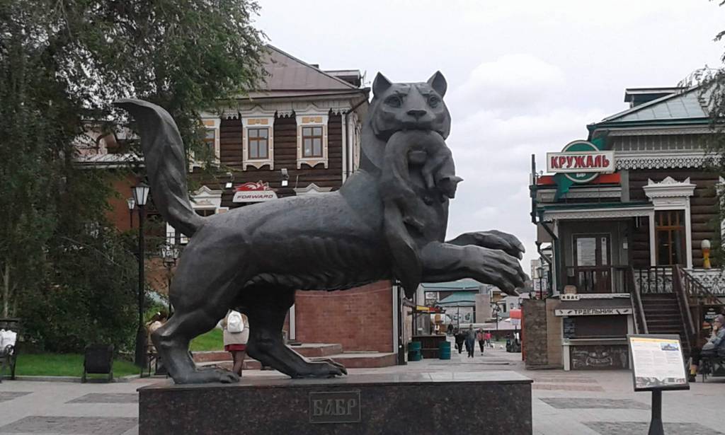
Endre, Katrine, Nataliya and Tom have recently been on a journey far removed from the ocean – although the location did hold a lot of fresh water…!

It really does look and feel a lot like an ocean…

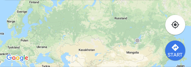
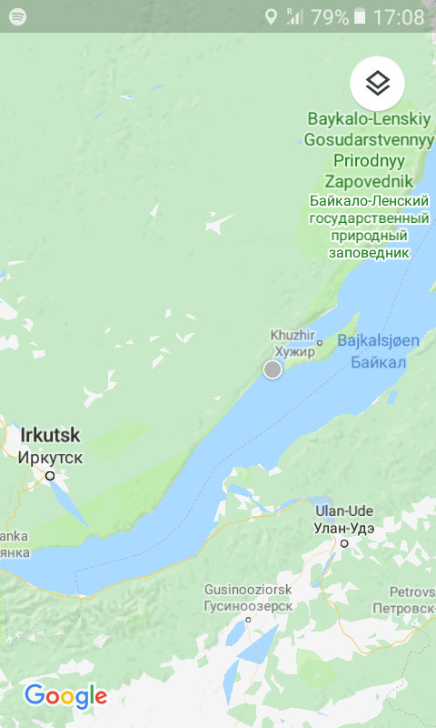
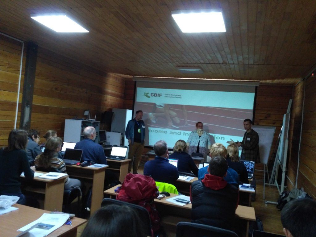
We have – together with Torbjørn Ekrem from the NTNU University Museum – been teaching DNA barcoding at the Russian-Norwegian course “Data mobilization skills: training on mobilizing biodiversity data using GBIF and BOLD tools”, which was held in Naratey on the western shore of Lake Baikal September 14-20, 2018!

The course consisted of two modules focusing on GBIF and BOLD tools. The GBIF part was taught by Dag Endresen from UiO, Laura Russell and Dmitry Schigel from the GBIF Secretariat.

It included both online preparatory work for the students and (mainly) onsite components. The online portion consisted of tasks that the students completed on GBIF’s eLearning portal.

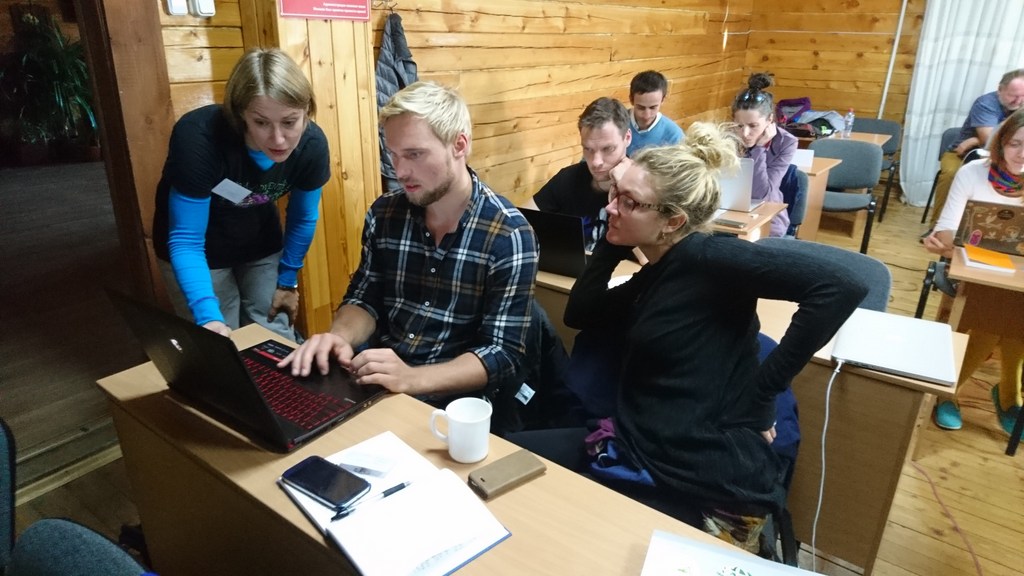
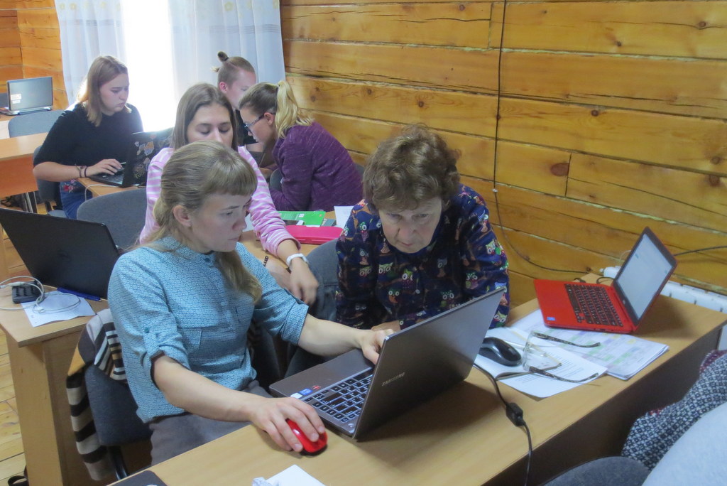
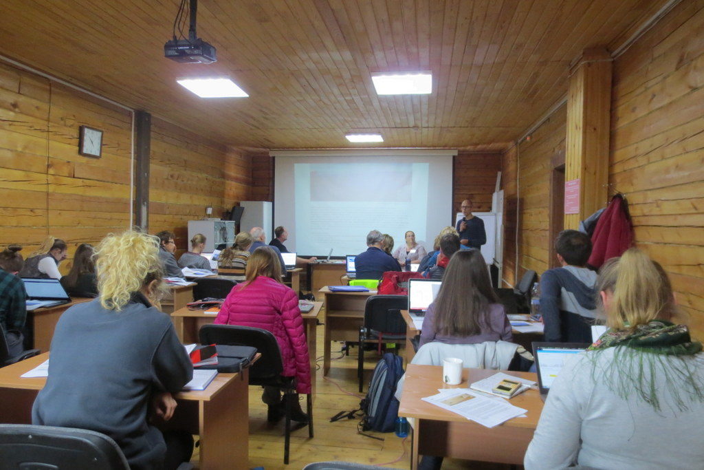
The onsite work was comprised of 20 different sessions of lectures and practical exercises, the latter with a significant component of group work.

16 students from Norwegian and Russian intuitions participated, and did a wonderful job of assimilating a lot of information in a short amount of time, and turning it into practical skills.

The two main platforms we used were GBIF and BOLD – two large depositories for different kinds of biodiversity data. The GBIF-part of the course focused on the technical aspects of data mobilization, such as data capture, and management and online publishing of biodiversity data in order to increase the amount, richness and quality of data published through the GBIF network.

Team GBIF getting set up Photo: N. Ivanova

BOLD; Barcode of Life Data Systems

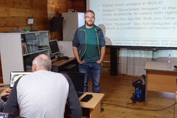
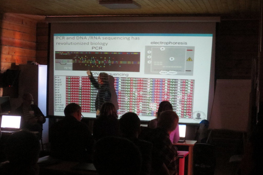
The barcoding part was aimed at both users and providers of barcoding data, and began with an introduction to the barcoding concept, and a case study of integrating data from BOLD and GBIF. This was followed by a session on the use of BOLD: creating projects and datasets, and the uploading of data, images, sequences and trace files. The students got to try all of this for themselves, and we were impressed by how well they worked together to find solutions and teach each other valuable tricks to solve the challenges.

Following the lessons on how to get sequence data into the database, we covered basics of sequence analysis, and gave an introduction to the free software MEGA X which can be used for sequence alignment, translation and phylogeny.

Working in MEGA Photo: k. Kongshavn

This was again followed by a practical session in MEGA on a given data set. We also had a session presenting the analytical tools in BOLD, with a practical session exploring a dataset from NTNU. Our lessons were very well received by the students, with an average score of 4.8 out of a possible 5 on the evaluations – nice feedback for the teachers!

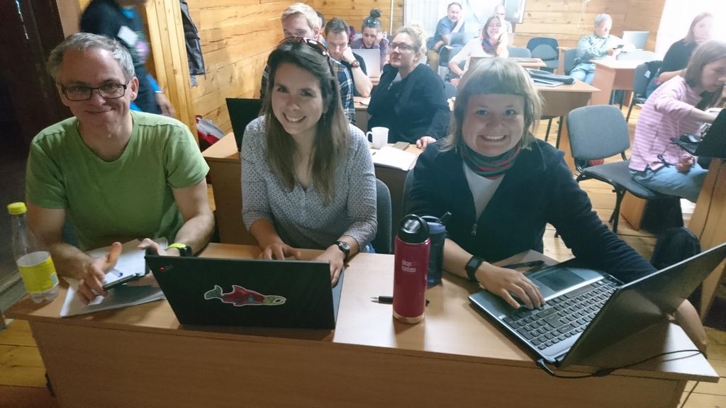
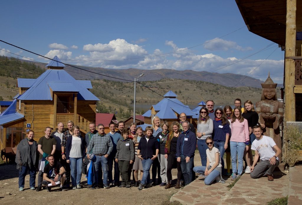
Students and teachers gathered in the Siberian sun
Photo: Dmitry Schigel, CC-BY-SA

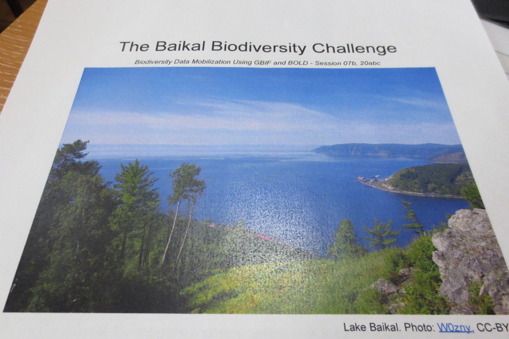
The final task for the students was to present their presentations for “The Baikal Biodiversity Challenge”, which they were presented with on the first day of the course.

The Baikal Biodiversity Challenge

The challenge was to develop a biodiversity inventory project to map and analyze the diversity of a selected animal group. To do so they would need to use available information in BOLD, GBIF and other sources to examine what was known and identify information that was missing, and come up with suggestions on how it could be solved. It was not the easiest of tasks, however all four groups gave excellent presentations.


Group selfie wearing the NorBOL buff scarves – #mydnabarcode! Photo by Laura Russell, CC-BY-SA

The course was arranged as collaboration between the University of Bergen, the Siberian Institute of Plant Physiology and Biochemistry Russian Academy of Sciences (SIPPB SB RAS), the Global Biodiversity Information Facility (GBIF) Secretariat. NorBOL (Norwegian Barcode of Life) supplied the teachers for the barcoding part of the programme, namely Endre, Torbjørn, Katrine and Tom. Funding came from the Norwegian Centre for International Cooperation in Education (previously SIU, now DIKU), GBIF and the Research School in Biosystematics (ForBio).

For those who might not know, ForBio is a teaching and research initiative coordinated by the Natural History Museum in Oslo, the University Museum of Bergen, the Tromsø University Museum and the NTNU University Museum. It is funded by the Norwegian Taxonomy Initiative and the Research Council of Norway. The Research School offers a wide variety of both practical and theoretical courses in biosystematics, and provides a platform for facilitating teaching and research collaboration between Nordic research institutes.  The course portfolio is likely to have something of interest to offer if you work with anything related to biosystematics –and is open (and often free) to you if you are student, researcher or staff at universities, institutes and consulting companies.

Most of the ForBio courses are arranged in Norway or other Nordic countries – but this course was the second of a total six that are arranged as part of the SIU-funded MEDUSA*-project (Multidisciplinary EDUcation and reSearch in mArine biology in Norway and Russia), which is coordinated by Nataliya. The six courses are

  1. ForBio and DLN course: Comparative Morphology Methods (Trondheim, Norway 2018)
  2. Data mobilization skills: training on mobilizing biodiversity data using GBIF and BOLD tools (Siberia, Russia 2018)
  3. Zooplankton communities – taxonomy and methods (Espegrend Marine Biological Station, UiB, Norway, tentatively May 2019)
  4. Systematics, Morphology and Evolution of Marine Mollusks (Vostok Marine Research Station, Institute of Marine Biology, Vladivostok, Russia, September 2019)
  5. Systematics, Morphology and Evolution of Marine Annelids (Espegrend Marine Biological Station, UiB, Norway, tentatively June 2020)
  6. Diversity and Evolution of Meiobenthos (White Sea Biological Station, Moscow State University, Russia, September 2020)

We enjoyed the opportunity to visit such a remote locality, and to get to know the students and teachers – thank you all for making this such a wonderful experience!

Thanks also to the BOLD support team for excellent help before and during the course!

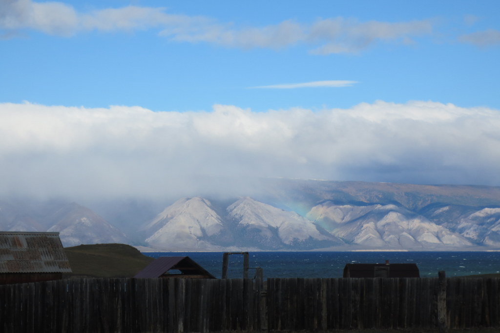
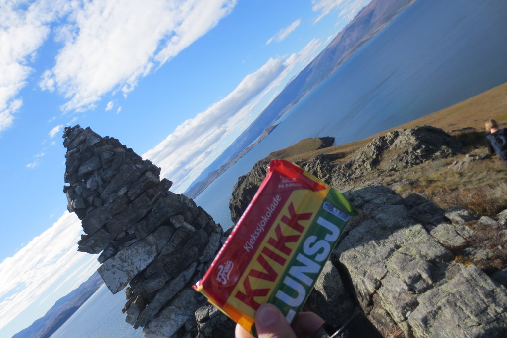
A few snapshots from the area – it was stunning! Photos by K. Kongshavn

A beautiful view from Olkhon Island (after the course), photo by K.Kongshavn

ps: we also tweeted using #ForBio_GBIF during the course

Exposing the Indian Ocean staggering diversity: fieldtrip to Nuarro, northern Mozambique (October 2017)

Pursuing our goal to understand the diversity, origin and biogeography of the Indo-West Pacific biota, we went back to Mozambique to continue the exploration of the reef systems of the country. After a first fieldtrip to the subtropical southern coast of Mozambique in 2014, and a second during 2015 to the tropical Quirimbas islands (Vamizi I.) not far from the border with Tanzania, we visited this time for two weeks between the 15–28 October 2017 the coastal pristine reefs and mangroves of Memba District in Nuarro, Nangata Bay, together with Prof. João Macuio from the University Lurio, Pemba and Dr Yara Tibiriça based at Nuarro Lodge, that kindly organized the expedition.

The staggering diversity of the tropical Indo-Pacific is well known and the area between the Philippines, Indonesia, and New Guinea (the Coral Triangle) is famous for hosting the highest marine diversity in the planet. However, for various reasons (political, economic, etc.) the Indian Ocean has been comparatively less surveyed, and much fewer studies are available for this region. Some recent publications are though showing that for corals and molluscs, at least, the region is extremely diverse, nearly comparable with the coral triangle.

We want to understand why is this region so diverse and whether the Indian Ocean harbours similar or a different faunal composition compared to the West Pacific Ocean. The traditional view is that most tropical species are broadly distributed across the Indo-West Pacific, but recent evidence from DNA studies suggests otherwise, but this is not yet well understood. If significant differences are found, then, understanding what may have driven speciation across both realms becomes a major quest.

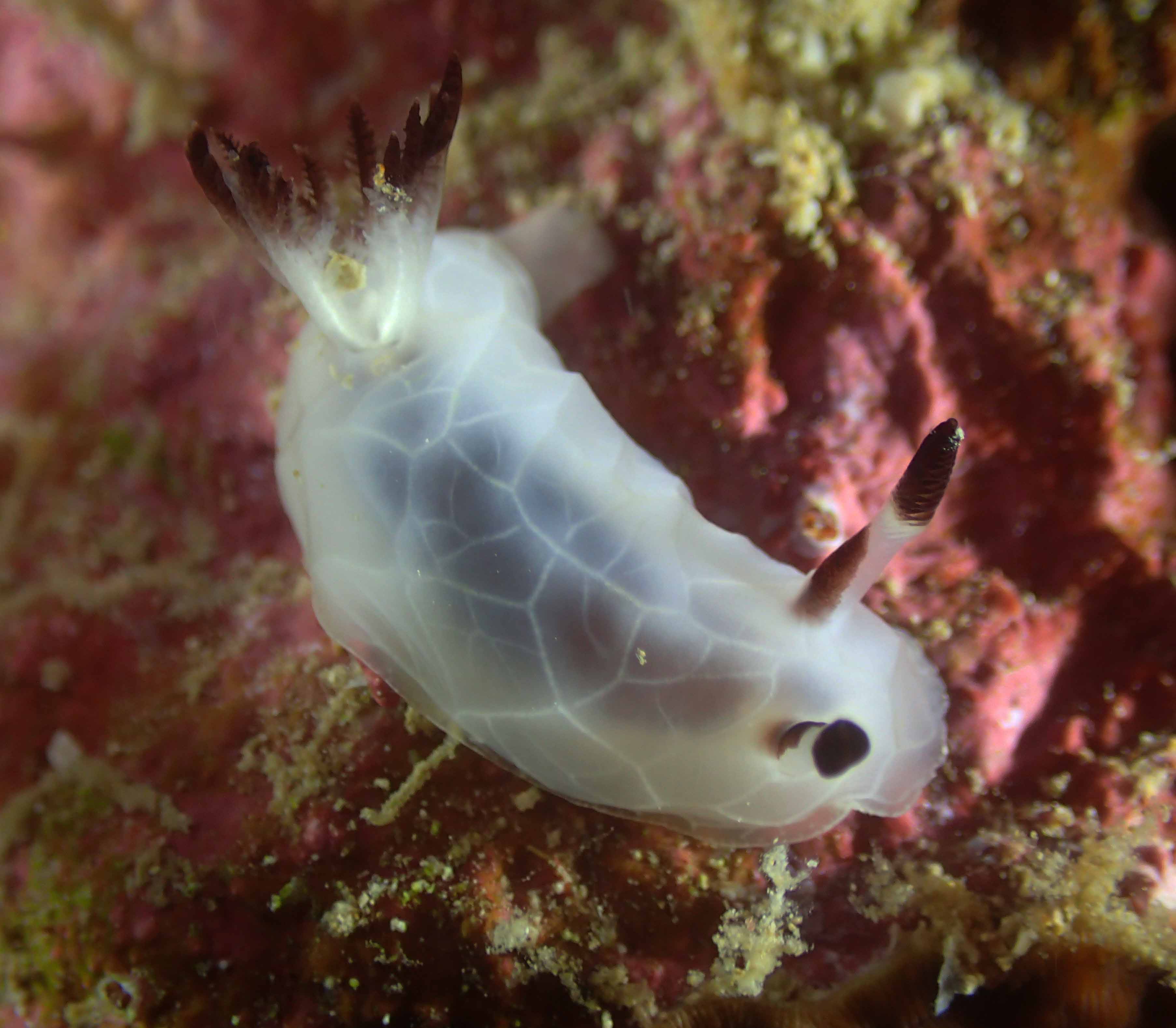
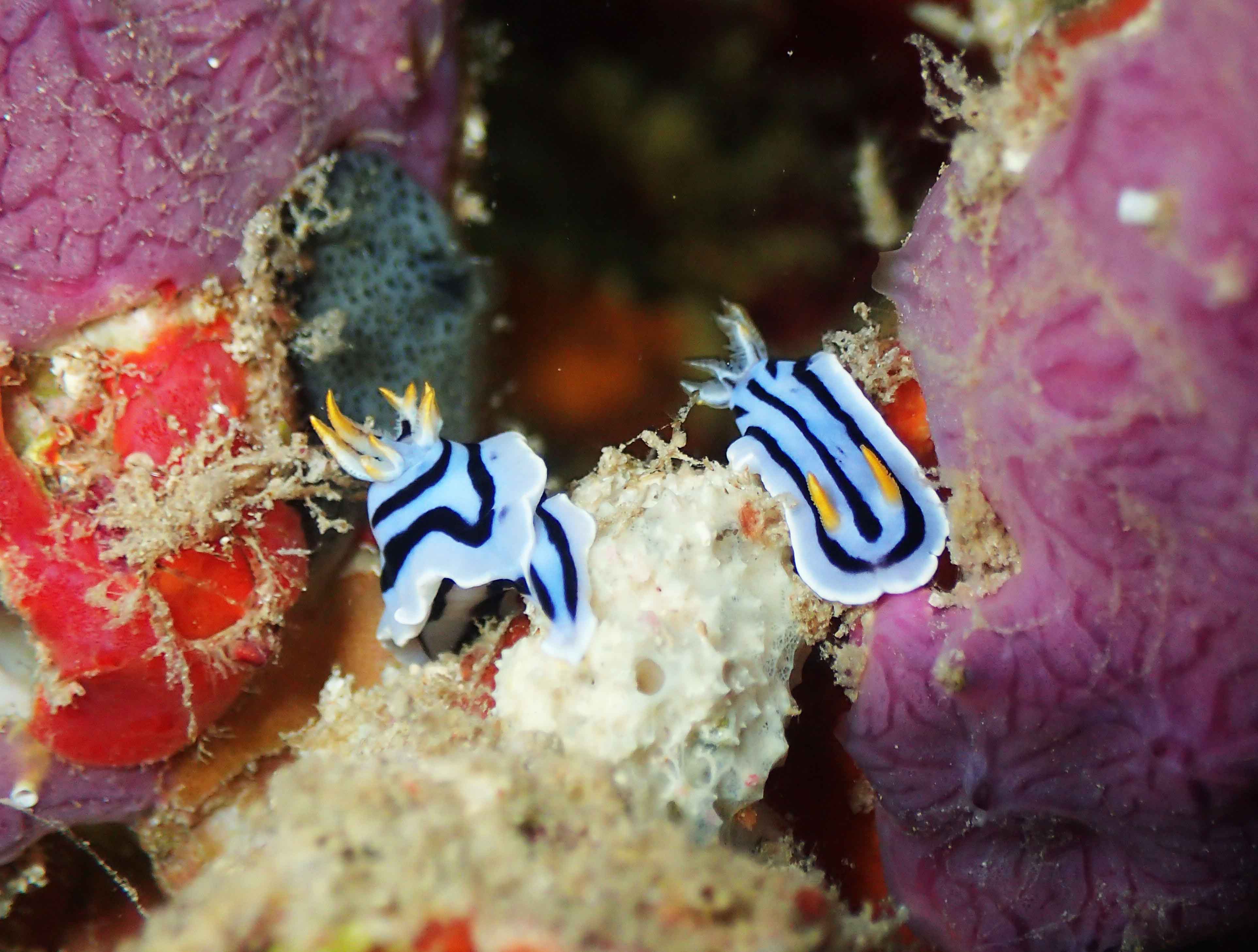
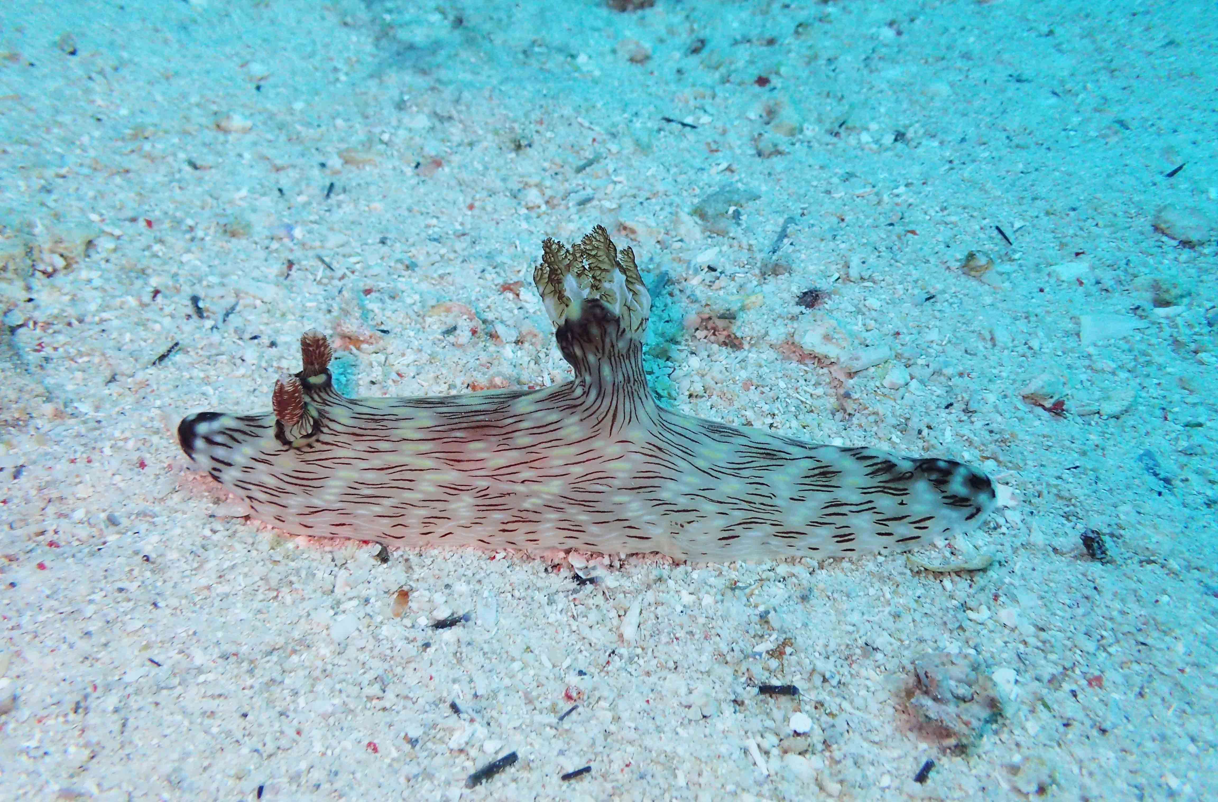
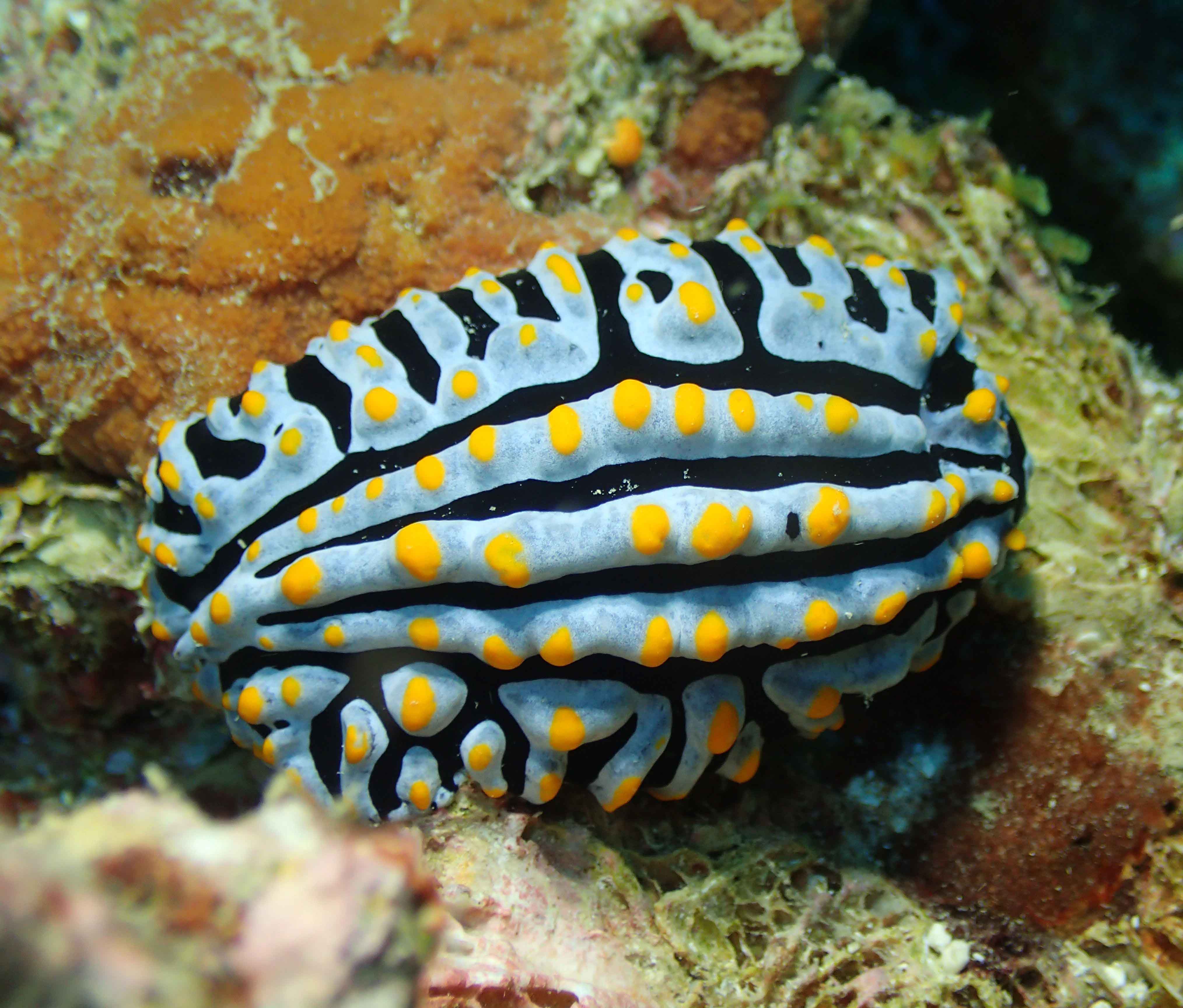
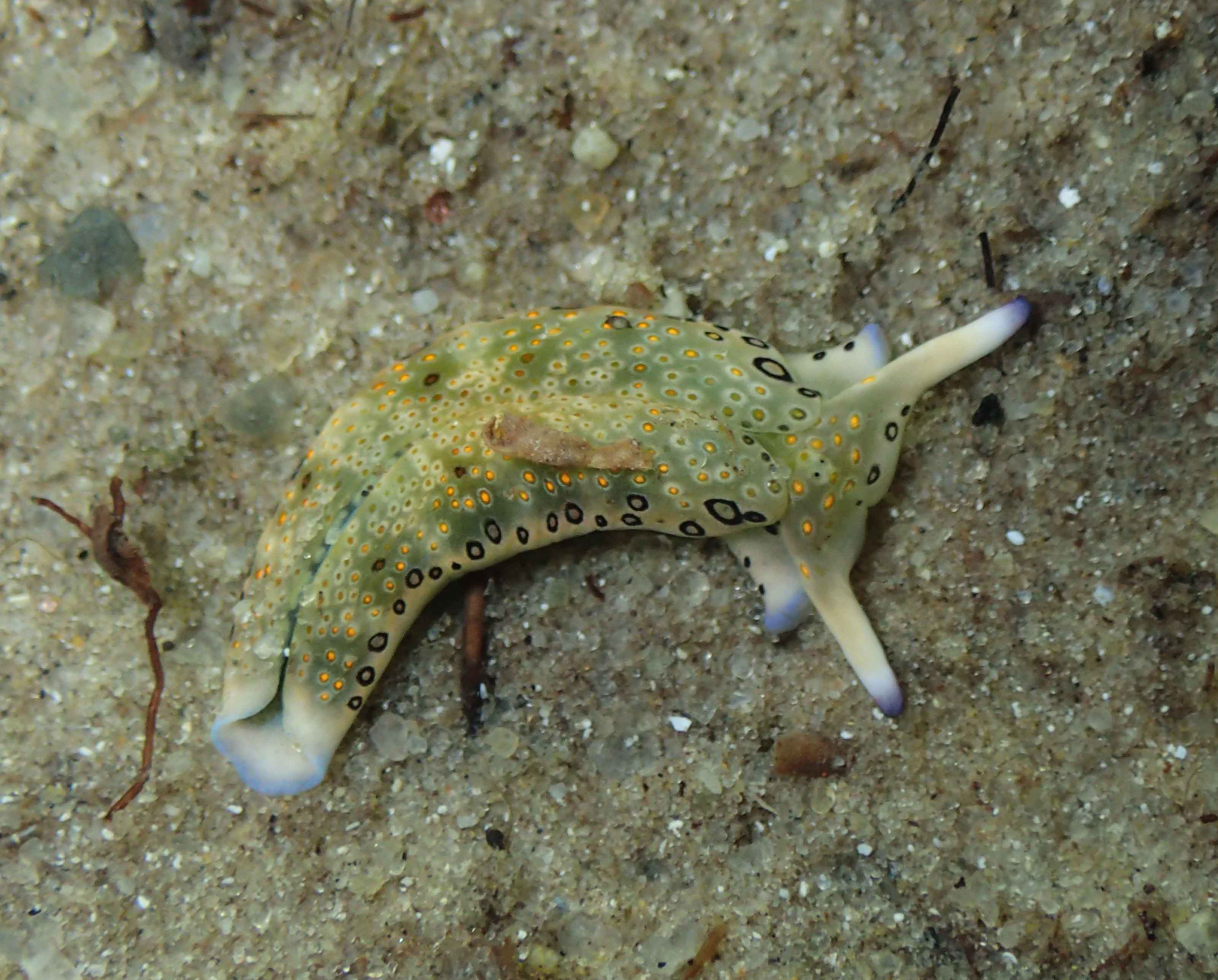
Jorunna rubescens

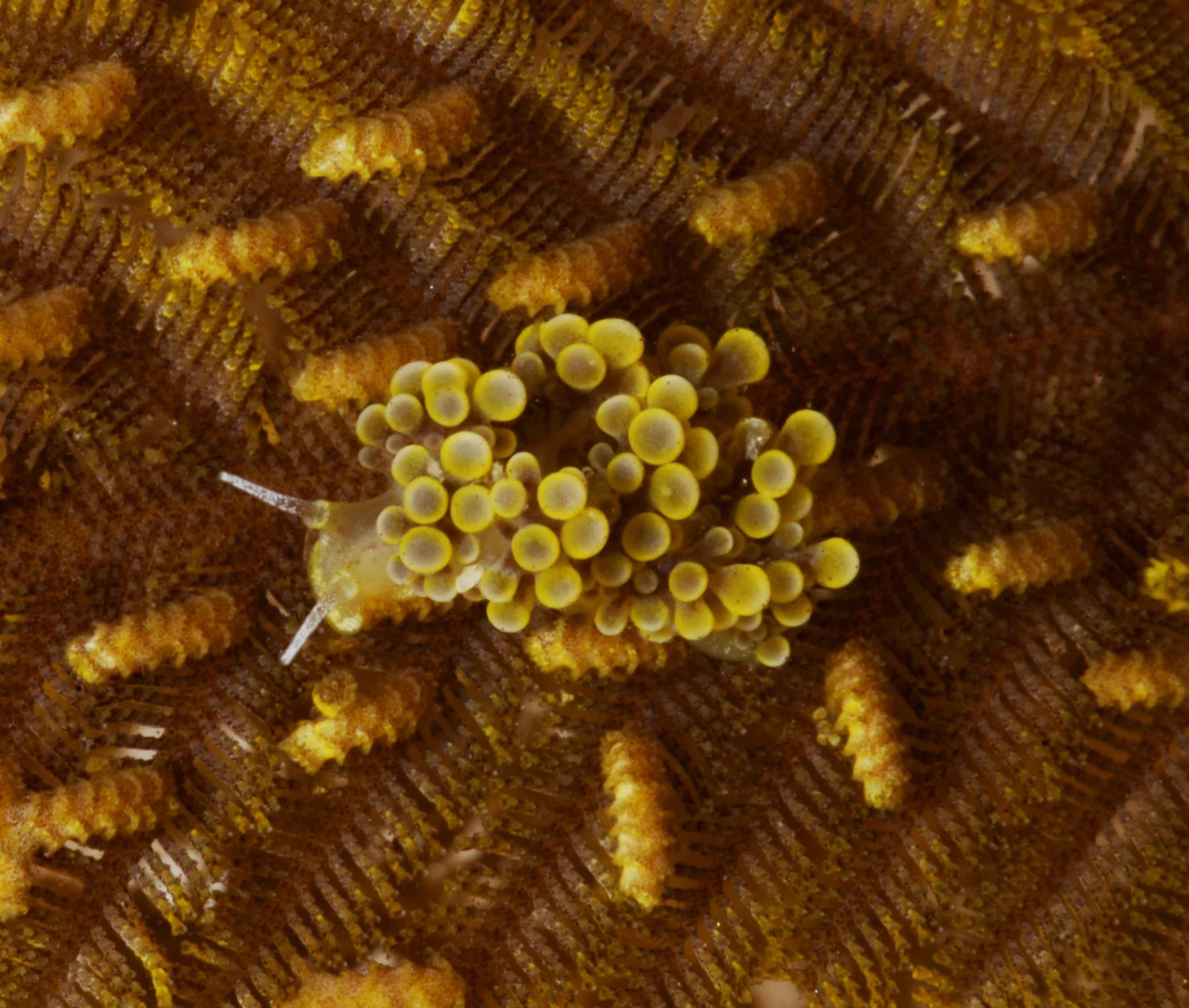
Phyllidia varicosa

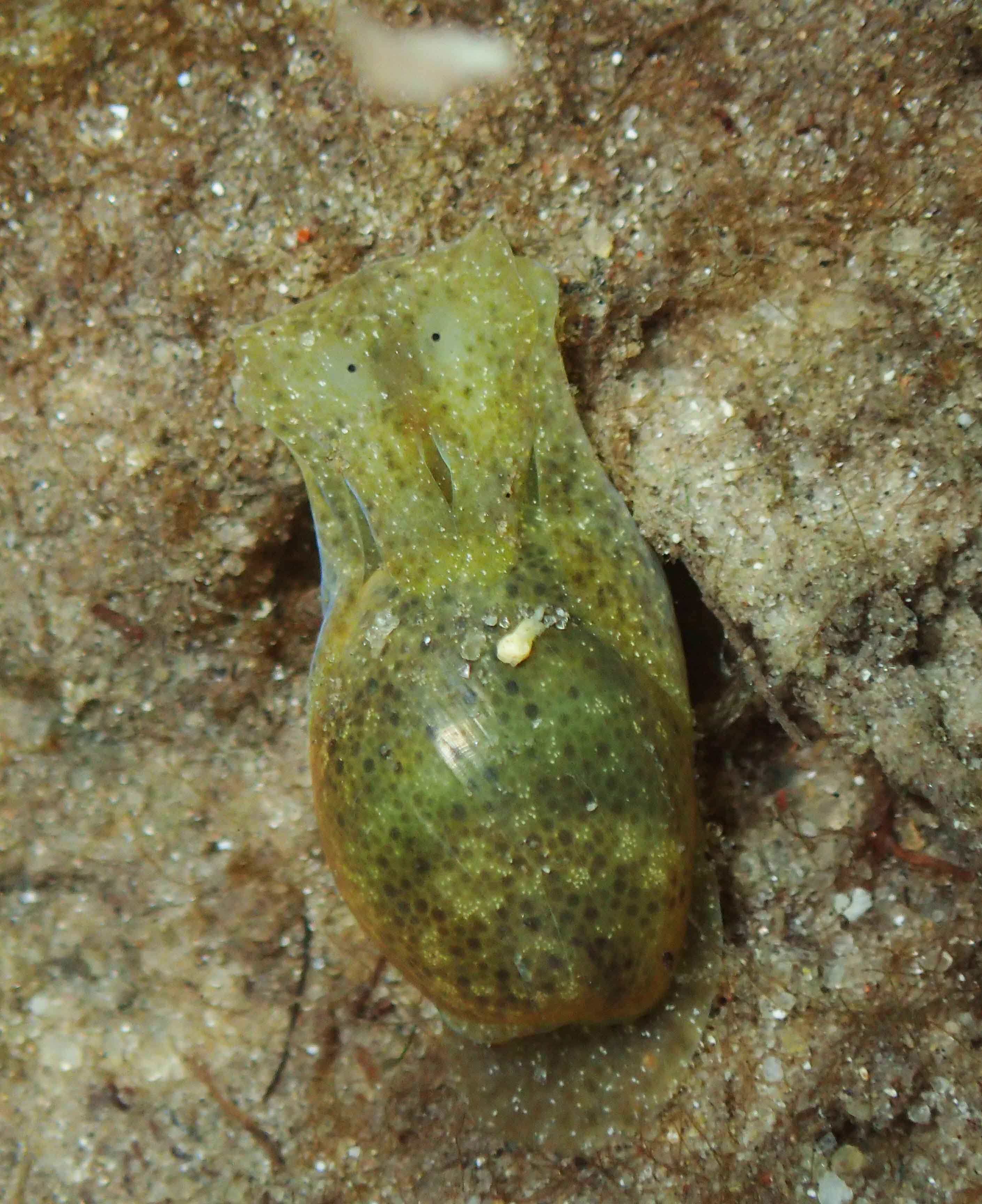
Plakobranchus sp.

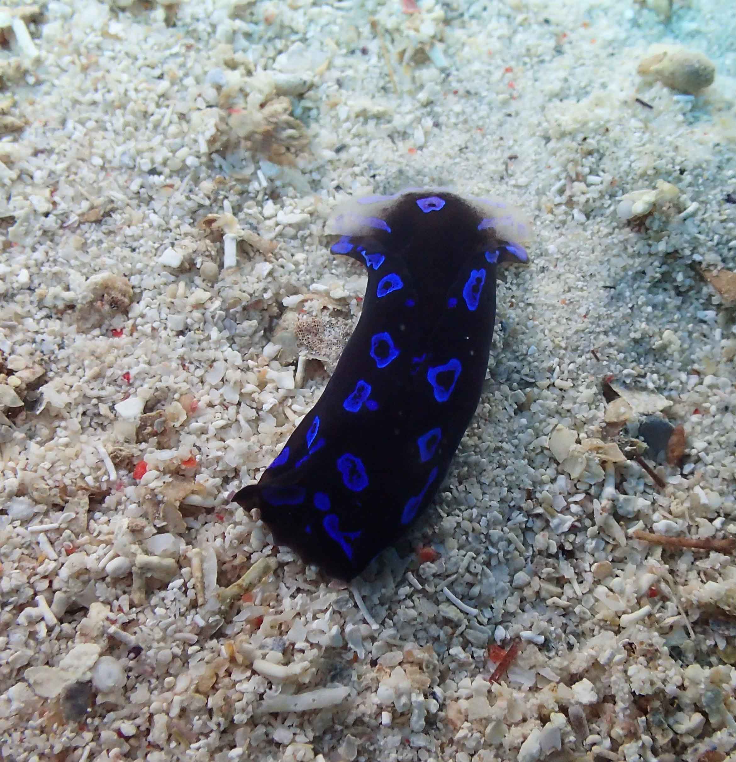
Reticulidia suzanneae

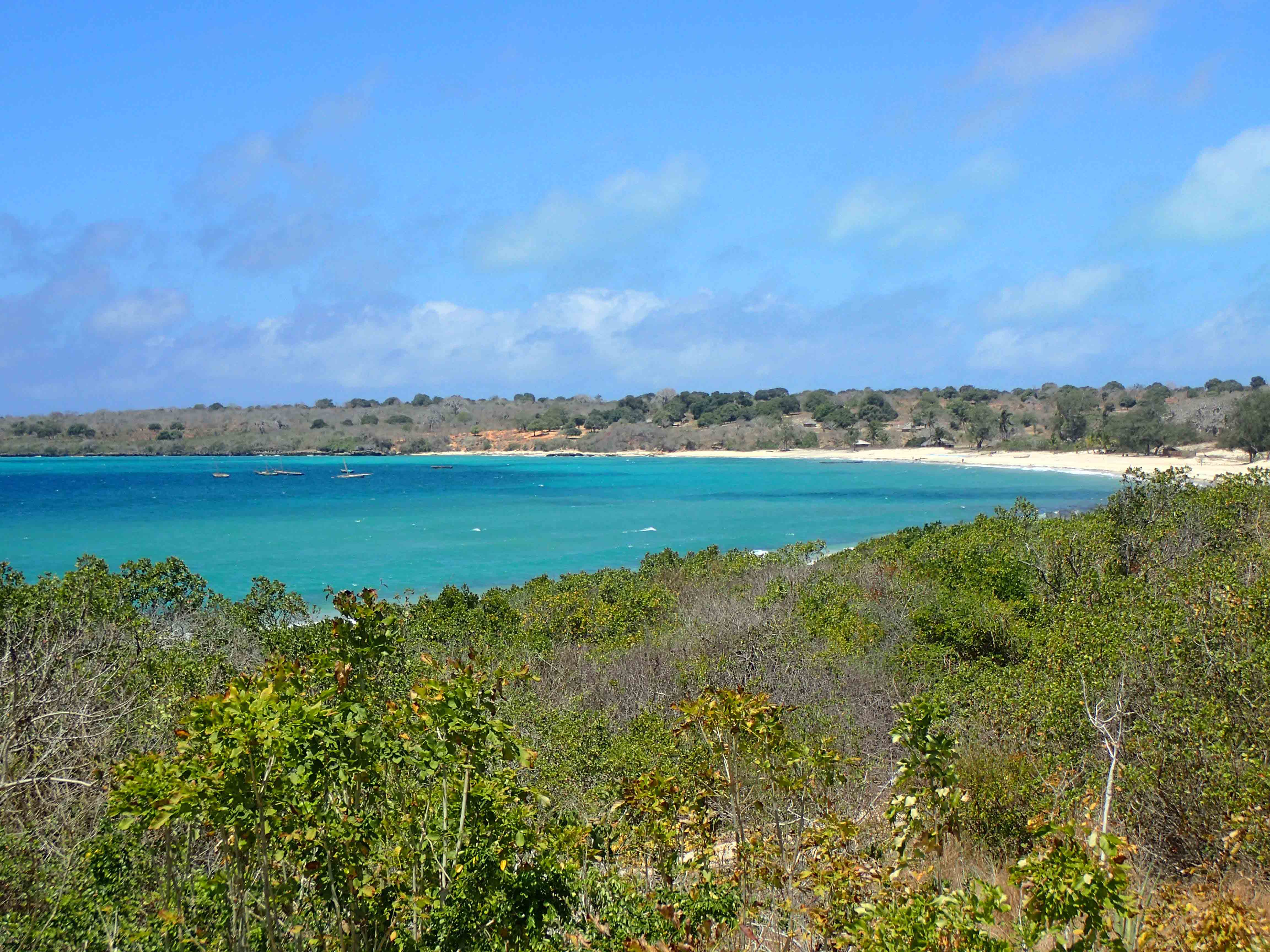
Nuarro is a remote place, four and half hours driving from Nampula international airport, half of them through earth roads. It is located in beautiful Nangata Bay lined by fine white sands and calm turquoise waters. The area is characterized by a large diversity of marine habitats like sand flats, coral bommies, seagrass meadows, coral reefs, and coralline drop-off walls plunging into the deep blue.

The expedition had its basecamp at Nuarro Lodge, an eco-lodge offering diving facilities and a research center. The lodge besides its hostel activities is engaged in social, educational, heath, and nature conservation programmes with the local communities helping to improve education standards for the younger generations, mitigate the impact of native diseases like malaria, and the sustainable use of nature resources. For this last goal a marine protected area was created in 2008.

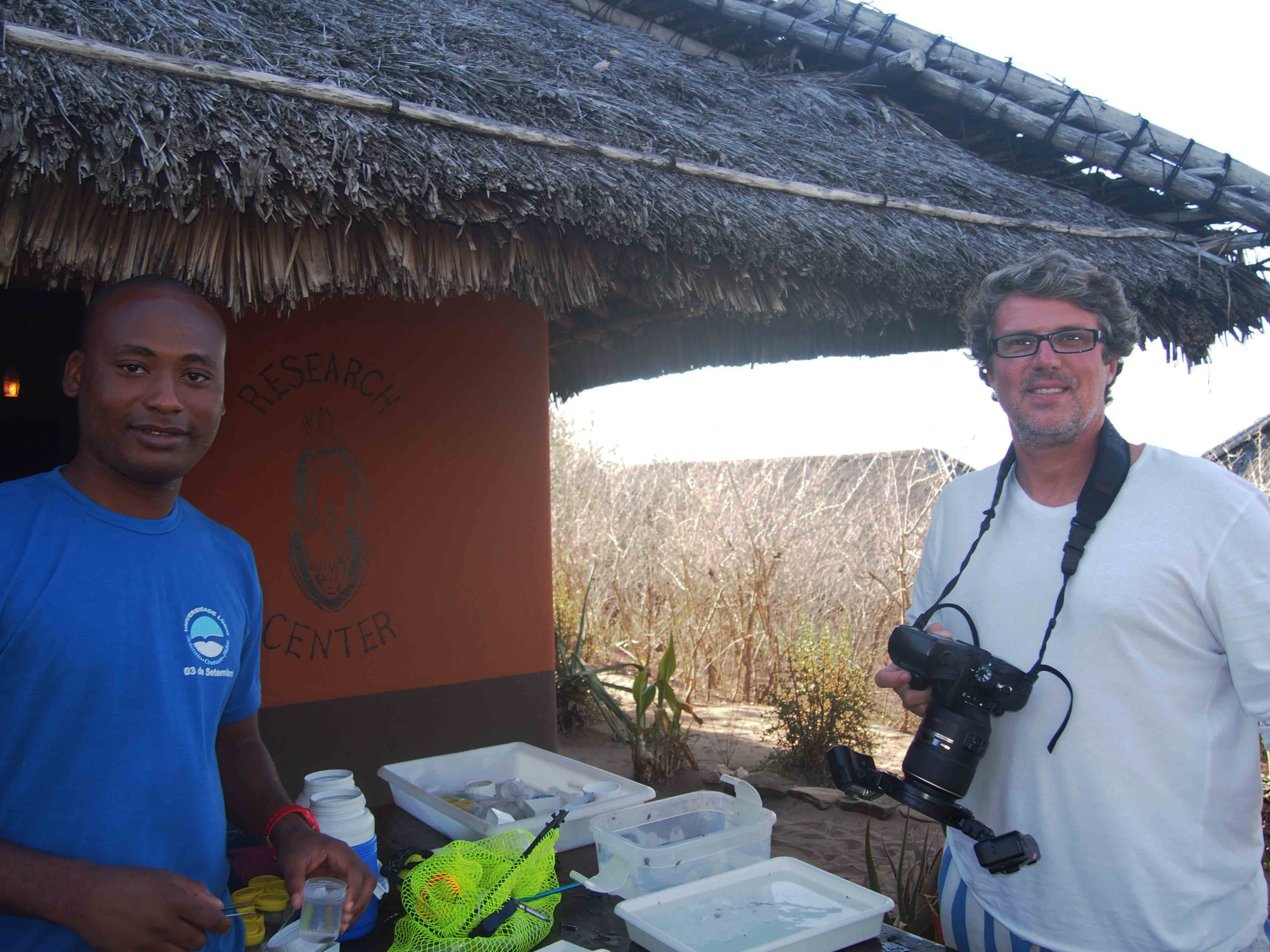
João Macuio and Manuel Malaquias during a nudi photo session

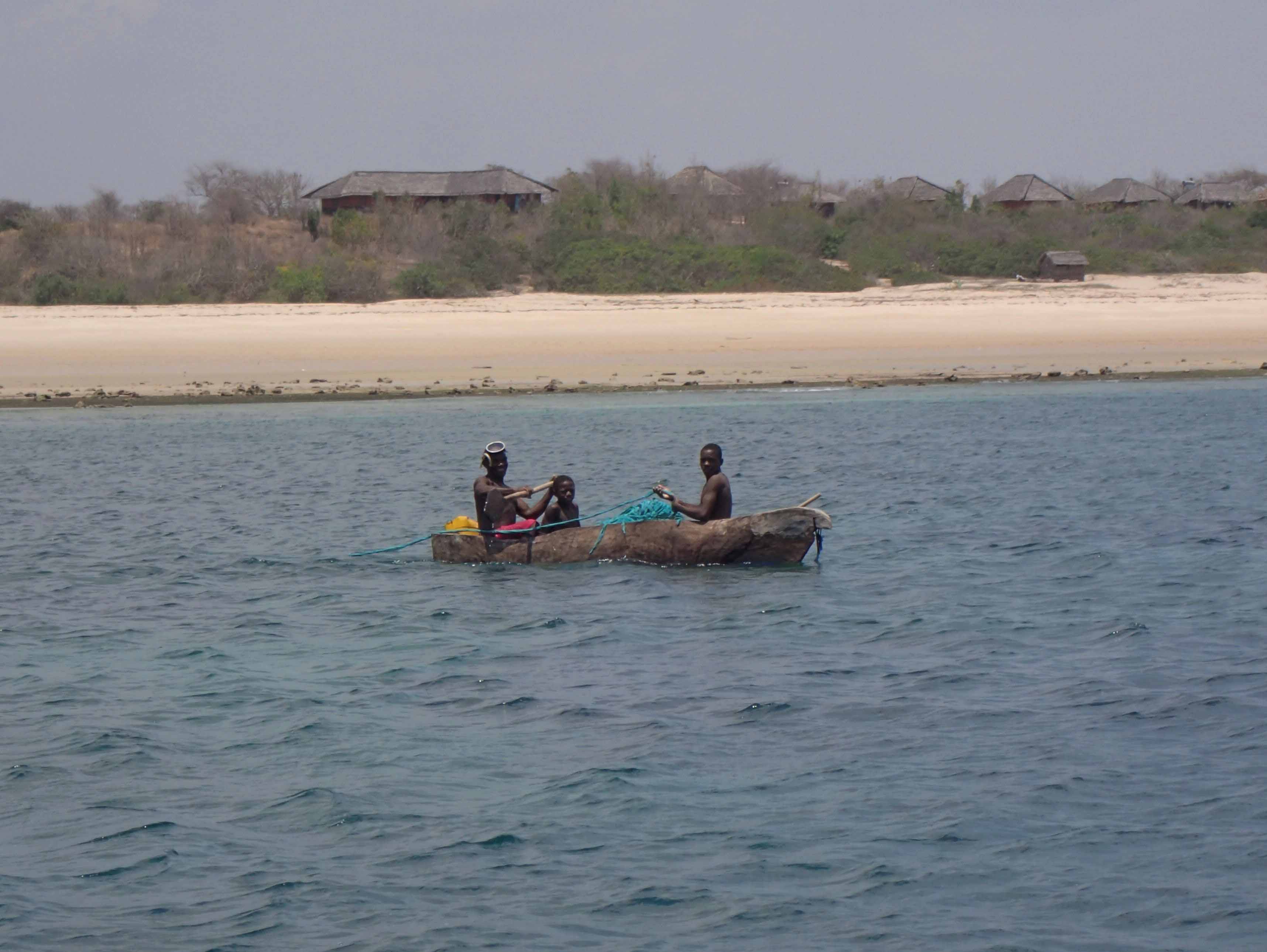
Local fishermen in a wooden canoe

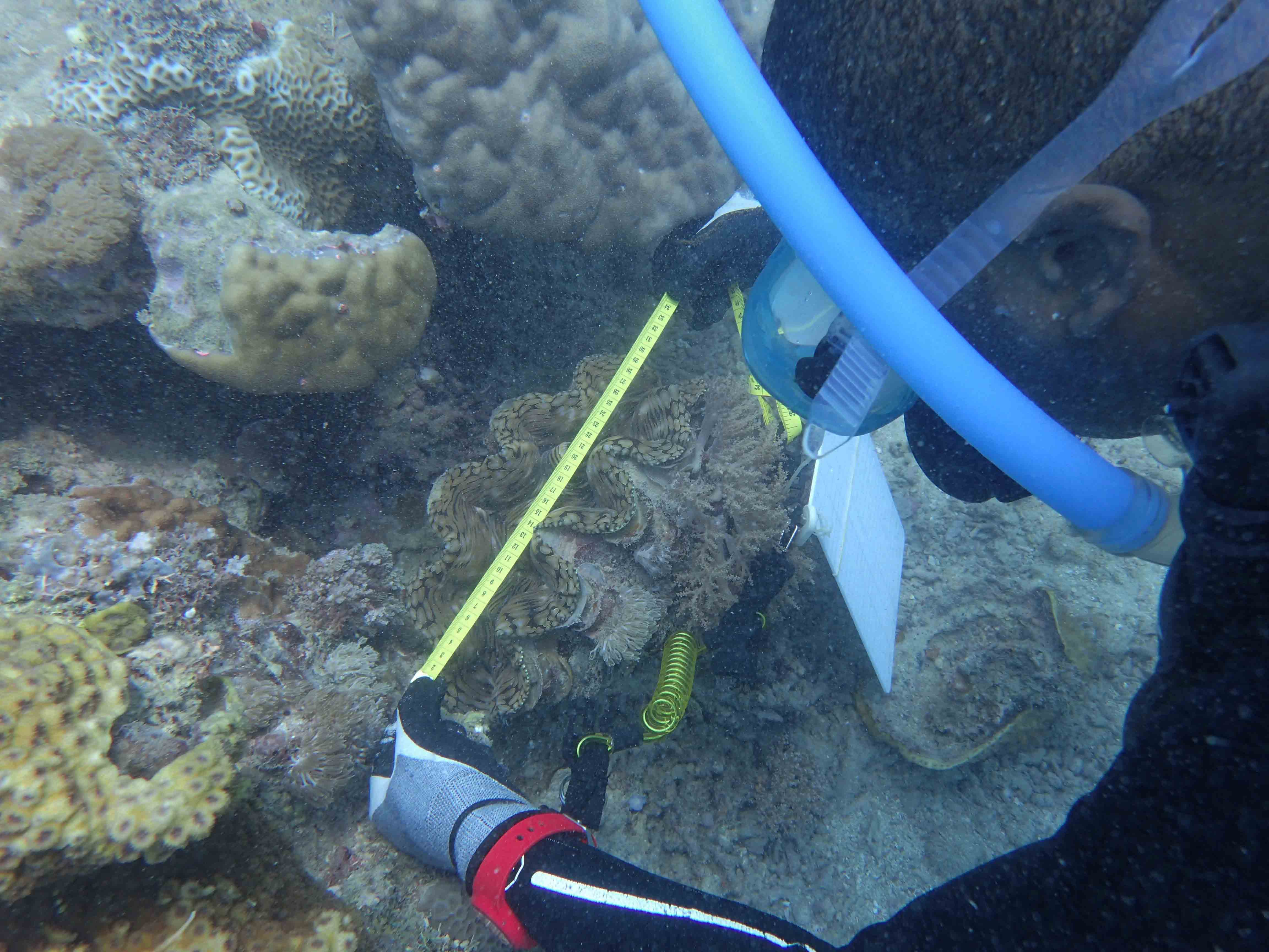
Measuring giant clams underwater

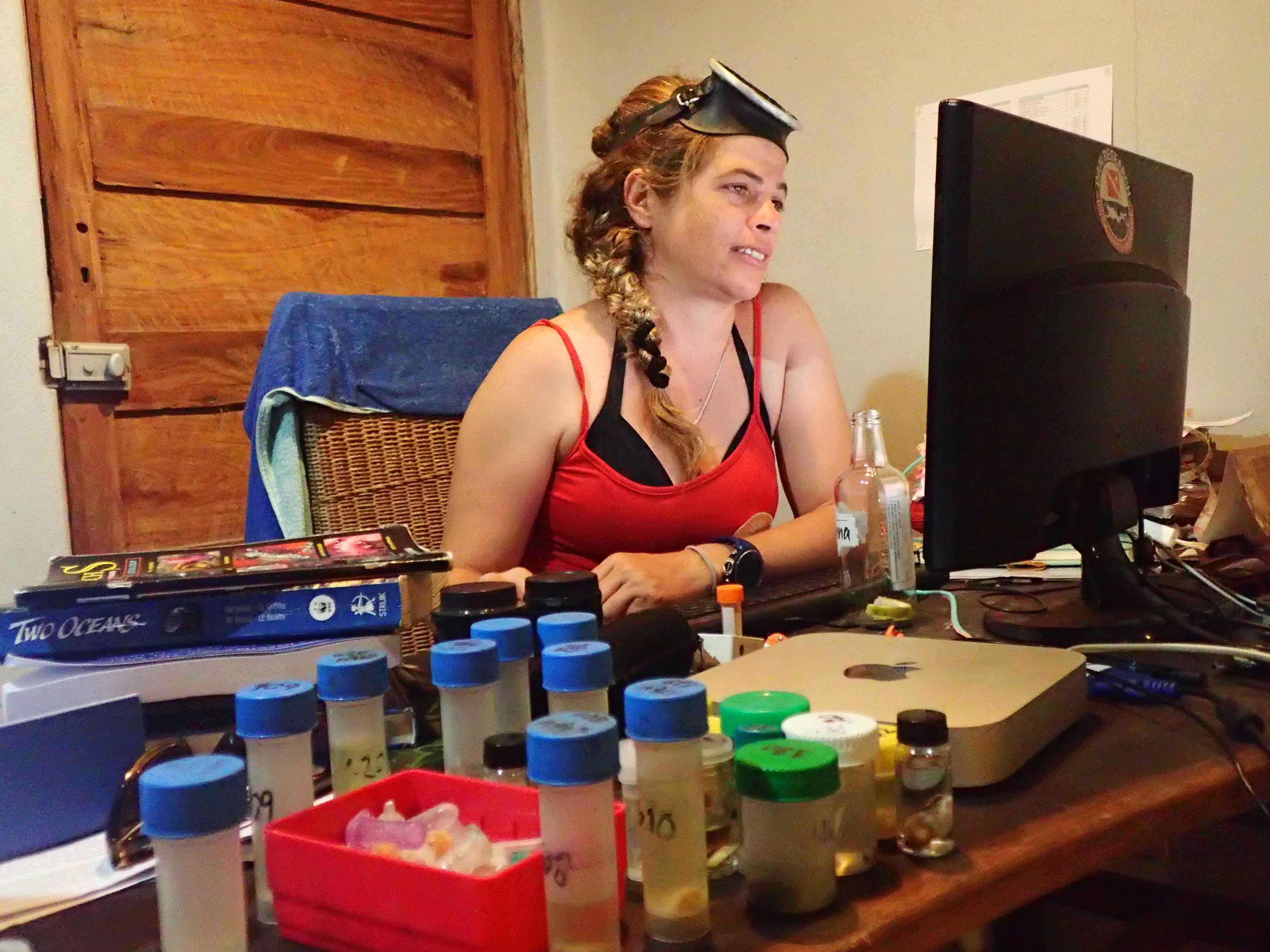
Yara Tibiriçá databasing the catch of the day

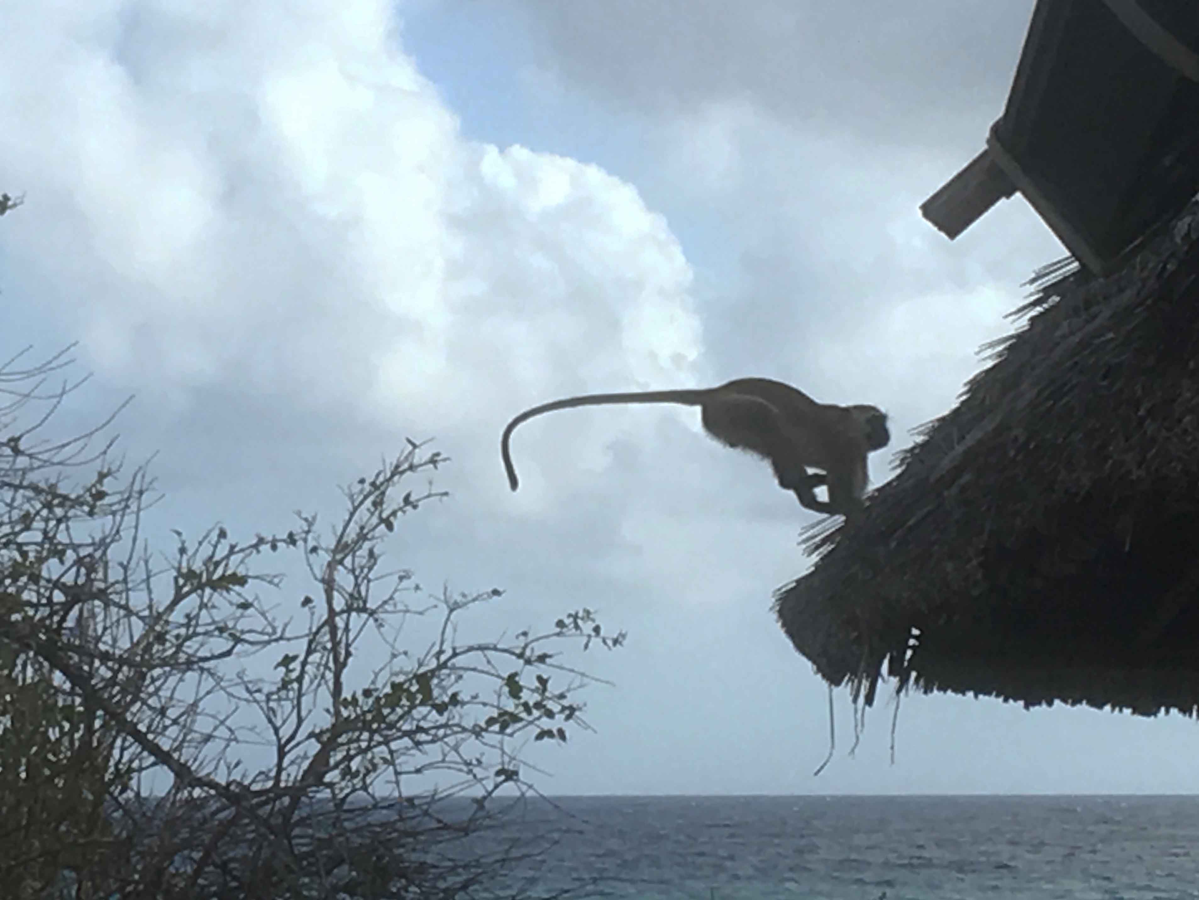
During two weeks we have surveyed the area for marine molluscs (sea slugs) and studied the impact of the marine protected area in the conservation of the highly sensitive and threatened giant clams (Tridacna spp.). The days at Nuarro began early, around 5.30–6am, with the sun already out and high in the blue sky. Waking up was not difficult, with the awesome strident ensemble of sounds from the many local different birds and the monkeys running and jumping on the thatched roofs of our housing! First sampling of the day around 8.30am, followed by a second one after lunchtime. The evenings were dedicated to study, photograph, identify, and database the catch of the day. We have collected approximately 68 species of sea slugs; 11 are new records to Mozambique and an additional 12 still undescribed. The conservation status of the populations of giant-clams was evaluated in areas under fishing pressure by the local communities and inside the marine reserve for comparison, and it was obvious the positive impact of the marine protect area.

Monkey jumping on the roof of our accommodation

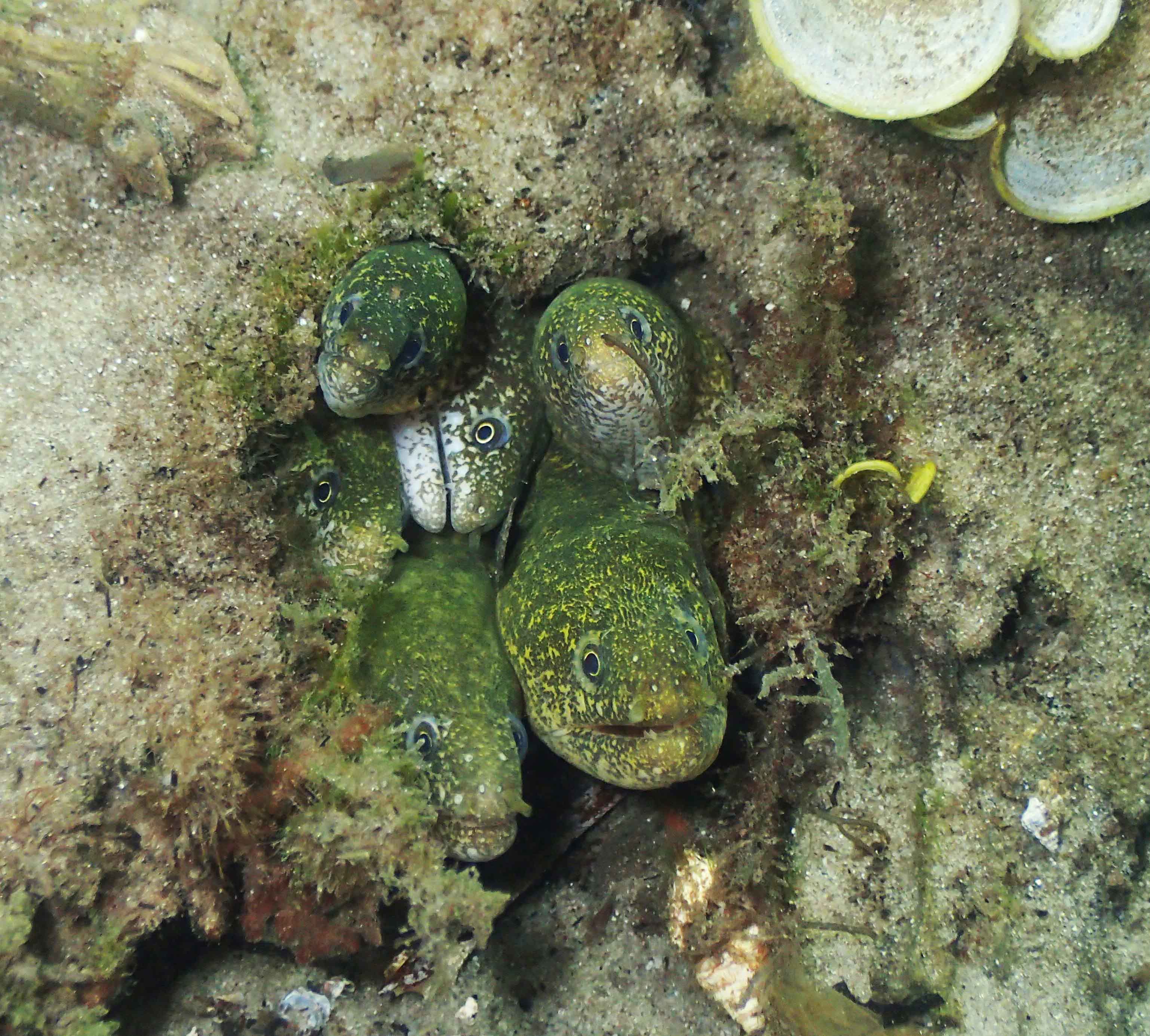
A “family” of moray eels

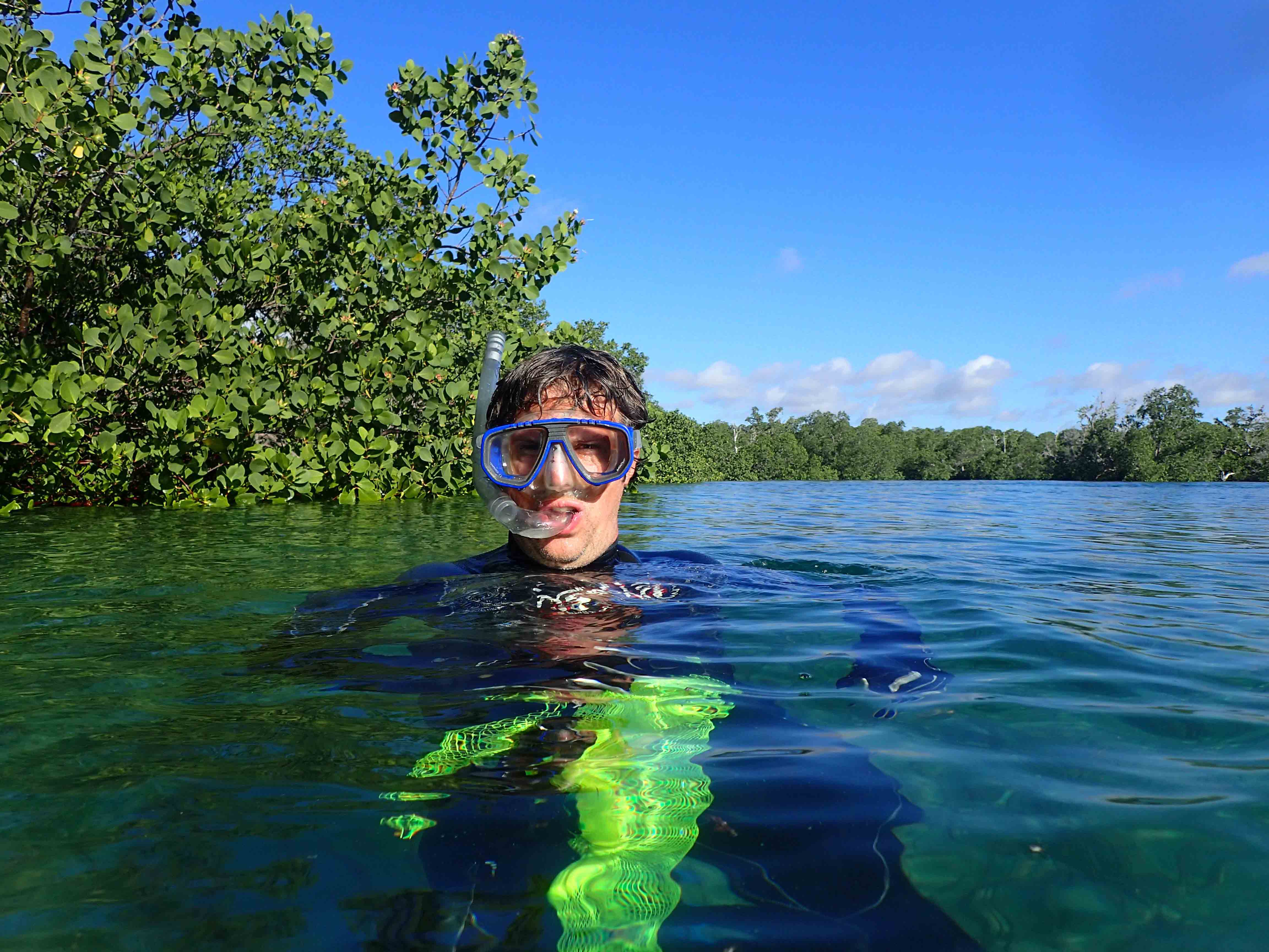
Collecting in the mangroves

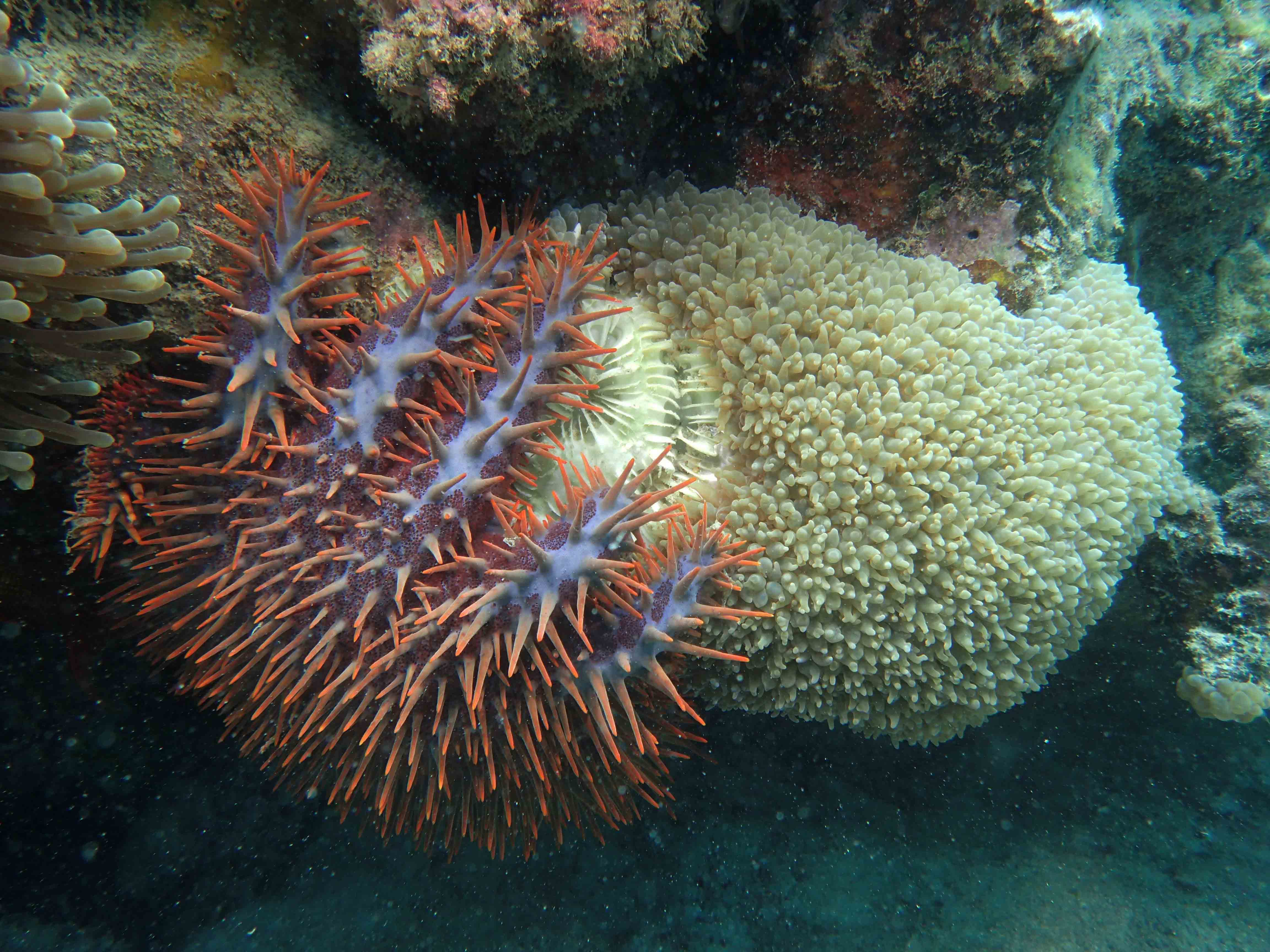
Crown-of-thorns starfish feeding on coral

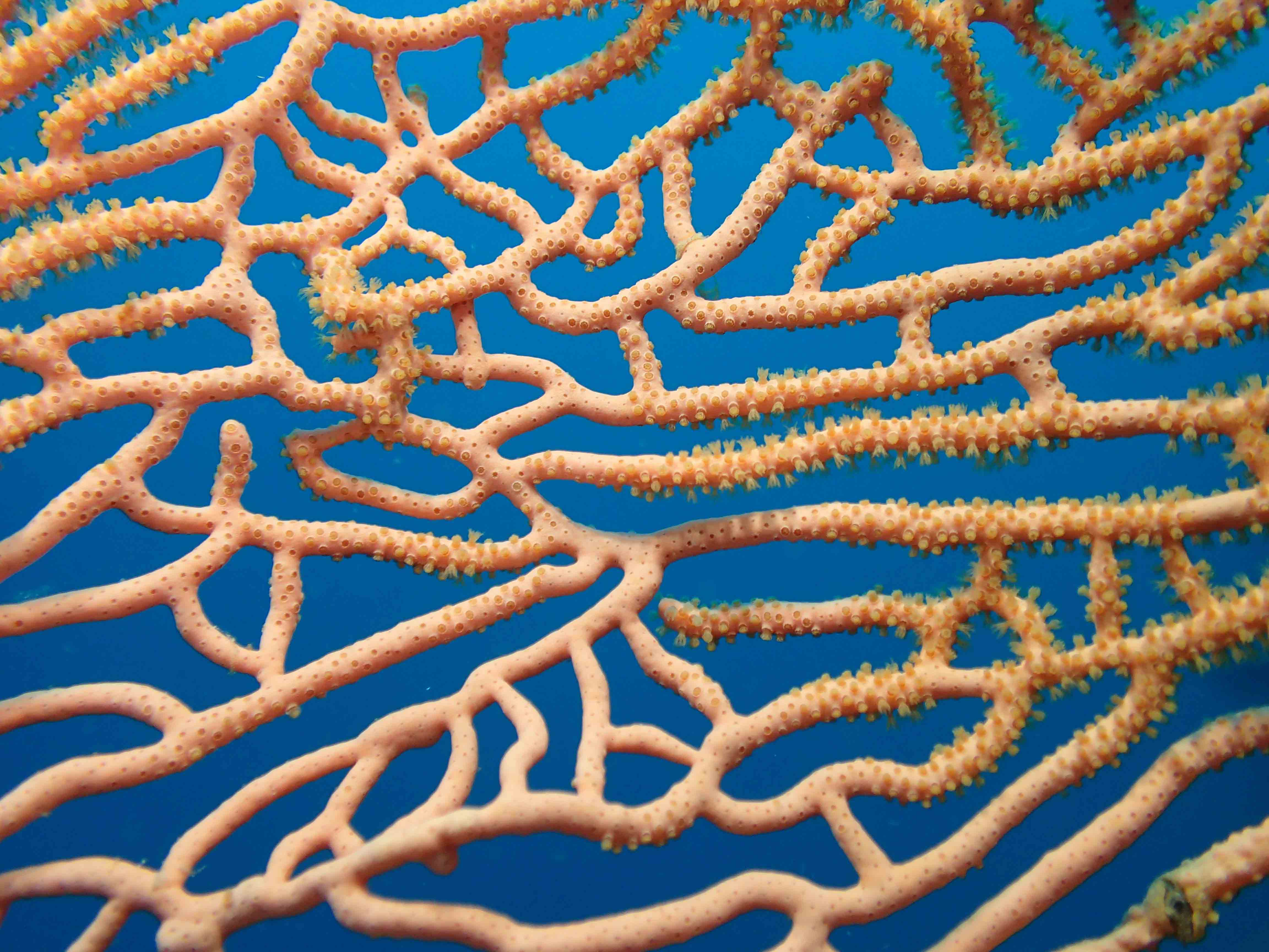
Detail of a gorgonian coral

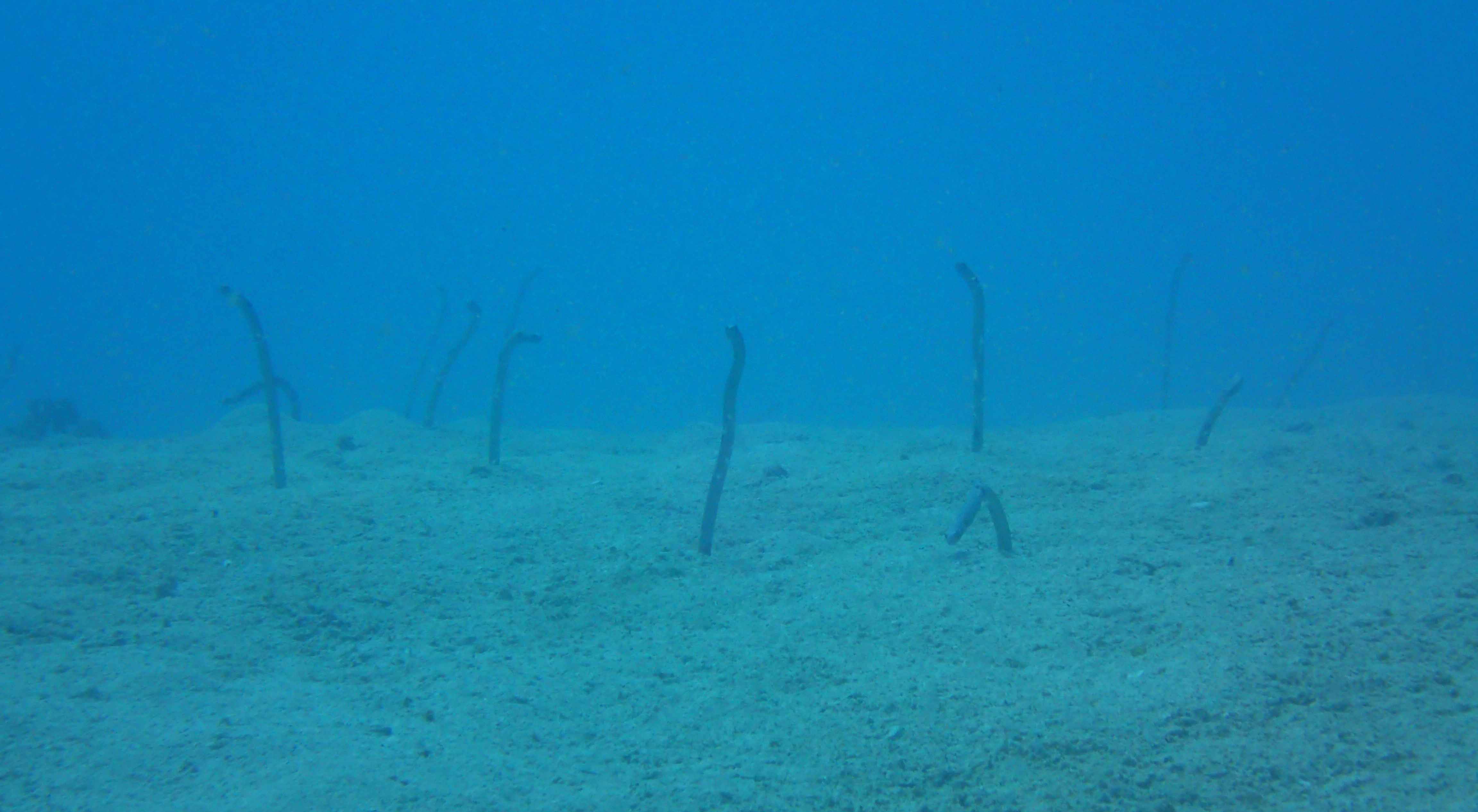
Garden eels

Nangata Bay, Nuarro

The flatworm Pseudoceros lindae

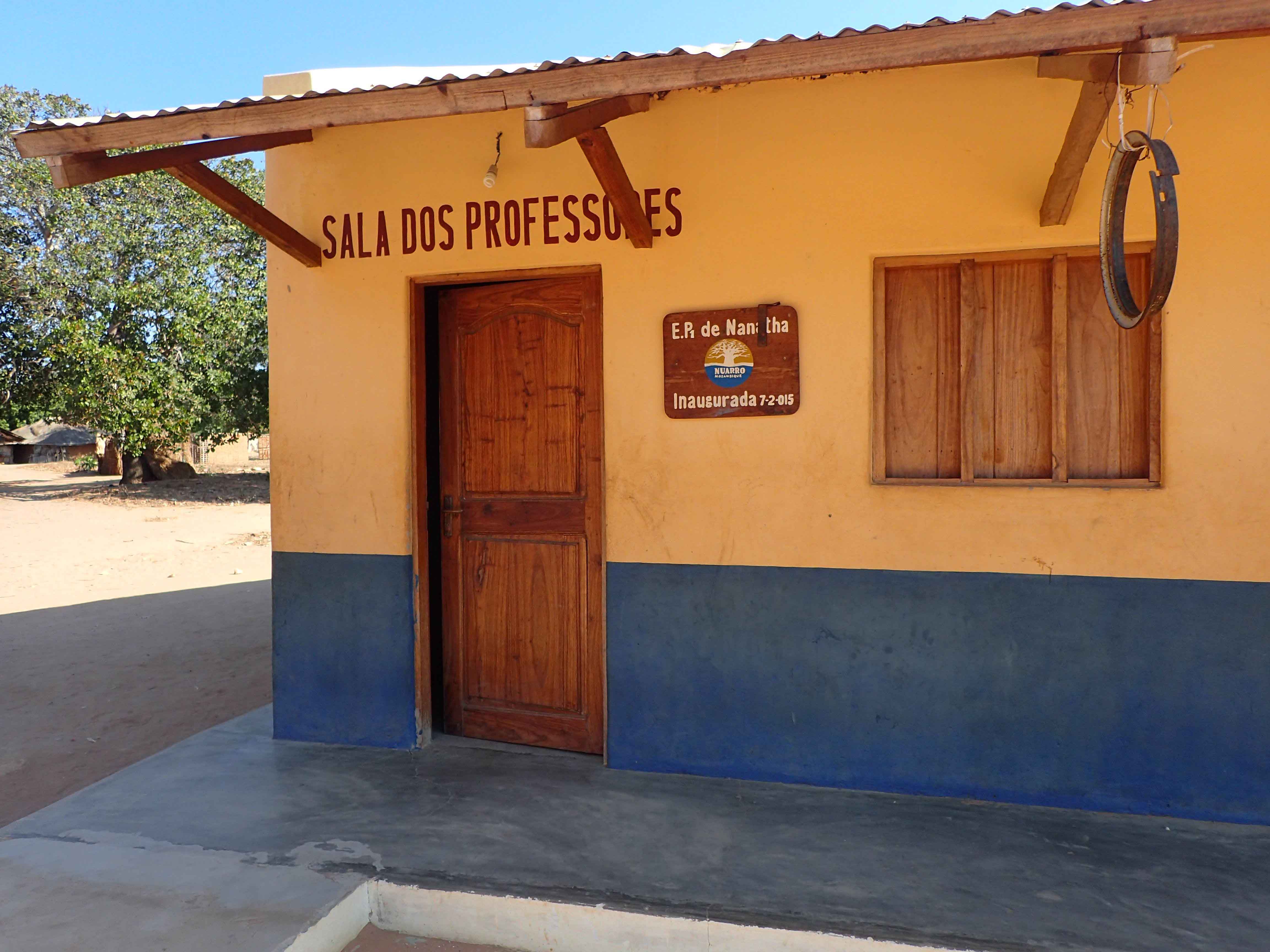
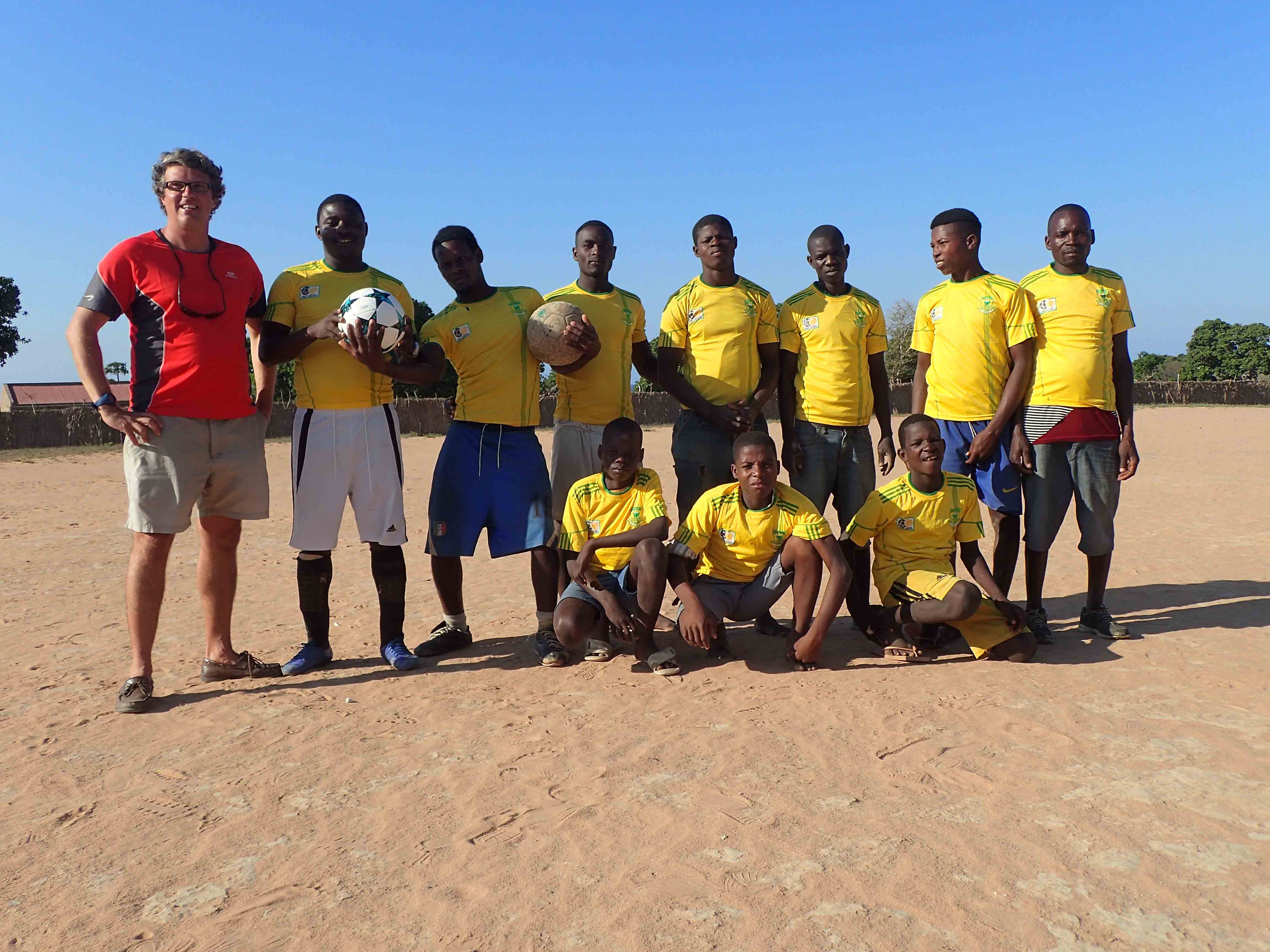
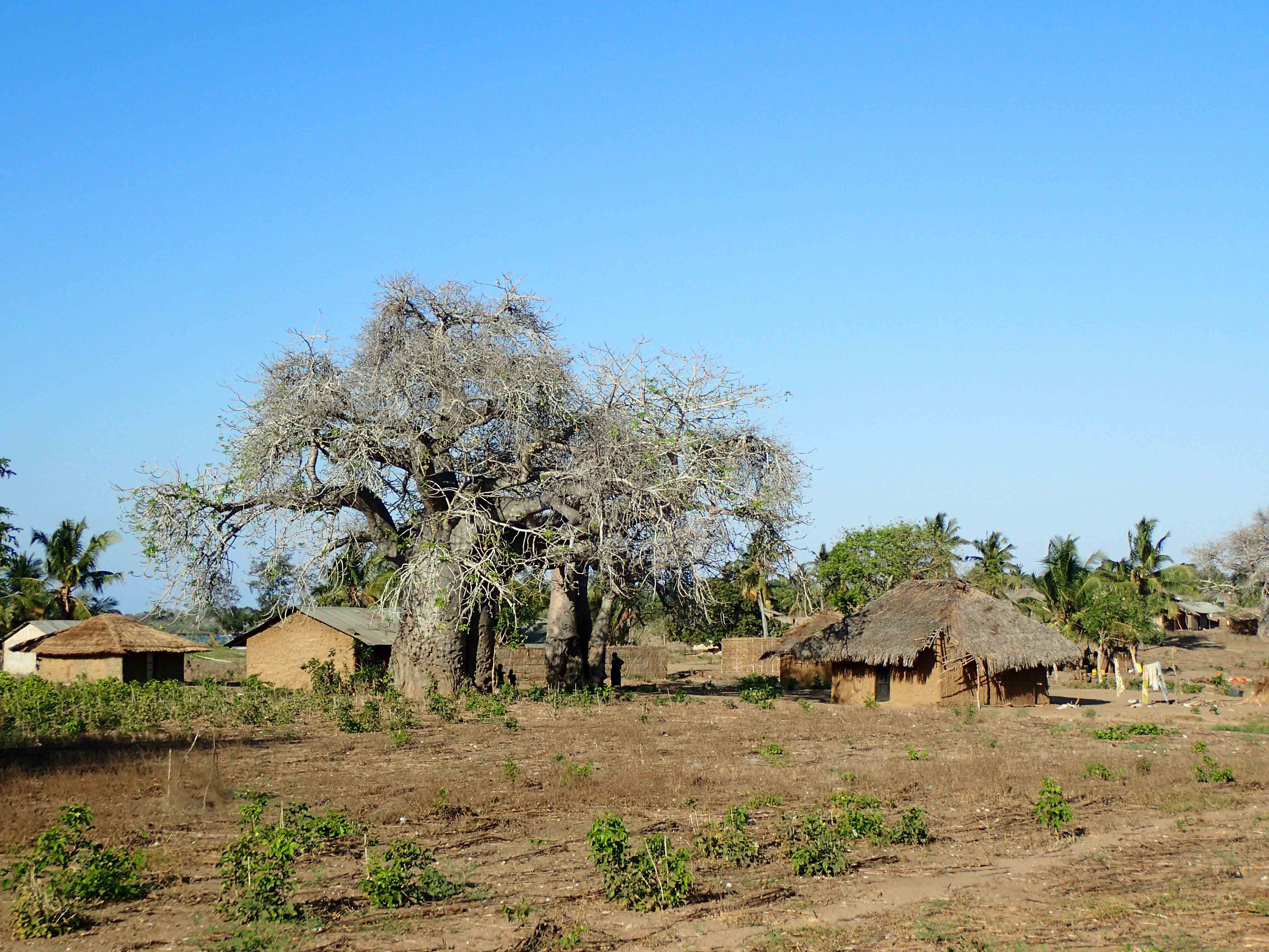
The days were busy and went far too fast, but there was time for a short social programme where we paid a visit to the local village and visited the primary school built by the lodge and met a group of local activists that work with the community to raise awareness for issues concerning hygiene, malaria, and other health matters, which regrettably still claim lives in the region far beyond acceptable numbers on the XXI century. Before the trip to Nuarro I was challenged to take a football ball with me for the local team. I have obviously eagerly done it, and it was a great delight to the see the joy of the players of the “Real Nangata” playing around with their new ball!

Primary school build by the lodge at Nangata village

Real Nangata football club

View of Nangata village with a baubau tree

The Nuarro expedition would not have been possible without the gratitude and support of the Nuarro Eco-Lodge to which I am deeply indebted.  I am also thankful to Prof. Isabel Silva from the University Lurio, Pemba and to the Fisheries Department of the Cabo Delgado Province from granting collecting and exporting permits.

Manuel António E. Malaquias, Associate Professor

Natural History Museum of Bergen, Norway