Category Archives: 2018 december calendar

Door #24: Happy Holidays!

And so we arrive at the final post of the 2018 edition of #InvertebrateCalendar. We have covered so many topics – I am always amazed at what people come up with!

Here is a recap of the posts in the 2018 edition, for previous years you can look behind door #1 here.

Door #1: Last Christmas…
Door #2: A glimpse of Hydrozoan anatomy
Door #3: Mollusc hunting around the world
Door #4: PSA: abstract submission for iBOL Conference is open!
Door #5: DNA-barcoding with BOLD
Door # 6: The key to the question
Door #7: New shipment of tissue samples for barcoding
Door # 8: The DNA-barcode identification machine
Door #9: To catch an Amphipod
Door #10: The Molluscan Forum 2018 in London
Door # 11: Animal rocks and flower animals
Door #12: Meet the chitons!
Door #13: The story you can find in a picture…
Door #14: Annelids from the deep Norwegian waters
Door #15: The eye of the beholder
Door #16: Basic anatomy of the sea slug
Door #17: Sea bunnies of Norway?
Door #18: The hypnotic adventure of the Norwegian jellyfish
Door #19: Photosynthetic vs solar-powered sea slugs
Door #20: The Hitchhikers Guide to the Ocean
Door #21: Barcode taxonomy and the “taxonomic feed-back loop”
Door #22: recommended reading for the holidays
Door #23: A model in the making
Door#24: Happy Holidays!

I hope our readers have enjoyed the calendar, and that you will check back in January for more of our blog posts. We have several new and exciting projects beginning in the new year, as well as the continuation of AnDeepNor and Seaslugs of Southern Norway – not to mention our ever growing invertebrate collections.

Then all that is left is to wish everyone all the best for the holidays!

From all of us to all of you..!

Door #23: A model in the making

The University Museum is being renovated and prepared for a grand new opening within 2019. The building has been put back in shape and looks great (more on that (in Norwegian), and images here), and we are now preparing new exhibits for everyone to enjoy.

As part of producing the new exhibits, we are working together with the excellent model-makers at 10 TONS in Copenhagen to be able to show large scale models of some of our beautiful friends. But how do we make a proper model of a small invertebrate? We want it to be a correct large-scale version of the animal we are portraying, not some half-good almost-look-alike…And if it happens to be strikingly beautiful as well, that is not a bad thing!

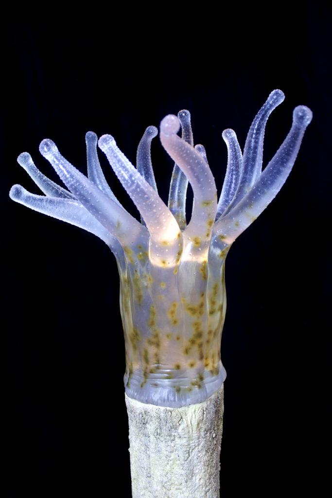
A gorgeous model of something too small to be observed properly with the naked eye; the zooxanthellae in the polyp of a coral. Read more here: http://www.10tons.dk/coralpolyp Photo: 10tons.dk

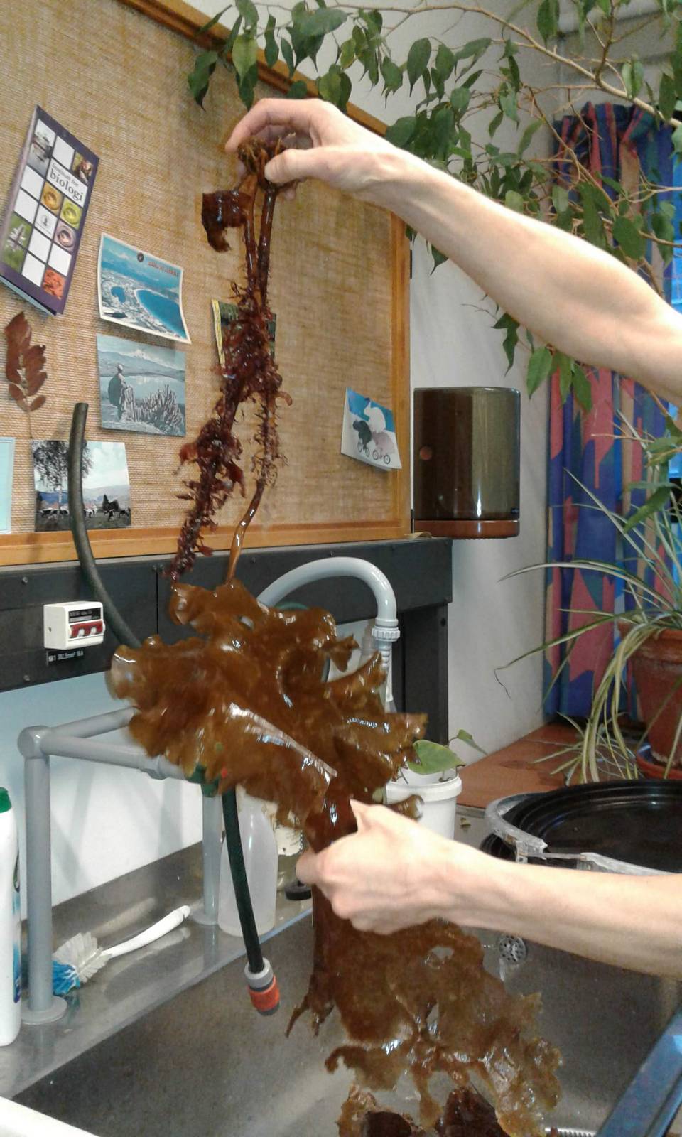
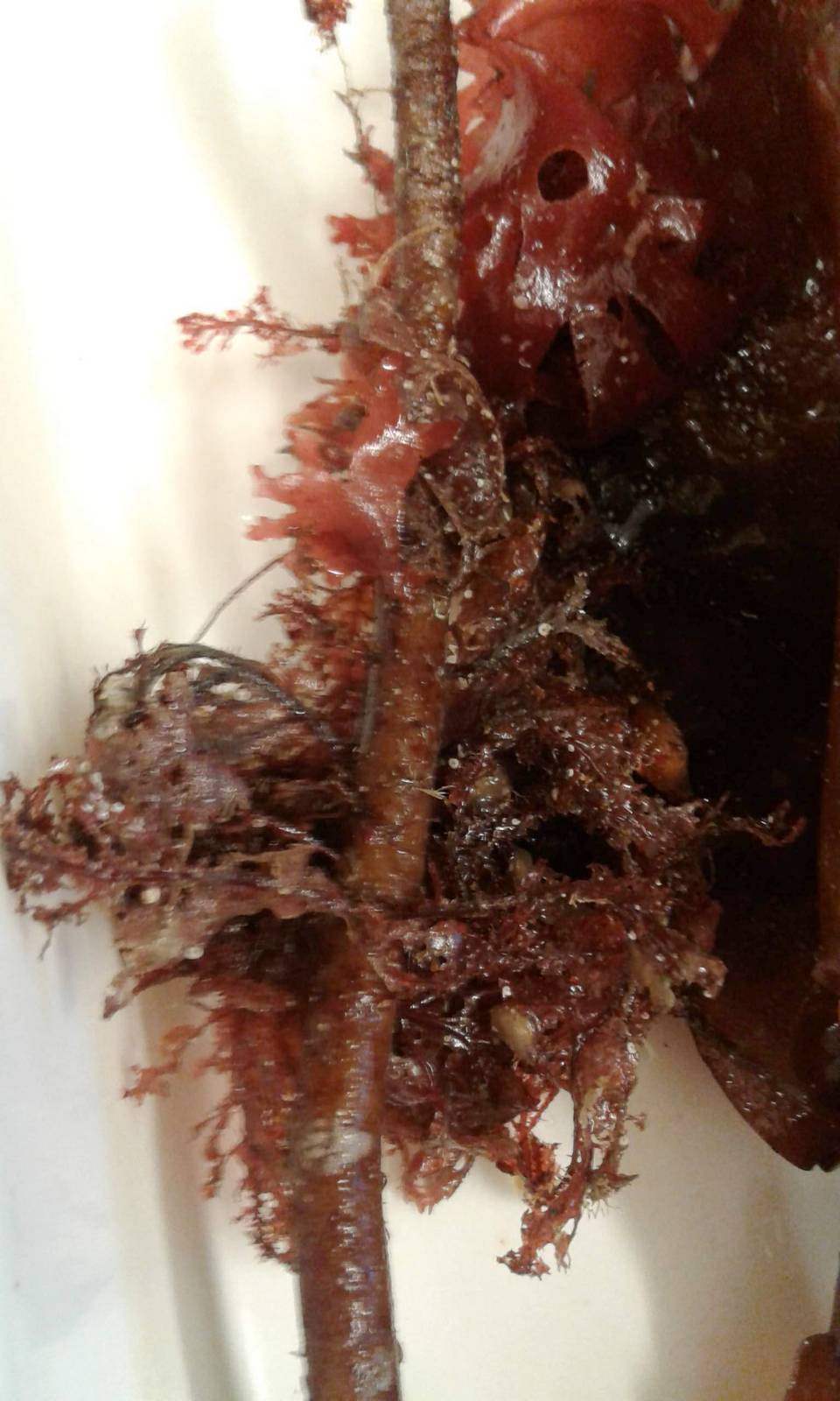
First of all, a 3-D computer model is made. At this stage, the guys at 10 tons work closely with us scientists to make sure that all details are correct – and we use a load of photos, films, SEM-photos and taxonomic drawings to make sure we have all things covered. Sometimes we even send them a specimen that they can scan. The models are passed back and fourth between the model-makers and the scientists, with indications of small corrections pointed out and performed until all parties are happy. You can be sure that the scientists have several minute details they want to change just a tiny little bit more, but we get there in the end…

Work in progress. Image: 10tons.dk

The next step is that the 3-D computer model is printed in the size that is going to be in the exhibits.

The printed out model is coated in a super-thin layer of wax to make it smooth, and then all the tiny details are added. Small notches in the epidermis or tiny plumose seta that have been separately made are added.

For a researcher who describes all the separate seta on the different mouthparts this is an amazing process to observe, and for everybody who later will see the model there can be an assurance that what you will see is actually how the species looks.

But this is not the end! The materials that have been used this far in the process will loose or change their colour when exposed to light. Therefore, a silicon mold (a “negative mold” or a cast of the outside of the model) is made from the finished first model. This mold is used to produce a new positive cast of polyurethane resin – and this is the model that will be shown in the exhibit. This material allows the model makers to add the right colour, translucence and texture to give the right look and feel of the finished product.

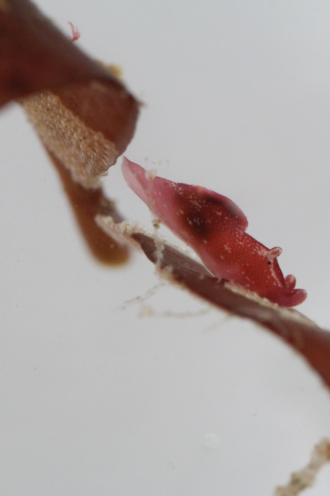
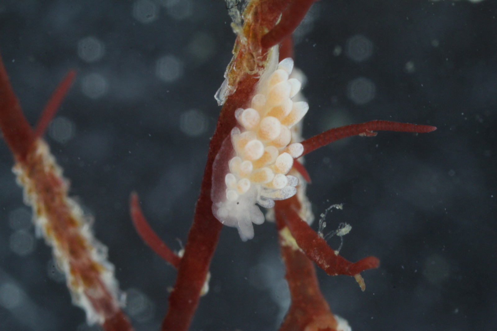
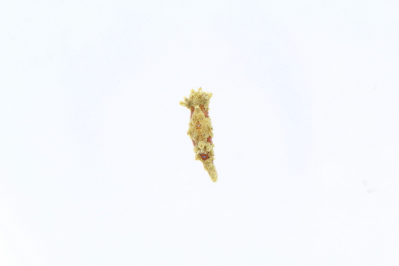
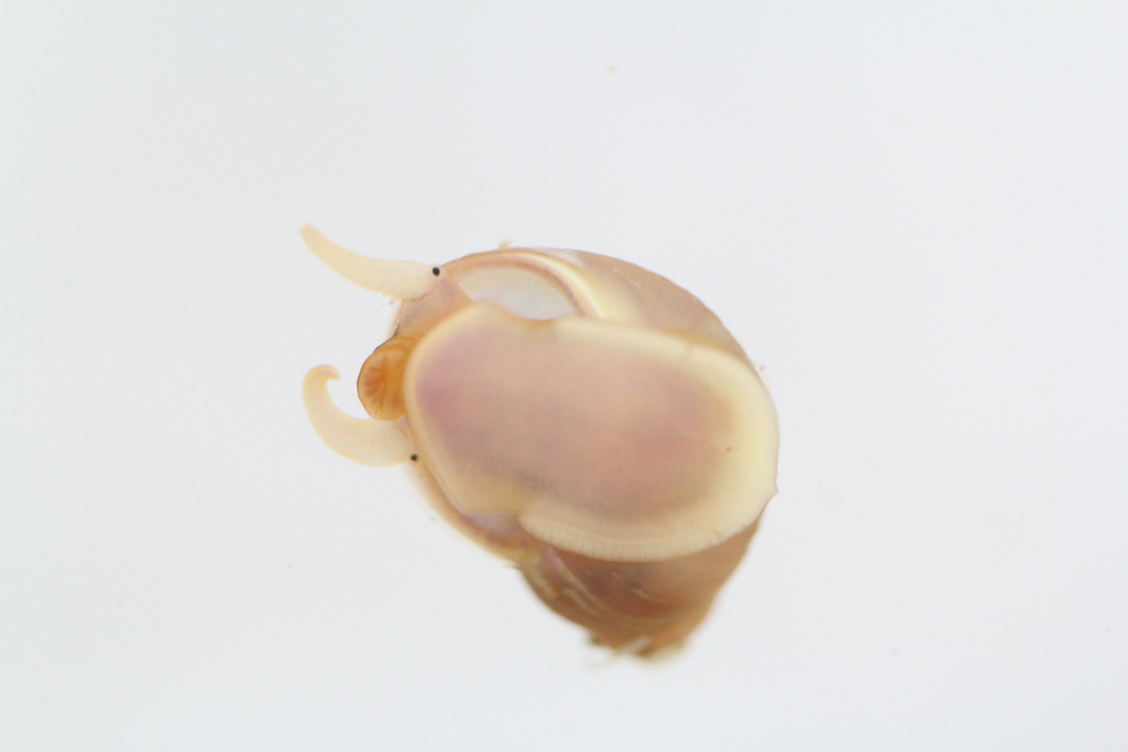
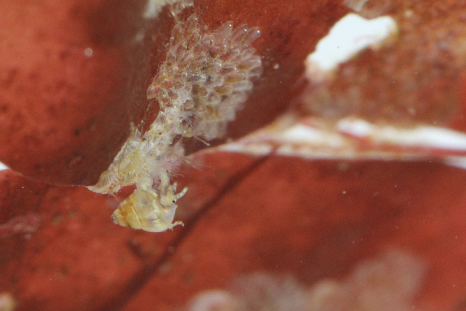
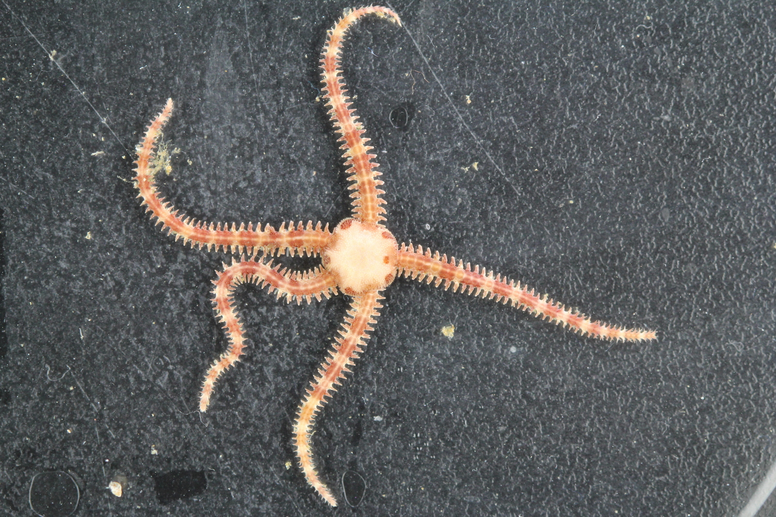
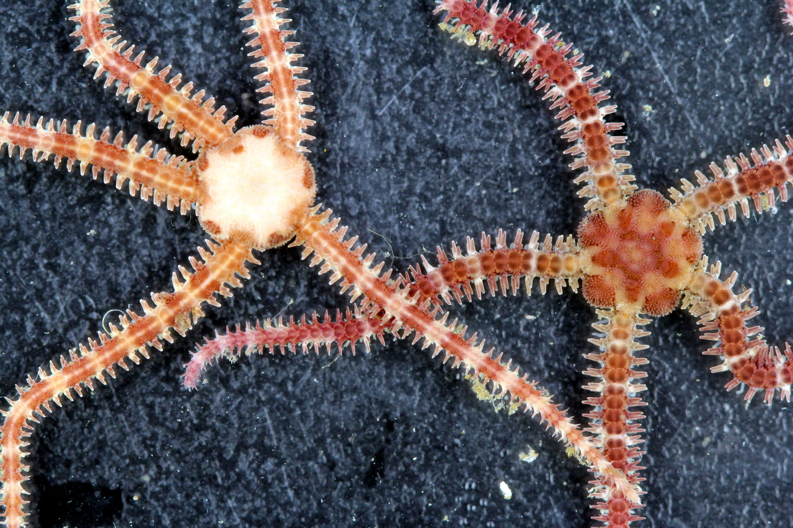
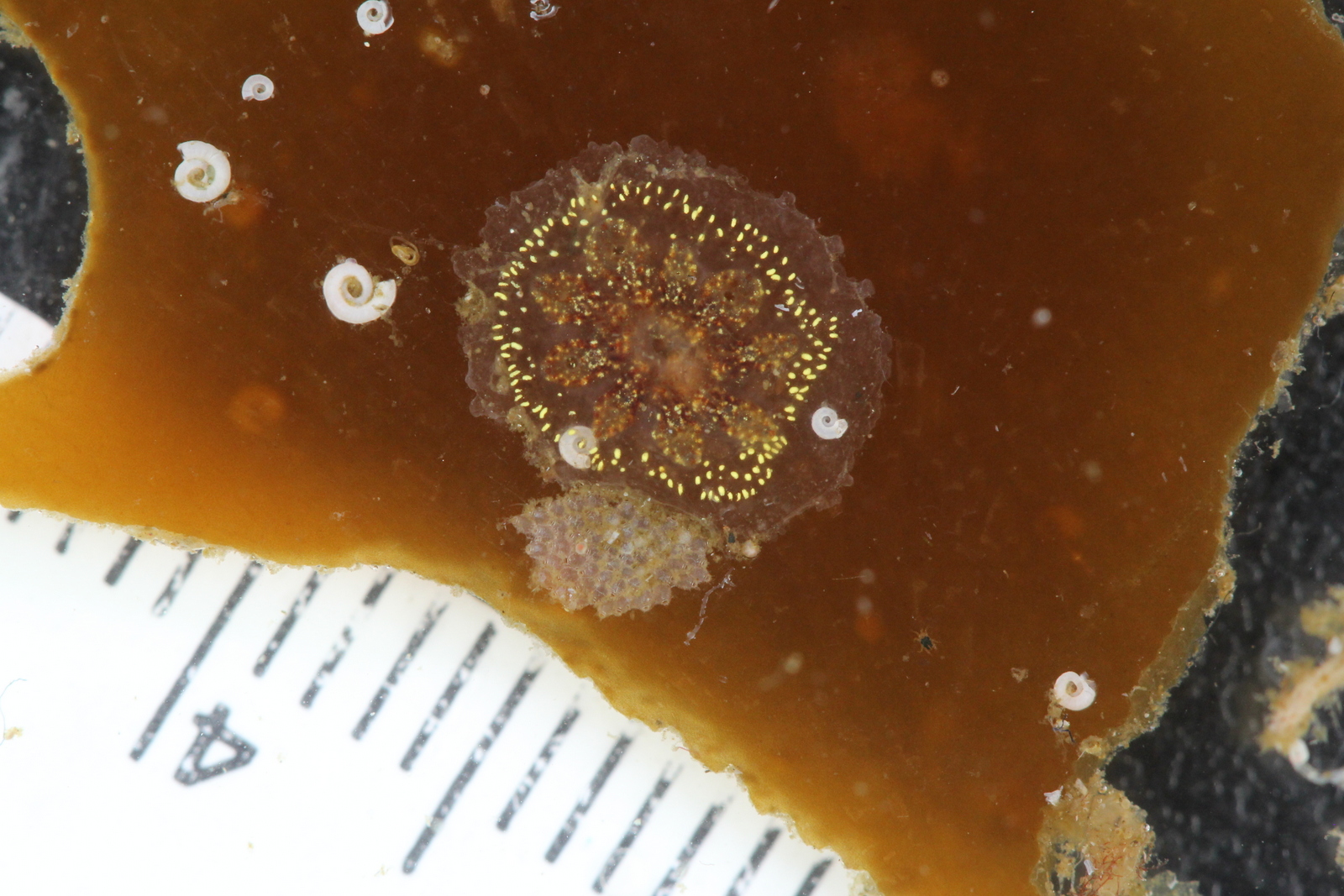
Here are a few of the scientific models, many more can be found here


Models are not made only of small animals – sometimes they are scaled 1:1, like this minke whale:

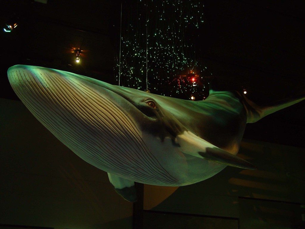
Balaenoptera acutorostrata, image 10tons.dk

Or *just a bit* bigger than what we could expect to find our in nature, like this crab (Cancer pagurus)

image: 10tons.dk

Here’s a video of how models are made – there are a few more videos here

We researchers are at the moment eagerly awaiting the models that will come to the University Museum – we have seen the 3-D models, and some of us have seen some photos of the models that are being made. We know that the models will look good, and we are looking forward to sharing them with everybody who comes to see the exhibits.

Now, what species will you be able to see models of, and in which exhibits will they be? That is for us to know now, and you to find out next year!

The holiday-season is a time for secrets to be kept, and this is one of those secrets. Come visit the University Museum when we reopen the building in autumn 2019 to see for yourself!

-Anne Helene

Would you like to know more about the process of making such models? This paper gives details and photos of a project 10 tons did with a paleontologist from the university of Lund in Sweden: Eriksson ME, Horn E (2017) Agnostus pisiformis – a half a billion-year old pea-shaped enigma. Earth-Science Reviews 173, 65-76. https://doi.org/10.1016/j.earscirev.2017.08.004

Door #22: recommended reading for the holidays

Today we’re offering you an early Christmas gift; the entire University Museum of Bergen 2018 yearbook is available online!

Provided that you read Norwegian, it offers an array of varied and entertaining tales of fieldwork in the context of the university Museum

You can find it here:

Årbok for Universitetsmuseet i Bergen 2018

The people of the Invertebrate Collections have written three of the papers in this year’s edition, with tales of fieldwork in Zanzibar, a journey in the footsteps of renowned taxonomist Michael Sars, and the story of the different methods we use to collect our animals in the field. We hope you will enjoy them!

Door #20 The Hitchhikers Guide to the Ocean

The sea is for most of its inhabitants a vast place where danger can get to you anywhere. This might be especially true when you are one of those small and mostly harmless species spending your life slowly swimming around, minding your own business (eating and reproducing), somewhere in the upper 200m or so of water. Because there are many big-mouthed and possibly big eyed animals out there that think you might be one of the best things there is to eat.

Hyperiella antarctica with Spongiobranchaea australis. Photo: C Havermans, AWI.

For the small pelagic (living in the open ocean and not close to the sea floor) amphipods in the suborder Hyperiidea this is one of the dangers of everyday life. The genus Hyperiella can be found in the Southern Ocean, and one of their main predators are the icefishes (Nototheniidae). So what do you do when you are a small and quite tasty animal that is not a very fast swimmer and there are a lot of fishes out there to eat you?

Don´t panic!

Hyperiella antarctica with Spongiobranchaea australis (a and b) and Hyperiella dilatata with Clione limacina antarctica (c). Figure 2 Havermans et al 2018.

Two of the three Hyperiella-species have found a quite ingenious solution. They hitchhike with a group of other small slow-swimming pelagic animals – pteropods. Pteropods (from the greek “wing-foot”) are sea snails (gastropods). Hyperiella australis pics up a life with Spongiobranchaea australis, and Hyperiella dilatata hangs out with Clione limacina antarctica. Both pteropods are from the group we call Sea Angels (Gymnosomata), and in a way they are saving angels for the amphipods: the ice fish don´t eat these strange couples. Why?

It seems the pteropods have developed a chemical protection against predation. They obviously taste extremely bad, for observations of icefish trying to eat the hitchhiking amphipods together with the pteropods result in them both being spit out again. Most times, the fish would see what it thought was good food, and then swim away when they discovered what they were almost eating. Not so very strange, then, that Hyperiella are holding on to their colleagues for their life!


Clione limacina antarctica. Photo C Havermans, AWI.

It might not be hitchhiking after all, but rather kidnapping – or brute force. The amphipods hold on to the pteropods with their to-three hindmost pairs of legs, and keep the sea angel on their back – much like a backpack. Observations are that they are repositioning them there all the time – almost like kids running with bumpy backpacks on the way to school. They don´t even let go when the researchers preserve them!

Hyperiella antarctica with Spongiobranchaea australis backpack. Photo: C Havermans, AWI

What this treatment do to the pteropods we still don´t know. But it does not seem they are able to eat very much when being held hostage as chemical defence-backpacks. That may not be the biggest problem in a short time-scale – their Arctic relatives have been shown to survive almost a year without food. What happens when they really get hungry we do not know. The amphipods are still able to feed, even though the pteropods can be up to 50% of the amphipod size. Maybe the pteropods do some of the swimming for the amphipods?

This behaviour is much more common close to the coast than in the open sea: close to the McMurdo area, 75% of the Hyperiella were seen hitching with a pteropod. Now we know that this pairing can be found in the open sea, and maybe is it more common that we think. It is not the first thing we have looked for so far when examining samples. When the University Museum of Bergen joins the Norwegian Polar Institute and the Institute of Marine Research to the Southern Ocean in the austral autumn this coming March, we will make a special effort to search for such collaborators.

Anne Helene


Literature

Havermans C, Hagen W, Zeidler W, Held C, Auel H 2018. A survival pack for escaping predation in the open ocean: amphipod-pteropod associations in the Southern Ocean. Marine Biodiversity https://doi.org/10.1007/s12526-018-0916-3

McClintock JB, Janssen J 1990. Pteropod abduction as a chemical defence in a pelagic Antarctic amphipod. Nature 346:424-426.


Door #19: Photosynthetic vs solar-powered sea slugs

I think we can all agree that sea slugs are amazing creatures. Some species contain toxins that are useful for cancer research and others are photosynthetic! There are a few species of sea slugs that have the ability to photosynthesize. But beware; the ability for them to do the thing that plants and algae are good at (photosynthesis), doesn’t always mean they are solar-powered. And I will explain you in the next few lines what I mean with that.

There are two families of sea slugs known to have a few species within them that can photosynthesize; it’s within the family of Facelinidae and the family of Sacoglossa.

Within the family of Facelinidae we have the genus Phyllodesmium; and all species in this genus are considered solar-powered, meaning that they get a part of their daily energy intake via photosynthesis. At the moment the theory goes that they are able to do so as they contain photosynthetic zooxanthella stolen from their feeding source (soft corals), and they continue to ‘farm’ these zooxanthellae in their own bodies. The species Phyllodesmium longicirrum masters this trade and is best known as the solar-powered Phyllodesmium

Solar-powered Phyllodesmium longicirrum by Jason R. Marks

The other family does things quite differently but nevertheless as impressive; those are the Sacoglossa.

A few representatives of the photosynthetic sea slugs within the Sacoglossa

Unlike Phyllodesmium, they don’t farm zooxanthella but they steal the photosynthetic cell organelles (chloroplasts or plastids) from the algae they feed on; also known as kleptoplasty (Kleptes (κλέπτης) Greek for thief). Approximately 140 years ago these sea slugs were first described by de Negri and de Negri, who discovered that these sea slugs were green colored due to foreign ‘bodies’ that were reminiscent to those known from plants. It took at least another 100 years before the ‘granule bodies’ were identified to be chloroplasts from the algae the slugs feed on. Sacoglossa are also known as sap-sucking sea slugs because of the way they eat their algae; at first, the sea slugs pinch a whole in the algae wall with their special teeth, called the radula. Then they suck out the cytosolic content of the algae (hence the name sap sucking sea slugs). Finally, the cytosol content is being digested in the digestive tract that perforates the entire body and within some species the chloroplasts are being sequestered and continue to photosynthesize in the animal’s digestive cells

Sap-sucking slug sequestering chloroplasts from the algae it feeds on (figure from Rauch et al. 2015)

Although the sea slugs are famous for their ability of stealing chloroplasts, they only represent a minority within the Sacoglossa. In fact, only 6 out of 300 described species, can keep the chloroplasts fully functional for long term, or long-term retention species; that is the chloroplasts are photosynthetic active for longer than 21 days after sequestration. The gross majority are either short term retention species (with functional chloroplasts up to 14 days or more) or cannot retain the chloroplasts at all (called non-retention species). This means that the chloroplasts are immediately being digested like the rest of the algae content.

So why are Sacoglossa photosynthetic but not solar powered like Phyllodesmium? Well this is because numerous studies observed how the sea slugs died as soon as they were being starved (meaning they couldn’t eat their algae food anymore) even though they had photosynthetic active chloroplasts in their cells. Besides, based on CO2 fixating measurements, it turned out that 99% of the sea slug has to live from normal ingested food, like all other animals do. This could very well explain why only 6 out of the 300 described species are able to sequester chloroplasts long-term, and apparently for reasons other than carbon fixation!

The take home message is that this case of photosynthetic and solar-powered sea slugs is a great example of how good science is about resisting the pull of easy conclusions. When something seems right at first, it should still be testified!

Furthermore
Interested in photosynthetic sea slugs? You can read more in the following papers that are also used as source for this blog:

  1. A sea slug’s guide to plastid symbiosis (2015) J De Vries, C Rauch, G Christa, SB Gould, Acta Societatis Botanicorum Poloniae 83 (4)
  2. Why it is time to look beyond algal genes in photosynthetic slugs (2015) C Rauch, J Vries, S Rommel, LE Rose, C Woehle, G Christa, EM Laetz (…), Genome biology and evolution 7 (9), 2602-2607
  3. On being the right size as an animal with plastids (2017) C Rauch, P Jahns, AGM Tielens, SB Gould, WF Martin, Frontiers in plant science 8, 1402
  4. Mitochondrial Genome Assemblies of Elysia timidaand Elysia cornigera and the Response of Mitochondrion-Associated Metabolism during Starvation (2017) C Rauch, G Christa, J de Vries, C Woehle, SB Gould, Genome biology and evolution 9 (7), 1873-1879
  5. The ability to incorporate functional plastids by the sea slug Elysia viridisis governed by its food source (2018) C Rauch, AGM Tielens, J Serôdio, SB Gould, G Christa, Marine Biology 165 (5), 82

Would you like to see pictures of sea slugs that you can find in Norway? Check out the Sea slugs of Southern Norway Instagram account,and don’t forget to follow us!
Become a member of the sea slugs of Southern Norway Facebook group, stay updated and join the discussion.

Explore the world, read the invertebrate blogs!

Door #18: The hypnotic adventure of the Norwegian jellyfish

The end of the year is always a crazy time full with work deadlines and holiday planning, but if one can find a few minutes in between last-minute shopping and gift-wrapping, the last days of December can also offer the opportunity to reflect on past and current experiences and what we have learned from them. It is also a special time for closure, so it feels natural that it coincides this year with the end of HYPNO, one of the projects of the Invertebrate collections funded by the Norwegian Taxonomy Initiative.

For the last three years, project HYPNO (Hydrozoan pelagic diversity in Norway) has been documenting and DNA-barcoding the diversity of hydromedusae and siphonophores in Norwegian waters. We have learned a lot about the distribution of these animals in the process, especially since we have come face to face (or rather face to tentacle) with more than 1100 specimens from all parts of the country.

Representative species of the groups within Hydrozoa that were the object of study of HYPNO (from left to right and top to bottom): Amphinema rugosum (Anthoathecata), Mitrocomella polydiademata (Leptothecata), Lensia conoidea (Siphonophorae Calycophorae), Agalma elegans (Siphonophorae Physonectae), Botrynema ellinorae (Trachymedusae), Bathykorus bouilloni (Narcomedusae). Pictures: Luis Martell and Aino Hosia.

Telling all these organisms apart was not always an easy task, but thanks to a combination of traditional taxonomy and genetic methods we have been able to identify more than 120 species, including several new records for Norway and a couple of species that had gone unnoticed to the world until today. The project was very successful in getting useful DNA sequences for the majority of these species, which will facilitate the identification of hydrozoans in the future, and that will be available for everybody to use through the online platform of the Barcoding of Life initiative. All the records gathered during the project will also be available to the public (through the interactive maps of Artskart) and a collection of specimens will be stored at the University Museum of Bergen as a reference for anyone interested.

A nice overview of some of the results of HYPNO as displayed in the webpage of BOLD systems.

Additionally, keep an eye open for HYPNO’s most striking results that can be found in several scientific articles (published and in various stages of preparation), and for the project’s last updates in social media.

Without a doubt, HYPNO owes much of its success to the combined efforts of all the individuals and institutions that have collaborated in the project in one stage or another. NorBOL, GooseAlien, NorAmph, and Arts- og naturtypekartlegging av Sognefjorden are only some of the projects with which HYPNO has shared samples, experiences, and field trips. Collecting the animals and curating the specimens and data would not have been possible without the help of the crew working in the research vessels that hosted us and the technical staff at the DNA lab and the invertebrate collections of the museum.

HYPNO included samples from all over Norway, but we particularly enjoyed our regular sampling trips in the amazing Western Norwegian coast. Pictures: Luis Martell, Joan J. Soto Àngel.

Many records for the project came from friends and colleagues working in other institutions and universities in Norway and abroad, all of whom were eager to share their knowledge and time with us in this hypnotic adventure of Norwegian jellyfish.

We had always a great time jelly-hunting in the sea! Pictures: Luis Martell, Joan J. Soto Àngel, Anne-Helene S. Tandberg.

HYPNO may now be coming to an end, but this doesn’t mean that our work on Norwegian hydrozoans is finished. On the contrary, the Invertebrate Collections have been granted a new project starting next year and focusing again on these fascinating animals. We hope that the new project NORHYDRO will be as rewarding as HYPNO, and we know it will be at least as fun: time flies by so quickly when jellyfish and good friends are involved!

-Luis Martell and Aino Hosia


Some suggested reading related to the project
Schuchert P, Hosia A, and Leclere L. 2017. Identification of the polyp stage of three leptomedusa species using DNA barcoding.

Martell L, Tandberg AHS, and Hosia A. 2018. The illusion of rarity in an epibenthic jellyfish: facts and artefacts in the distribution of Tesserogastria musculosa (Hydrozoa, Ptychogastriidae). Helgoland Marine Research 72(1):12.

Door #17: Sea bunnies of Norway?

Some years ago, in 2015, the internet was taken a storm by the sudden rise of the so-called sea bunnies. It all started with a video taken the year before by a SCUBA diver in Japan who filmed the little creatures crawling around the seabed:

The species seen in the video is called Jorunna parva, and are since then worldwide unofficially known by the adorable pet name ‘sea bunny’ as it has a ‘fur’ like exterior with tiny upright ‘ears’ and a fluffy tail like bunnies do. The ‘fur’ is actually created by bunches of tiny rods, called the caryophyllidia. The caryophyllidia are arranged around little knots that are often dark coloured, which create the illusion of black dots on the animals. The seemingly ears are the animals rhinophores, that function as chemical receptors that make the animal able to detect its environment in search of food and other sea bunnies. Its tail on the back are actually its gills to extract oxygen molecules from the surrounding water, the ‘fluffier’ it is, the bigger the surface area, the easier it is to diffuse oxygen from the water. This sea bunny is small, often less than 2.5 cm, and can be found throughout the Indo-Pacific; from South Africa to Central Pacific. They have, like many sea slugs do, a high degree of colour polymorphism in the species, with colours varying from white with black dots, to yellow and even bright orange:

Different colours of Jorunna parva, aka the sea bunny (photo credits on image)

Unfortunately, our sea bunny J. parva is only short-lived and just lives from a few months up to a year, but at least during its short life it doesn’t need to worry about predators. They are very toxic, because of the food they eat, which are sponges. All dorid nudibranchs (the group of slugs J. parva belongs to) are toxic because of their diet, and these toxins are often used in cancer treatments for people. Who would have thought that sea bunnies would be lifesavers, besides  being cute? But is the word sea bunny only referring to this particular species called J. parva? A quick search on the internet definitely tells us otherwise, it seems people refer to sea bunnies when they talk about any other dorid nudibranch with a fluffy and round appearance.

So, the question remains, do we have any sea bunnies in Norway? And the answer is yes, we do!

Sea bunnies of Norway (click to enlarge!)

And they are absolutely great and adorable to encounter underwater. Let us make a list of the sea bunnies of Norway, so we know what species we are talking about. Sea bunnies of Norway are; Doris pseudoargusGeitodoris planataJorunna tomentosaRostanga rubraCadlina laevis, Aldisa zetlandicaAdalaria loveniAdalaria proximaOnchidoris muricataOnchidoris bilamata,
Onchidoris pussilaOnchidoris depressaAcanthodoris pilosaDiaphorodoris luteocincta and I personally would draw the line at Goniodoris nodosa, as the other Goniodoridae don’t resemble that much the typical sea bunny characteristics. What do you think? Which species do you think are missing in this list, or which species should be left out?

I think it is time to take over the internet with our sea bunnies of Norway!


Furthermore
You want to see more beautiful pictures of sea slugs of Norway!

Check out the Sea slugs of Southern Norway Instagram account; and don’t forget to follow us.
Become a member of the sea slugs of southern Norway Facebook group, stay updated and join the discussion.

Explore the world, read the invertebrate blogs!

-Cessa

Door #16: Basic anatomy of the sea slug

Haminoea sp, photo by M. Malaquias

“Sea slugs” include both the by far most famous nudibranchs, and groups such as the Sacoglossa (sap-sucking slugs, more about these later in the calendar!) and Cephalaspidea (the bubble snails), amongst others. These latter ones often do have shells – but reduced ones, too small for the animals to completely retreat into, like this Haminoea:

Nudibranchs, however, are the “naked” snails: Their name “nudibranch” comes from the Latin nudus, naked, and the Greek βρανχια, brankhia, gills. They don’t have a shell, but this wasn’t always the case. In their early larval life stage, they actually have a shell, but when settling down and transforming from zooplankton into adults, they lose the shell. The loss of the shell in adults might be responsible for the amazing diversity we see in body forms present in sea slugs.

So, in this basic anatomy of sea slugs we will focus mostly on the body forms of nudibranchs, but all other sea slug orders are not far off from this anatomy, if you know the basics of nudibranchs, you can extrapolate to the other orders as well. So here we go!

Nudibranchs are roughly divided in two type of body forms; the dorid nudibranchs and the aeolid nudibranchs.

Two basic body types found within the Nudibranchia; the dorids and the aeolids. (Illustration: C. Rauch)

The dorid nudibranchs have a thick mantle that extends over their foot. In some species the surface of the mantle is covered with tubercles than can vary in different sizes, numbers and shapes. This gives them often a rigid body that offers some protection.  In most of the dorids it is the mantle that contains toxins to defend themselves, the toxins are extracted from their food sources.

Aeolid nudibranchs have mantles that are covered with finger-like extensions called cerata. The cerata are very special as they contain branches of the digestive tract, and in some species, this is also visible! The tips of the cerata contain special organs called the cnidosacs. Cnidosacs store stinging cells (called nematocysts). These are obtained from their food source which are often cnidarians like hydroids, sea-anemones and soft corals. The cnidosacs are activated when the nudibranchs feel threatened and the stinging cells will be discharged!

All sea slugs have rhinophores. On the head of all sea slugs you can find a pair of sensory tentacles called the rhinophores. They detect smell and taste and in most of the dorid nudibranchs the rhinophores can be retracted into a basal sheath. Sea slugs know all kind of shapes of rhinophores which are a very important tool for identification of the species.

Diversity of sea slug rhinophore shapes

Besides a pair of rhinophores many nudibranchs also have a pair of oral tentacles, one on each side of the mouth. They are most likely involved in identifying food by taste and touch.

Sea slugs also need to breathe oxygen. They do this via the surface of their entire bodies, but their main reparatory organ are their gills. Dorid nudibranchs have often a feather like structure encircling their anus on the back of their body (the branchial plume). Some dorid species can also retract their gills into a pocket. Within the aeolid nudibranchs the cerata act like gills by diffusing oxygen from the surrounding water. The cerata are sometimes branched in order to increase the surface area, also here, like the rhinophores, different species can have different forms of cerata.

Diversity of aeolid cerata shapes

-Cessa

Door #15: The eye of the beholder

It’s funny to see the different reactions to fresh material that comes in to the museum;  the exhibition team had  received some kelp that will be pressed and dried for the new exhibitions (opening fall 2019), and I ducked in to secure some of the fauna sitting on the kelp before it was scraped off and discarded. For the botanists, the animals were merely a distraction that needed to be removed so that they could deal with the kelp, whilst I was trying to avoid too much algae in the sample as it messes up the fixation of the animals.

I chose the right shirt for the day- it’s full of nudibranchs! (photo: L. Martell)


I then spirited my loot into the lab, and set up camp.

Count me in amongst the people who stare at lumps of seaweed.


Who’s there? The whole lump is ~12 cm.

How many animals do you see here? Which ones appeal to you?

I have made a quick annotation of some of the biota here:

Note that these are just some of the critters present…! (photo: K. Kongshavn)

Let’s go closer on a small piece of algae:

Now, what do you see? (photo: K. Kongshavn)

For Luis, the first thing to catch the eye was (of course) the Hydrozoa

Hydrozoans (the christmas light looking strings), encrusting bryozoans (the flat, encrusting growth on on the algae – you might also know them as moss animals), and some white, spiralling polychaete tubes  (photo: K. Kongshavn)

Did you spot the sea hare (Aplysia punctata?) Look a bit above the middle of the photo of the tiny aquarium with the black background. Do you see a red-pink blob?

Hello, Aplysia punctata! (photo: K. Kongshavn)

There were also several other sea slugs that I have handed over to Cessa for inclusion in the sea slugs of Southern Norway project, here are a few:

Then there were the shelled gastropods:

The brittle star from the earlier image – this is a Ophiopholis aculeata, the crevice sea star (photo: K. Kongshavn)

In fact, they both are Ophiopholis aculeata (in Norwegian we call them “chameleon brittle stars” – they live up to the name!), one of the very common species around here. (photo: K. Kongshavn)

One of the colonial ascidian tunicates (and some of the ever present bryozoa just below it) (photo: K. Kongshavn)

Most of these animals will be barcoded, and will help build our reference library for species that occur in Norway. I also hope that they may have helped open your eyes to some of the more inconspicuous creatures that live just beneath the surface?

2019 will see the start of a new species taxonomy project where we will explore the invertebrate fauna of shallow-water rocky shores, so there will be many more posts like this to come!

-Katrine