The University Museum is being renovated and prepared for a grand new opening within 2019. The building has been put back in shape and looks great (more on that (in Norwegian), and images here), and we are now preparing new exhibits for everyone to enjoy.
As part of producing the new exhibits, we are working together with the excellent model-makers at 10 TONS in Copenhagen to be able to show large scale models of some of our beautiful friends. But how do we make a proper model of a small invertebrate? We want it to be a correct large-scale version of the animal we are portraying, not some half-good almost-look-alike…And if it happens to be strikingly beautiful as well, that is not a bad thing!
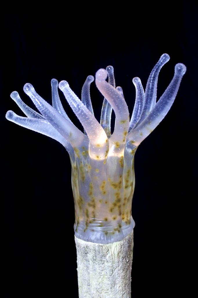
A gorgeous model of something too small to be observed properly with the naked eye; the zooxanthellae in the polyp of a coral. Read more here: http://www.10tons.dk/coralpolyp Photo: 10tons.dk
First of all, a 3-D computer model is made. At this stage, the guys at 10 tons work closely with us scientists to make sure that all details are correct – and we use a load of photos, films, SEM-photos and taxonomic drawings to make sure we have all things covered. Sometimes we even send them a specimen that they can scan. The models are passed back and fourth between the model-makers and the scientists, with indications of small corrections pointed out and performed until all parties are happy. You can be sure that the scientists have several minute details they want to change just a tiny little bit more, but we get there in the end…

Work in progress. Image: 10tons.dk
The next step is that the 3-D computer model is printed in the size that is going to be in the exhibits.
The printed out model is coated in a super-thin layer of wax to make it smooth, and then all the tiny details are added. Small notches in the epidermis or tiny plumose seta that have been separately made are added.
For a researcher who describes all the separate seta on the different mouthparts this is an amazing process to observe, and for everybody who later will see the model there can be an assurance that what you will see is actually how the species looks.
But this is not the end! The materials that have been used this far in the process will loose or change their colour when exposed to light. Therefore, a silicon mold (a “negative mold” or a cast of the outside of the model) is made from the finished first model. This mold is used to produce a new positive cast of polyurethane resin – and this is the model that will be shown in the exhibit. This material allows the model makers to add the right colour, translucence and texture to give the right look and feel of the finished product.
Here are a few of the scientific models, many more can be found here
[slideshow_deploy id=’3128′]
Models are not made only of small animals – sometimes they are scaled 1:1, like this minke whale:
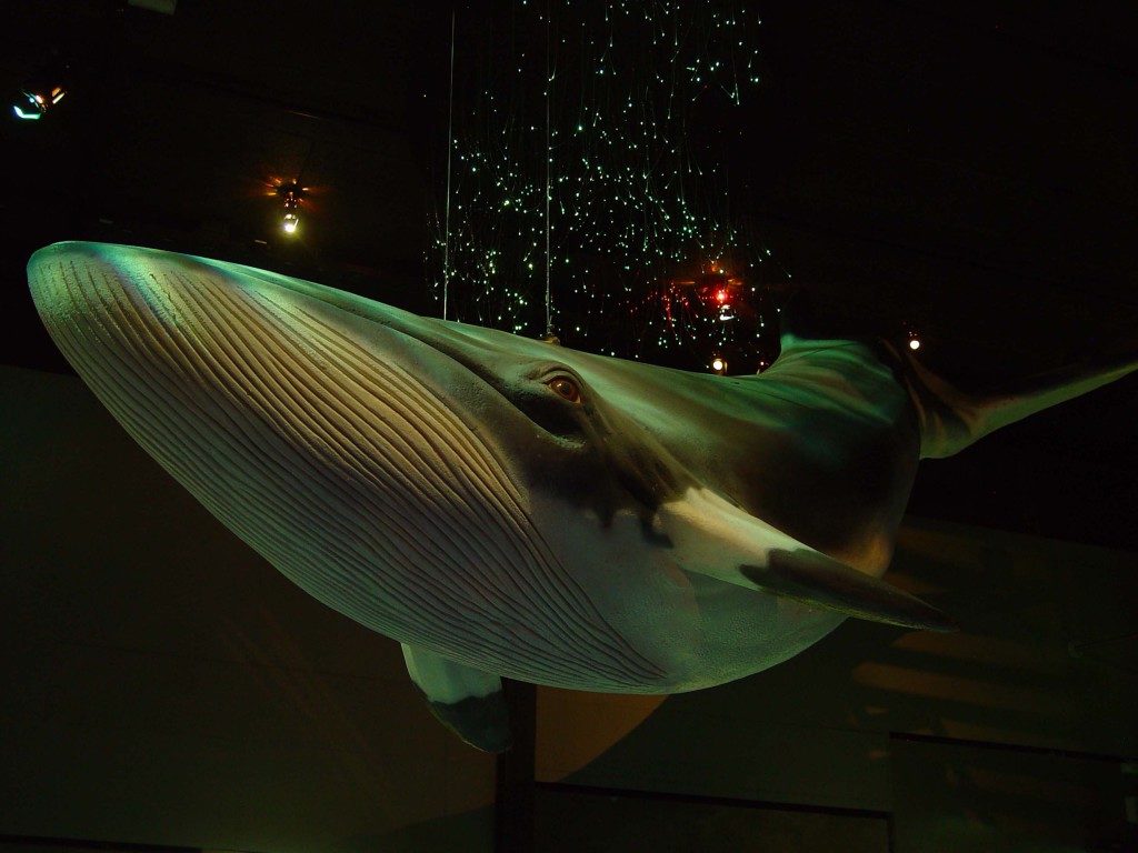
Balaenoptera acutorostrata, image 10tons.dk
Or *just a bit* bigger than what we could expect to find our in nature, like this crab (Cancer pagurus)

image: 10tons.dk
Here’s a video of how models are made – there are a few more videos here
We researchers are at the moment eagerly awaiting the models that will come to the University Museum – we have seen the 3-D models, and some of us have seen some photos of the models that are being made. We know that the models will look good, and we are looking forward to sharing them with everybody who comes to see the exhibits.
Now, what species will you be able to see models of, and in which exhibits will they be? That is for us to know now, and you to find out next year!
The holiday-season is a time for secrets to be kept, and this is one of those secrets. Come visit the University Museum when we reopen the building in autumn 2019 to see for yourself!
-Anne Helene
Would you like to know more about the process of making such models? This paper gives details and photos of a project 10 tons did with a paleontologist from the university of Lund in Sweden: Eriksson ME, Horn E (2017) Agnostus pisiformis – a half a billion-year old pea-shaped enigma. Earth-Science Reviews 173, 65-76. https://doi.org/10.1016/j.earscirev.2017.08.004
