10th International Workshop of the Hydrozoan Society

Earlier this year, the Cnidaria and Ctenophora group at UMB organized the 10th International Workshop of the Hydrozoan Society. The event was a success, and we asked visiting researcher Marta Gil to tell us a little more about it.

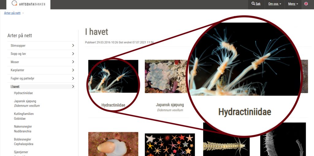
This is what Marta, who is currently collaborating in our Artsdatabanken projects ParaZoo and NOAH, has to say:

Hello to everyone!

I’m Dr Marta Gil, a researcher at the association Ecoafrik and a member of the Marine Zoology Group at the University of Vigo, Spain. This spring I began a research stay at the University Museum of Bergen to collaborate with Dr Luis Martell on project PARAZOO. I have visited the museum several times, but this visit had a special meaning for me. This year, in May, the 10th Workshop of the Hydrozoan Society was celebrated, organised by the Cnidaria and Ctenophora Group at the Department of Natural History, so I took advantage of my stay in the city and decided to attend.

Three images together: one showing the (taxidermied) birds in the staircase of the museum exhibition, one showing participants studying the exhibits in the whale hall - a whale skeleton can be seen over them, and a photo from the reception where the society president is welcoming everyone

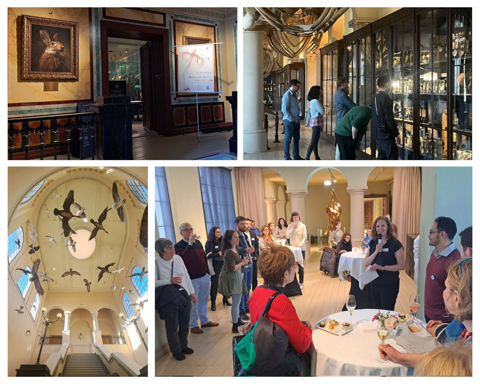
Welcome and icebreaker at the University of Bergen Museum of Natural History

This was my first time at this event and I must say it was a very rewarding experience, I don’t think I could have enjoyed it more. We were welcomed by the organisers at the public collections of the Natural History Museum, which was open only for the occasion, creating a fantastic atmosphere to meet the participants and many of the hydrozoan experts.

Over the next three days, we listened to talks from students who are just starting as well as presentations from established researchers who have been working with hydrozoans for years. We have had great speakers such as Dr. Nicole Gravier-Bonnet, Prof. Carina Östman, and Dr.Gill Mapstone; and how to forget the enthusiasm of Prof. Paul Bologna’s group, all the questions and remarks from Dr. Dhugal Lindsay, and the sampling demonstration from our Japanese colleagues Gaku Yamamoto and Aya Adachi? The discussions that took place after each talk and the engaging poster session made this workshop more than a simple scientific meeting, it was a group of friends talking about their favourite animal group: hydrozoans!

A collage of images showing participants presenting at the venue

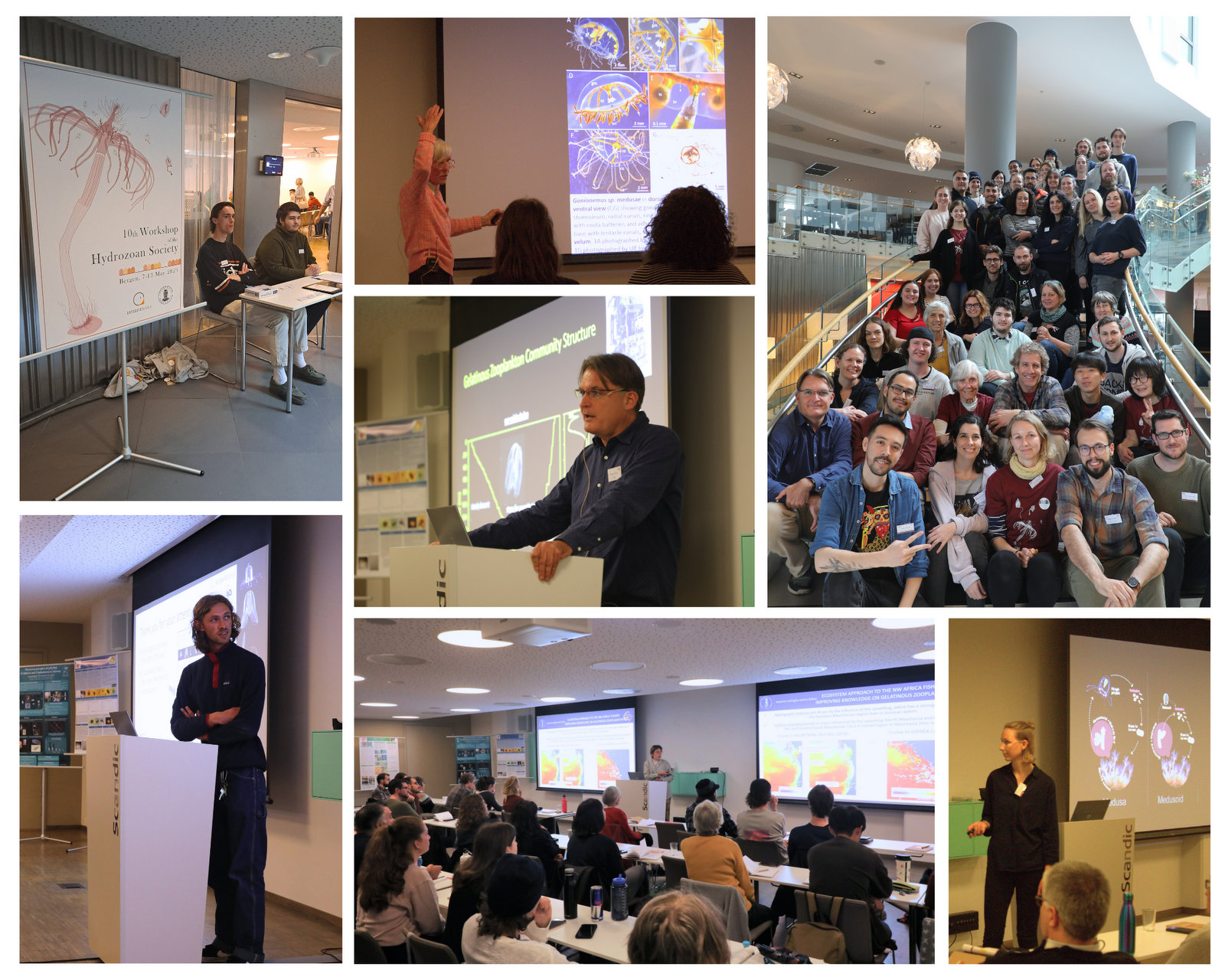
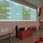
The conference part of the Workshop brought many interesting talks from a wide array of topics, together with fruitful and interesting discussions and networking.

Several images showing people studying large (A0) scientific posters presenting results of their research and projects.

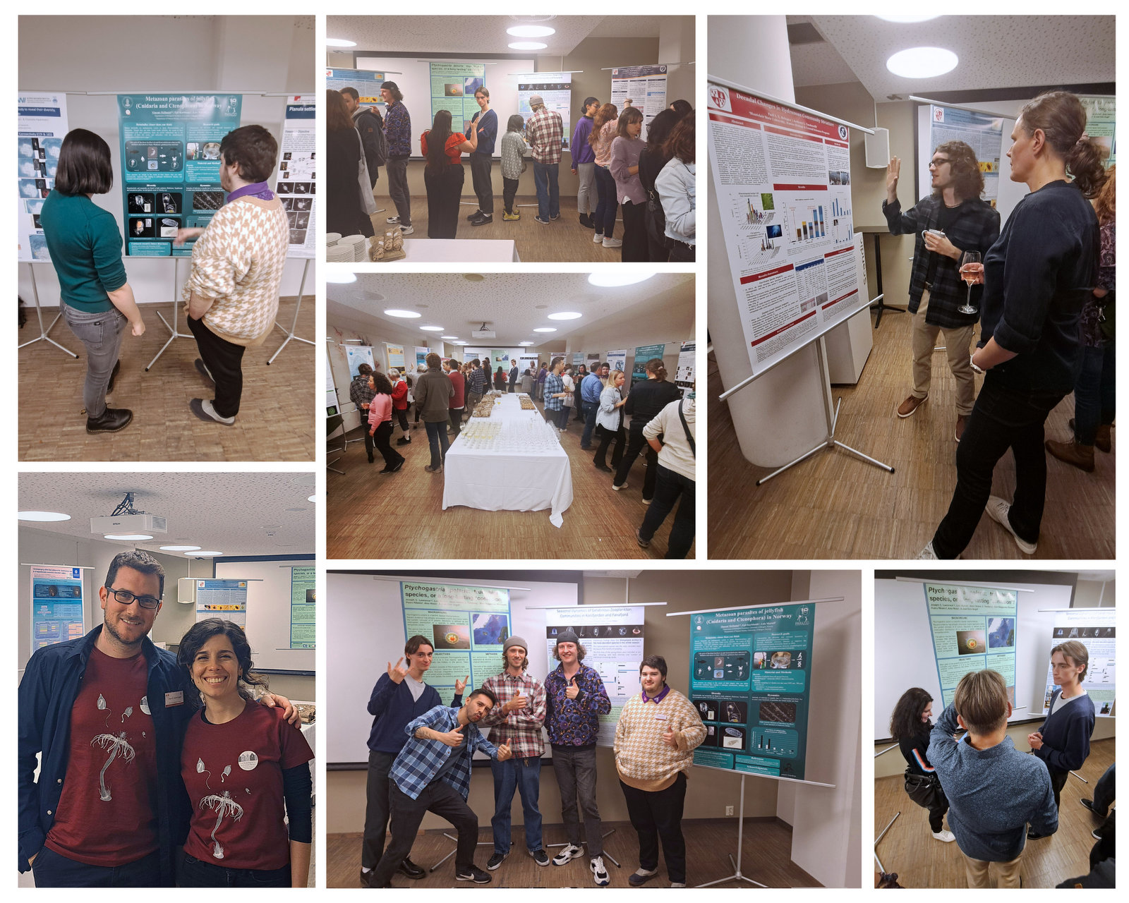
There was a dedicated poster session (and catering) to further discuss about hydrozoans in a more relaxed environment. Many great projects and preliminary results were presented!

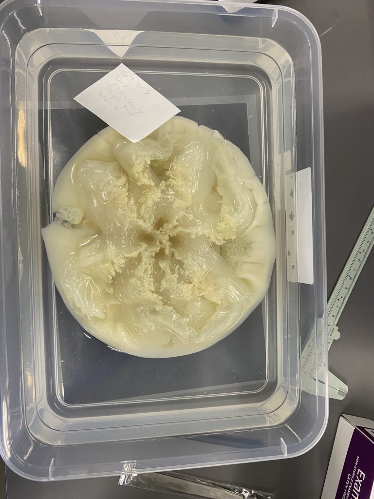
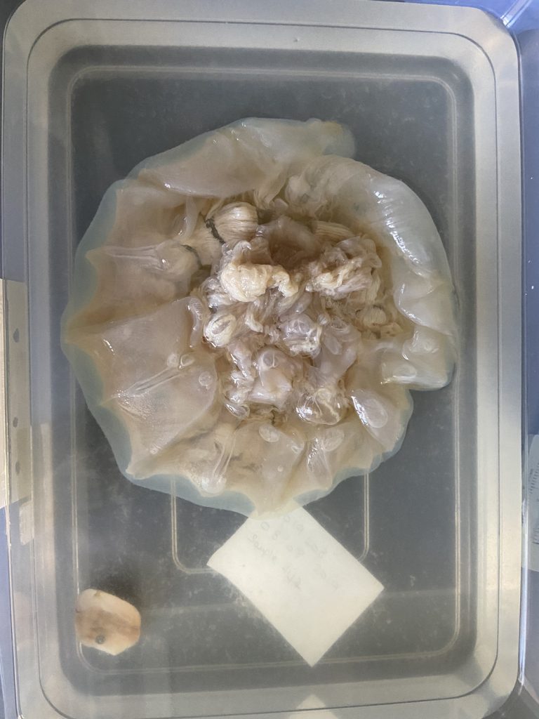
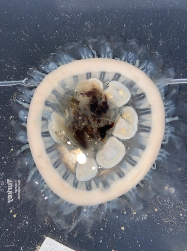
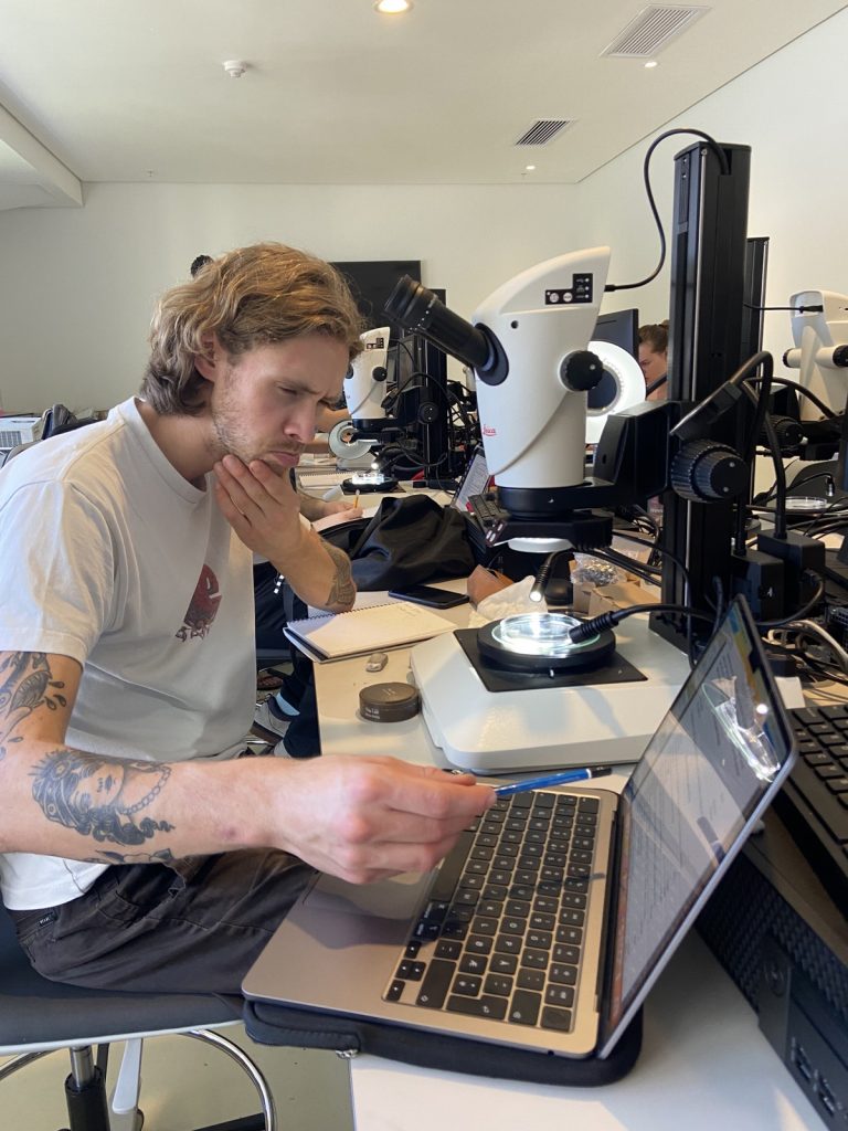
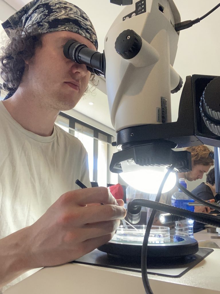
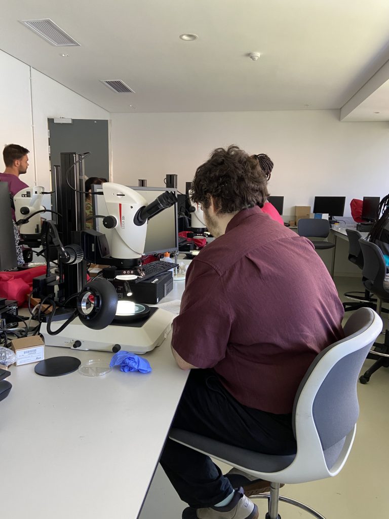
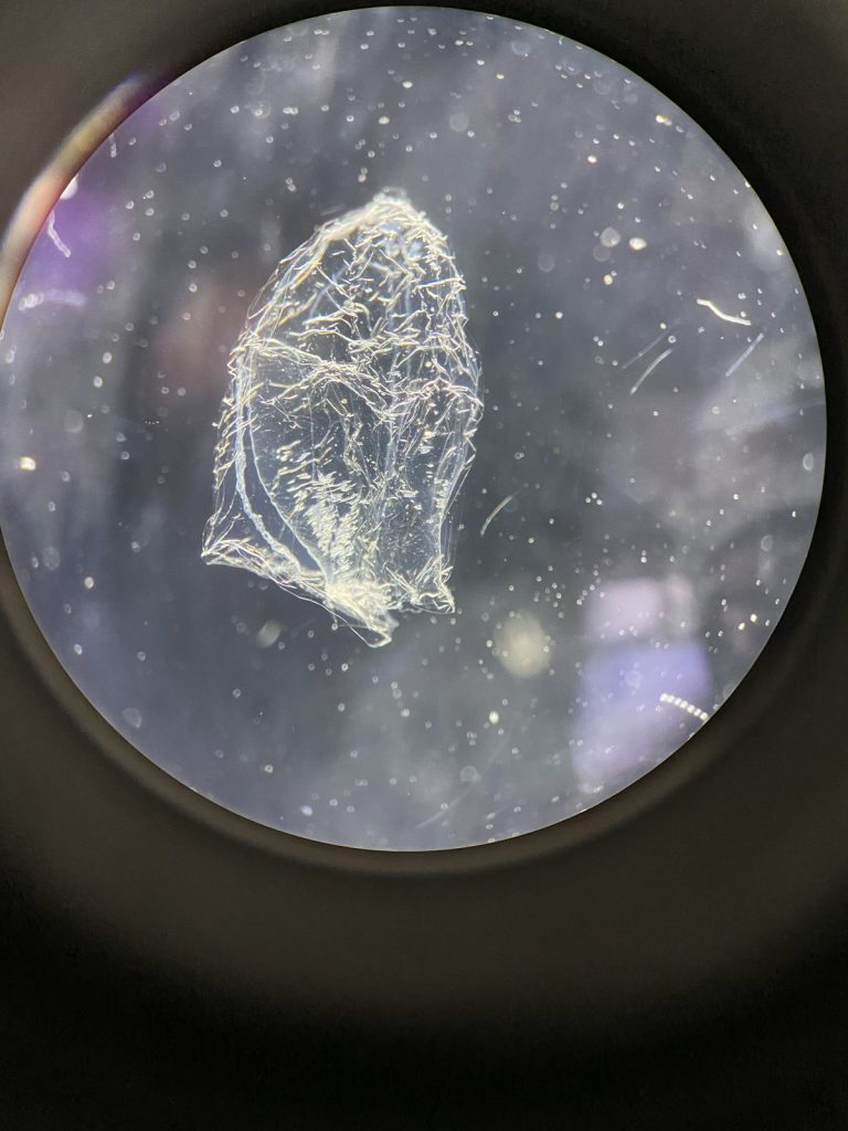
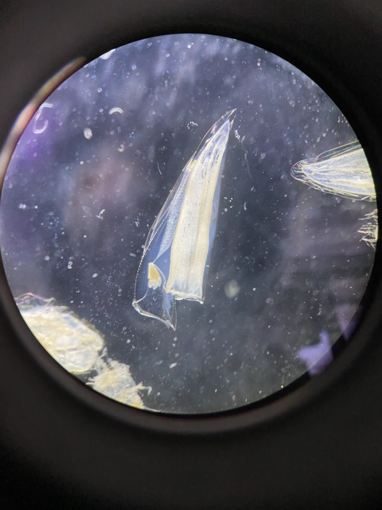
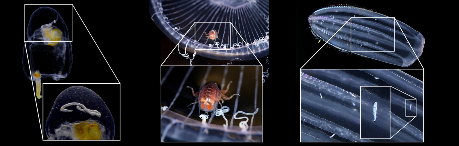
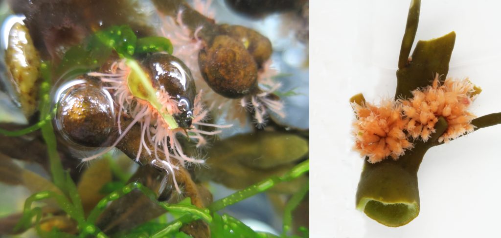
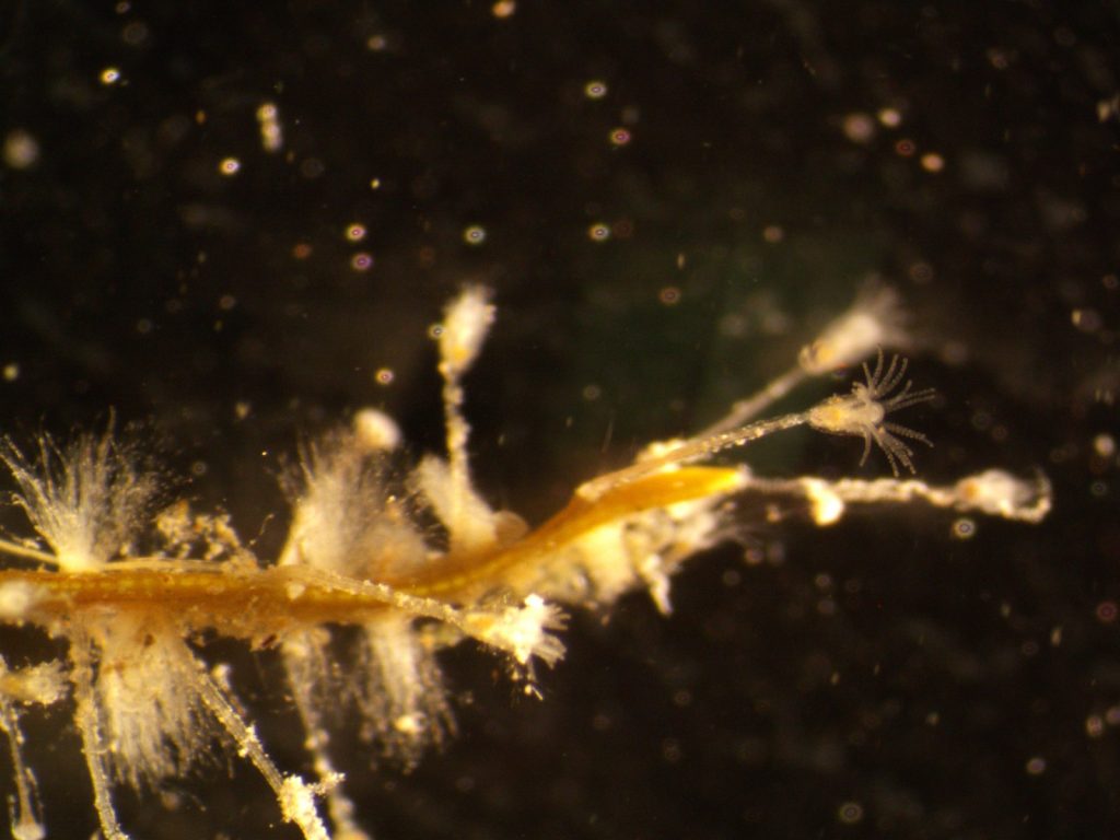
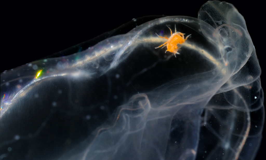
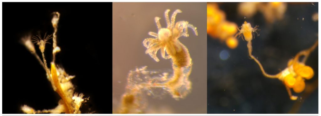
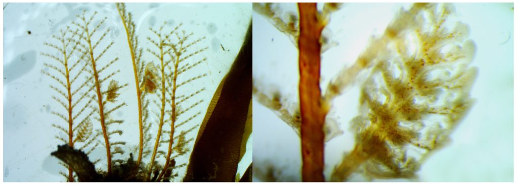
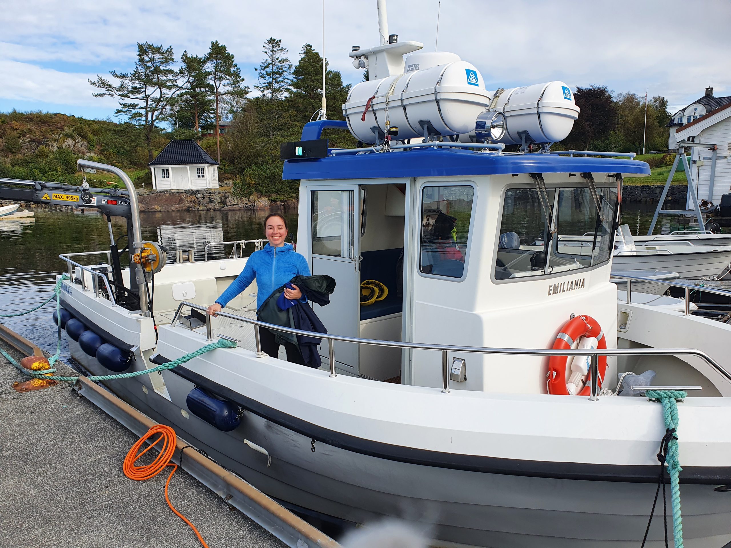
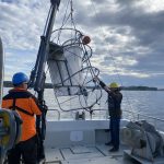
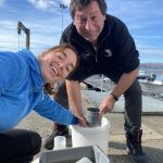
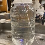
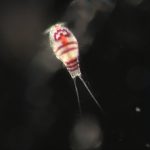
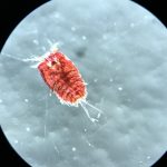
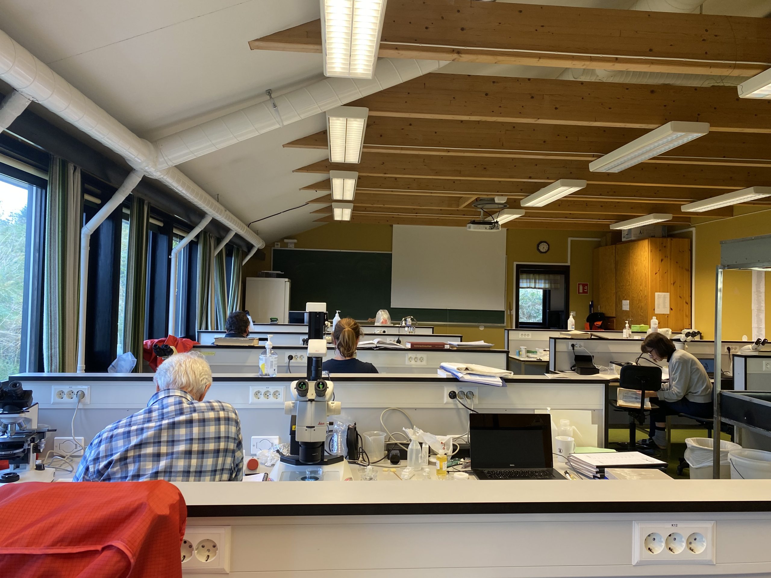
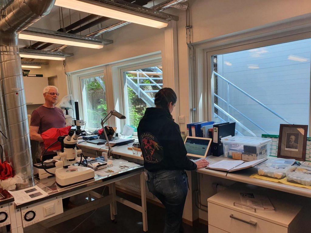
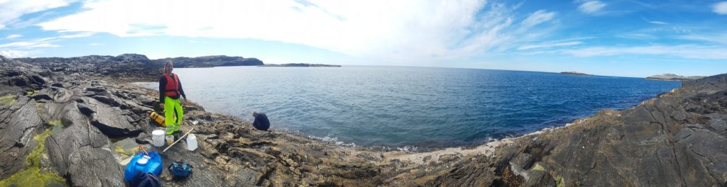
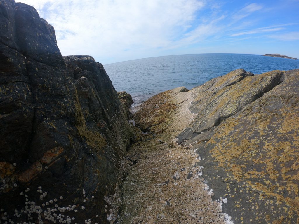
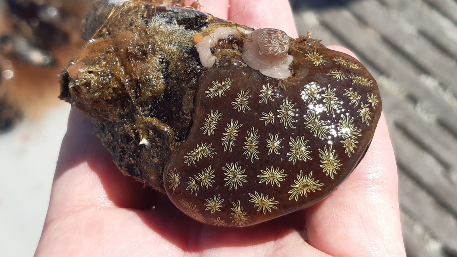
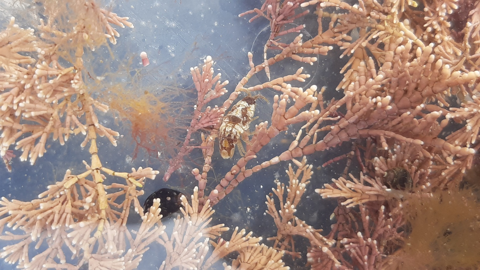
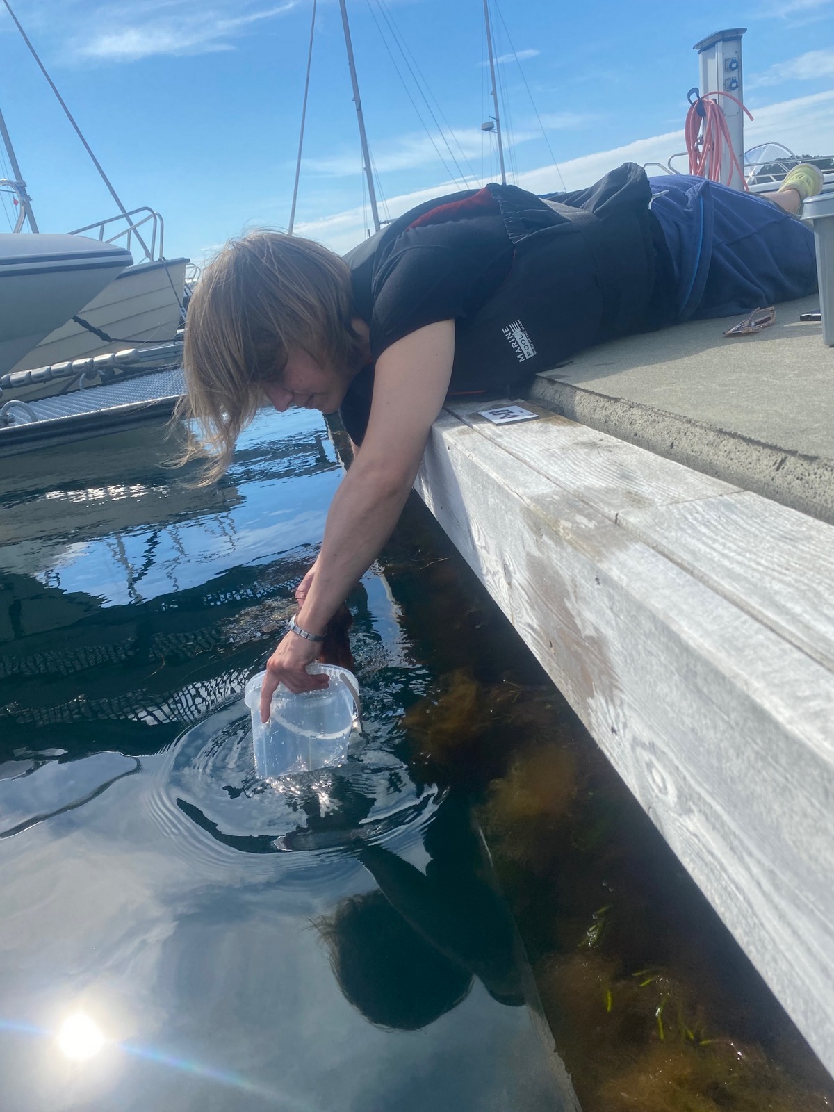
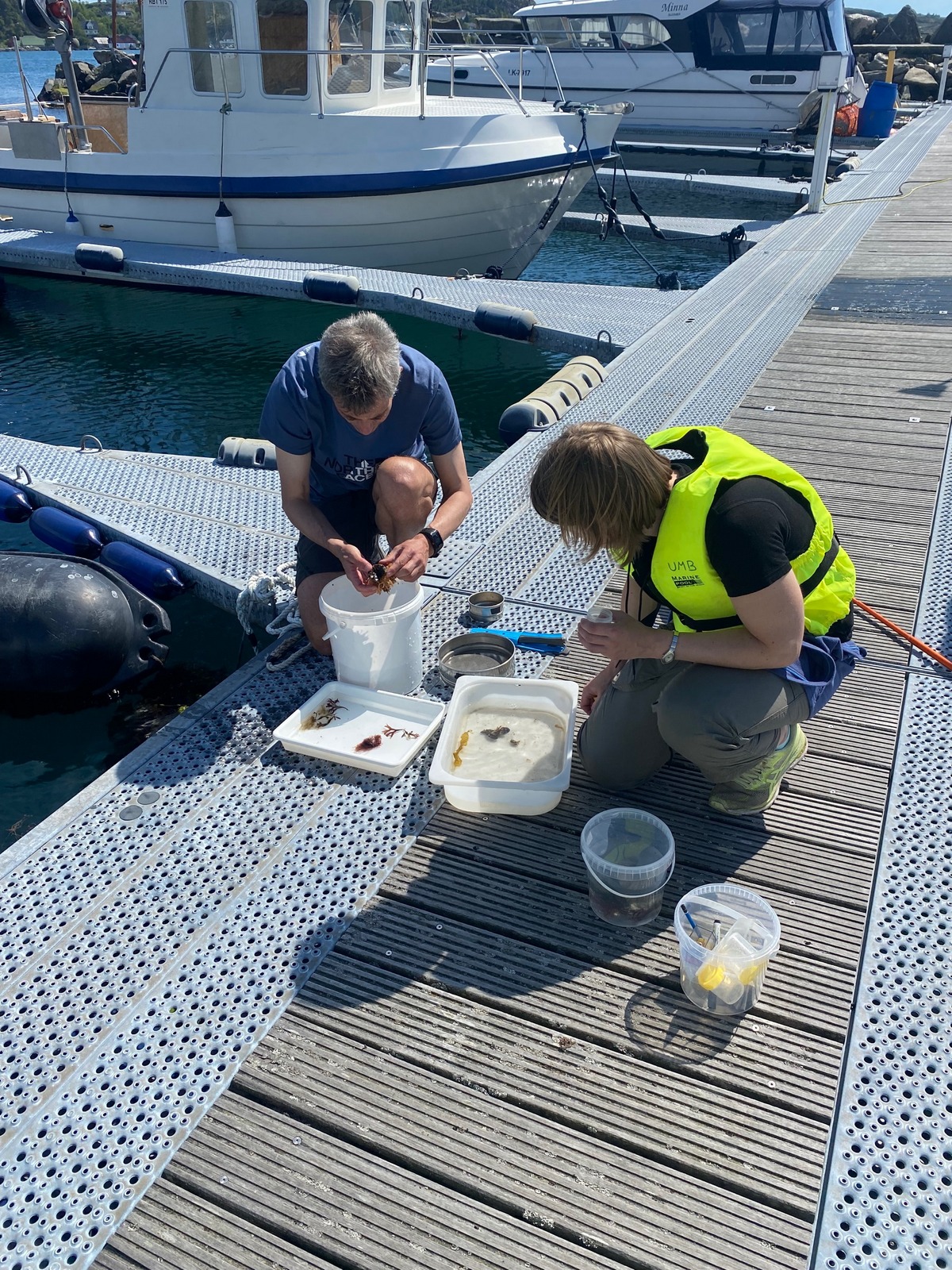
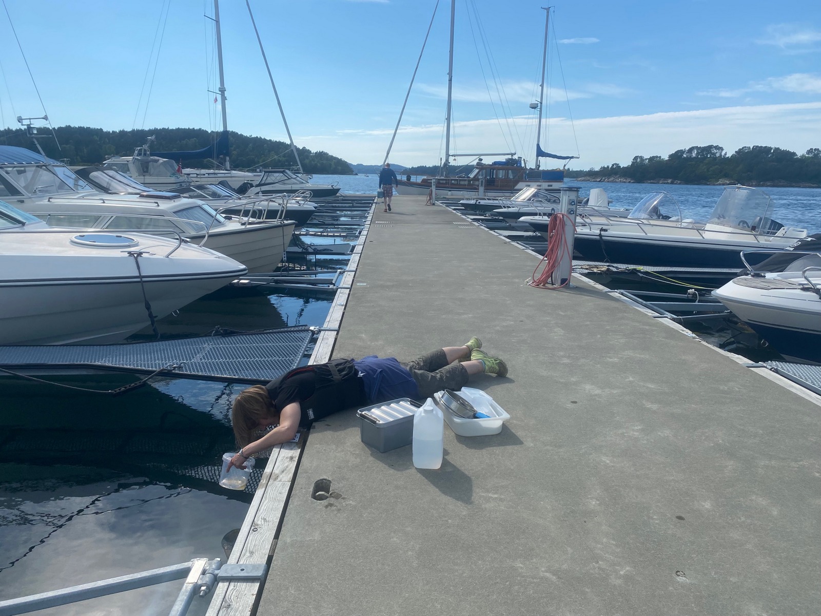
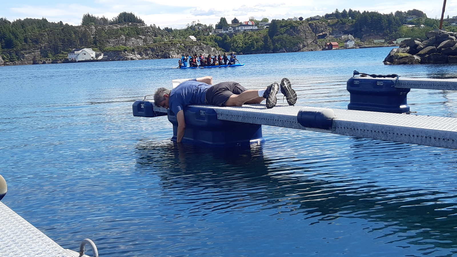
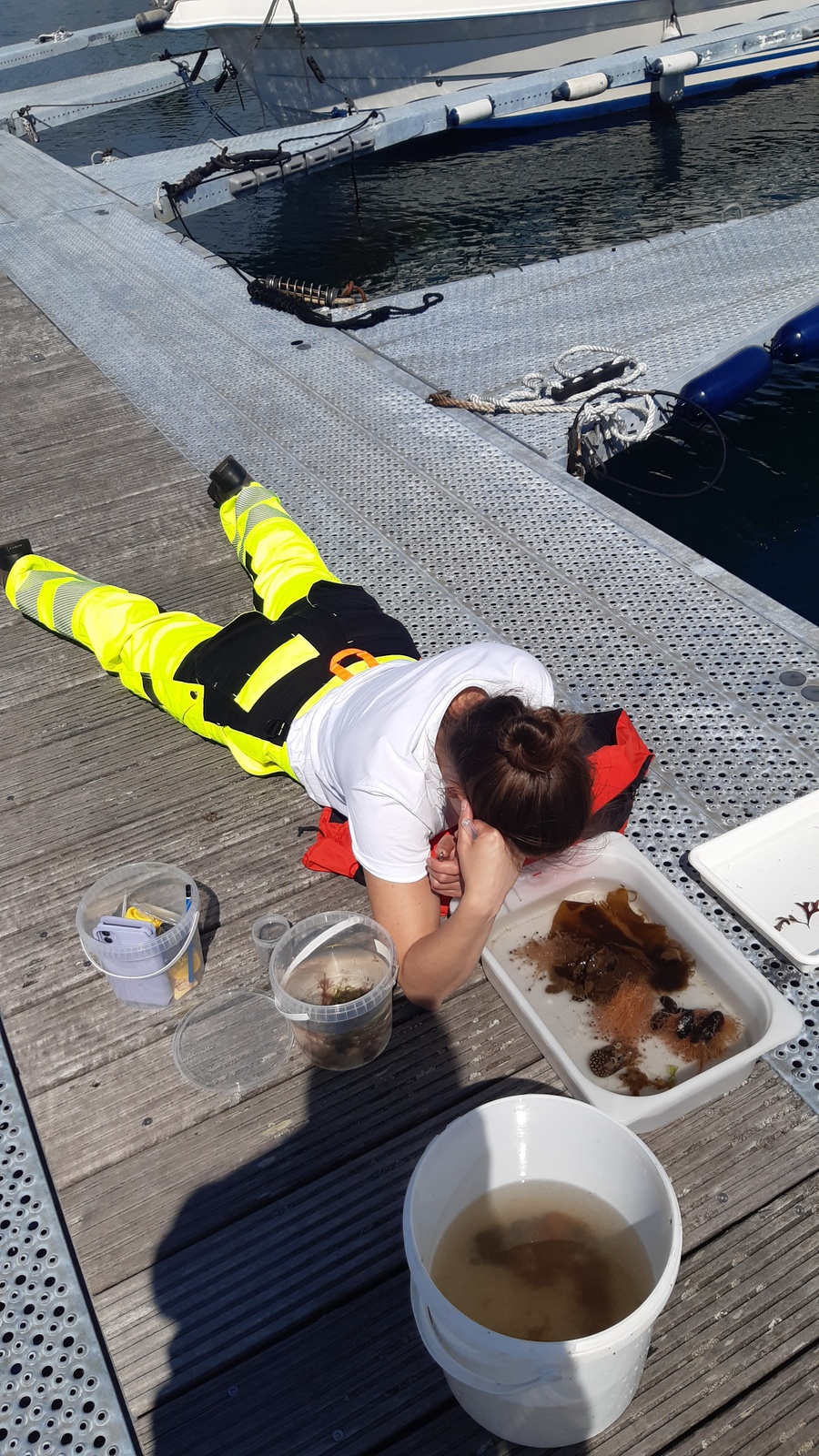
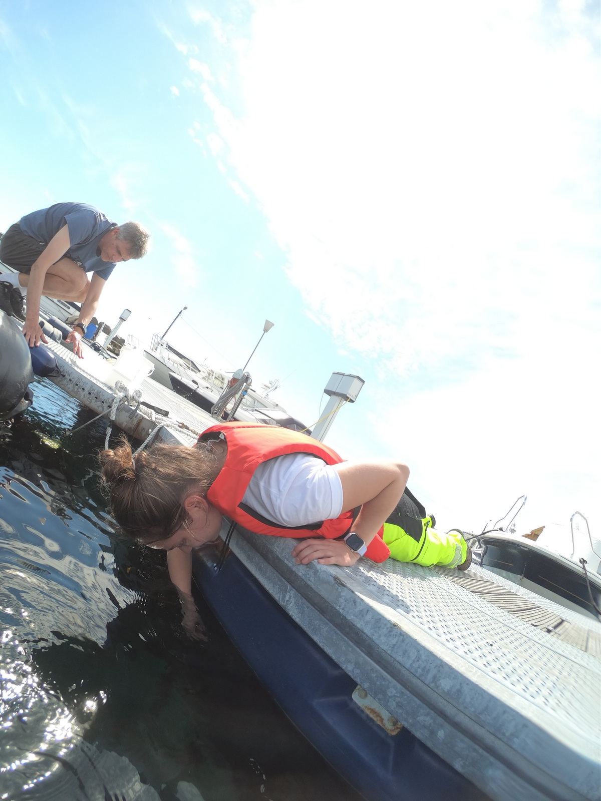
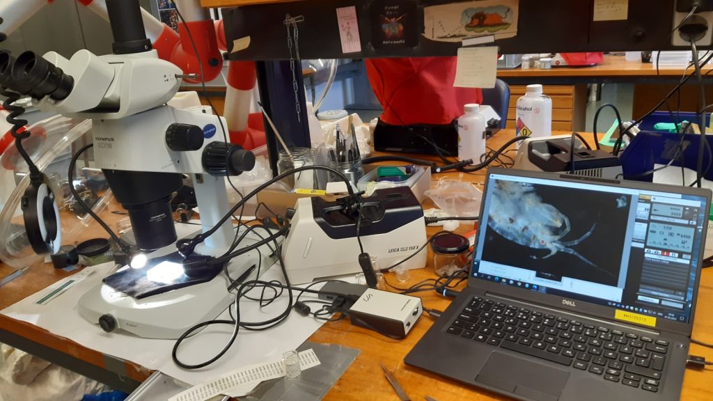
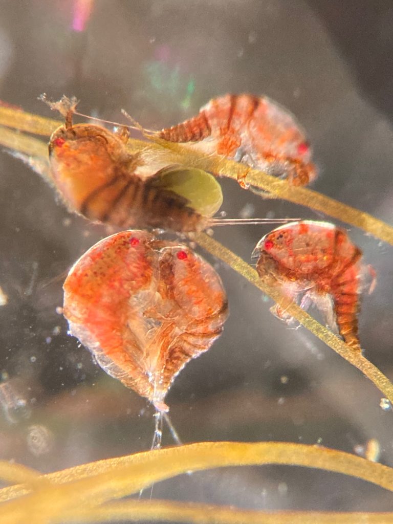
After the talks and presentations were over, the sampling part started, which was the most fun for me! We were able to sample benthic hydroids and pelagic hydromedusae and siphonophores on board the Emiliania and the R/V Prinsesse Ingrid Alexandra, and we later studied them at the Espegrend Marine Biological Station. This part gave us the opportunity to learn and share knowledge, techniques and very useful tips from other experts to study hydrozoans throughout their life cycle.

Snapshots from fieldwork, where people are collecting plankton and benthos from ship and shore

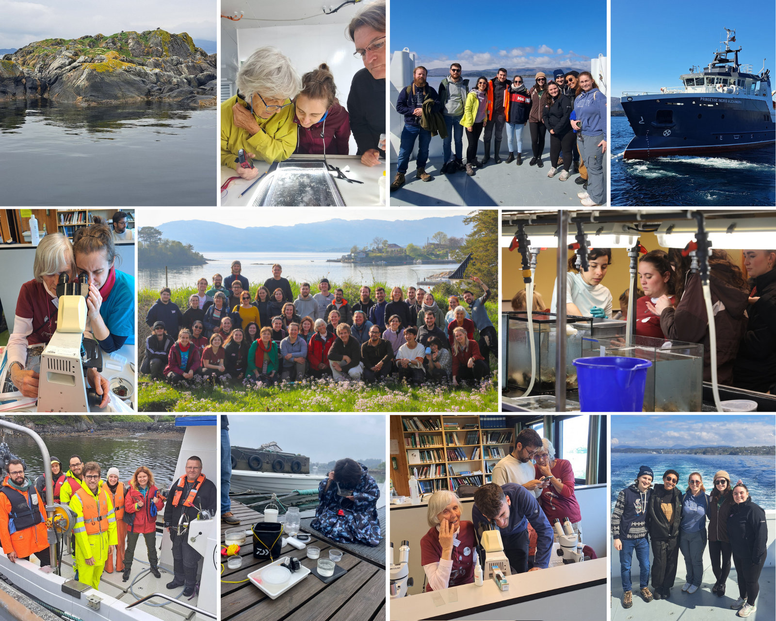
Two busy samplings days followed the conference part, where we all exchanged our expertise on sampling, handling and culture techniques. This were a couple of very productive sessions.

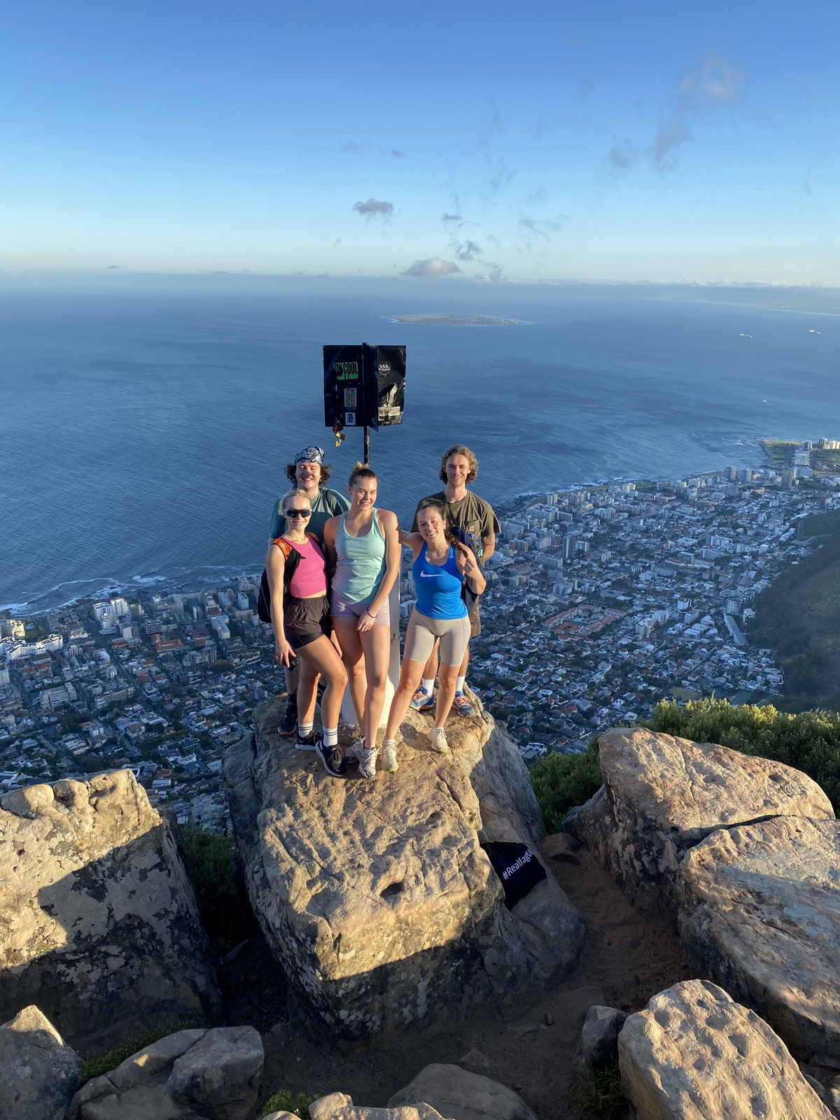
To round off an amazing week, on Saturday morning we visited the UNESCO World Heritage Site at Bryggen, where we learned about the history and traditions of Bergen. At sunset, we took the cable car up to Mount Ulriken and, after enjoying the incredible views, we held the award ceremony and the election of the new president of the Hydrozoan Society, who was unanimously elected: Dr. Lucas Leclère!

Snapshots from social activities throughout the event; shows people in historical Bryggen, as well as up the city mountains around Bergen

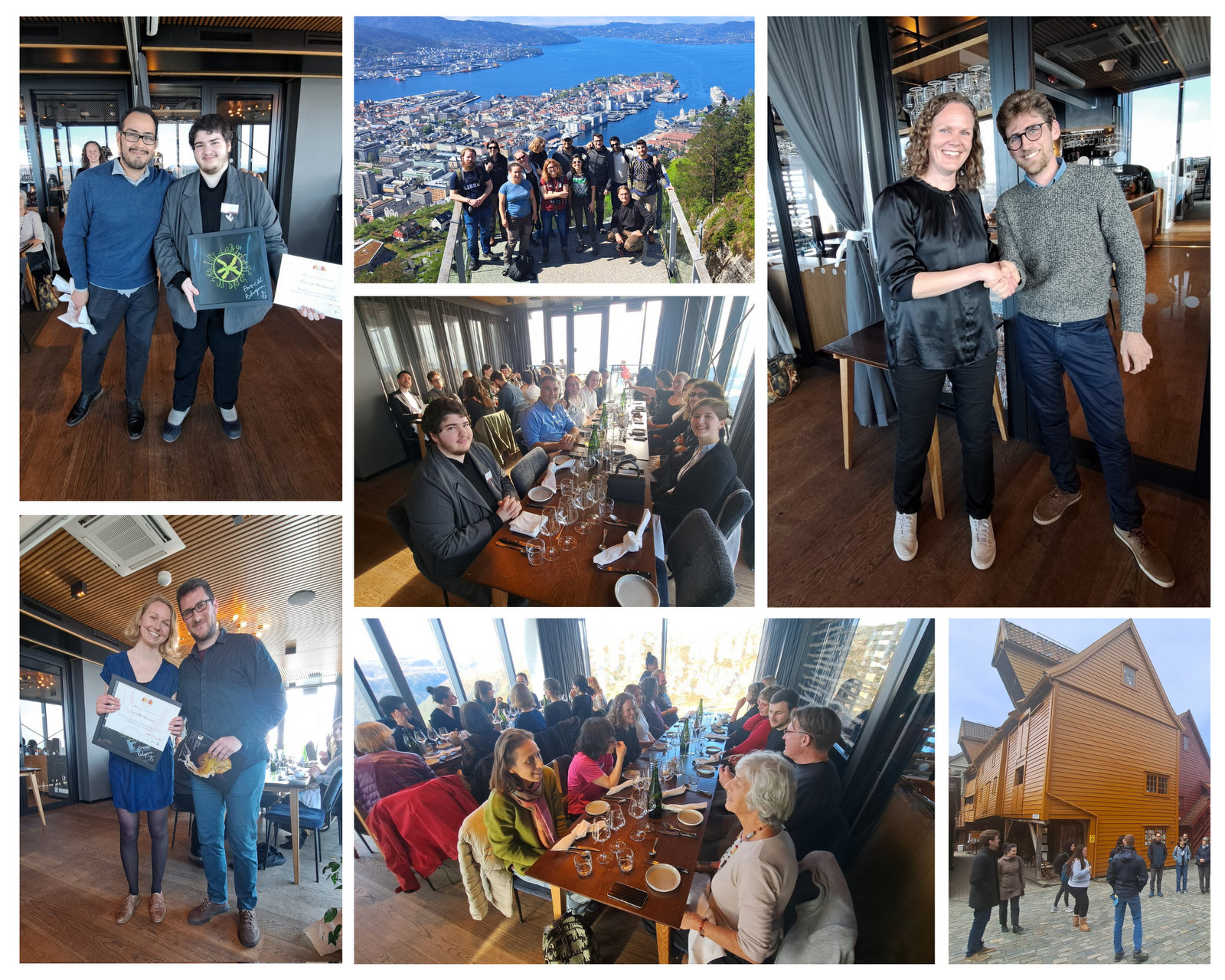
The social activities included a visit to Bryggen, an excursion to Fløyen, and the conference dinner in the top of Mount Ulrikken. The best student presentation awards were given and the new President of the Hydrozoan Society was elected. Exciting times ahead!

Group photo of the participants up in the mountains, with the city of Bergen in the background

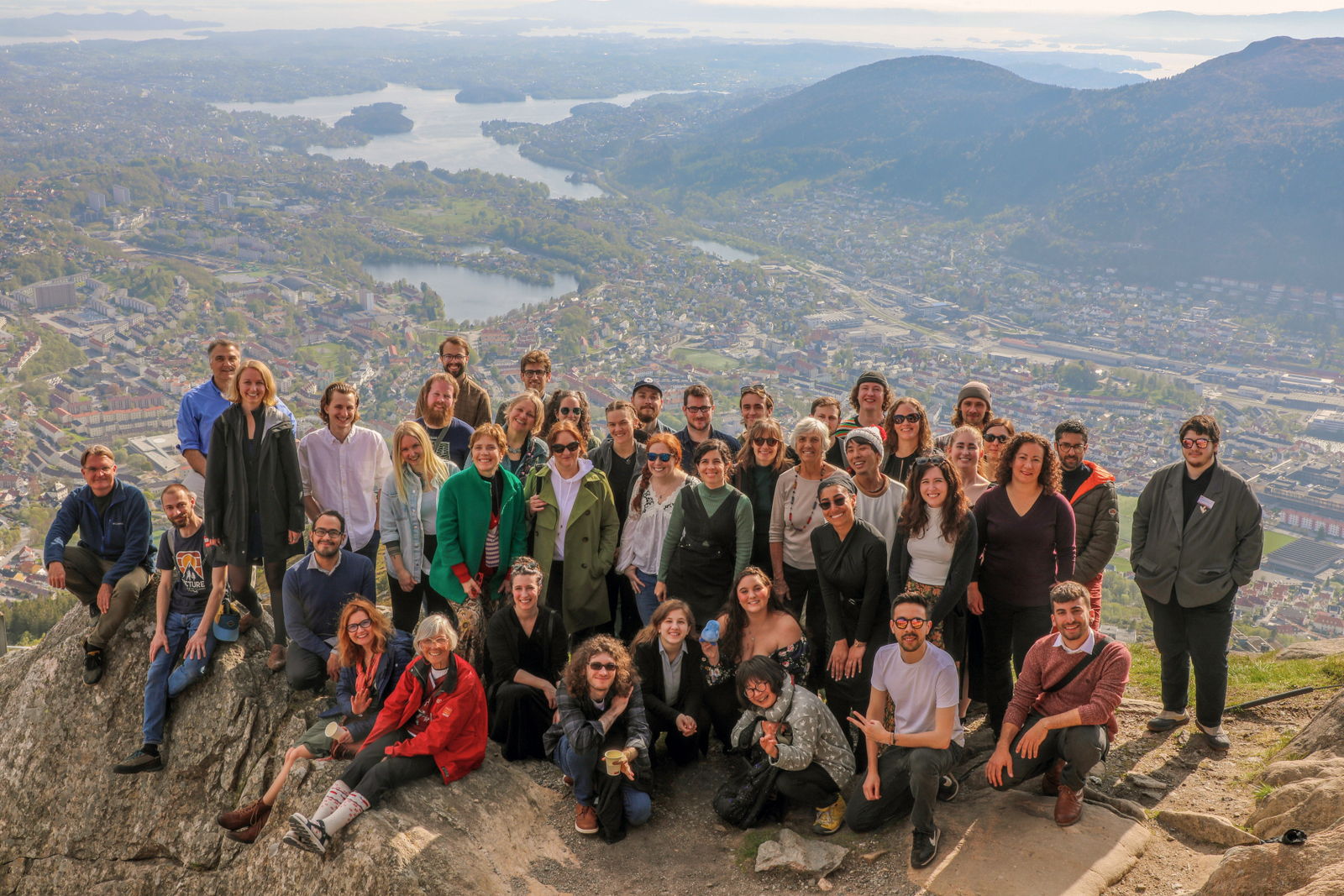
The third group picture we took in just a few days, this time with an incredible view of the city of Bergen as a background.

I would like to close this chronicle by thanking UMB’s Cnidaria and Ctenophora group: Aino Hosia, Luis Martell and Joan Soto, for their incredible work in coordinating the presentations, transfers and field work and for hosting such a rewarding event. I would also like to mention the great work done by all the students of the group, who helped organize the meals on the sampling days and other various activities. Thanks to all of them, the rest of us simply enjoyed the week.

It was definitely an experience worth repeating. I’m already looking forward to the next workshop in France!

-Marta Gil

The Cnidaria+Ctenophora Research group at the PRIMALearning Jellyfish Workshop in South Africa

On the 12th of February at the crack of dawn, we had the amazing opportunity to go to Cape Town to attend a Jellyfish workshop. The “we” in question are the three authors of this blog post: Vincent, Vetle, and Håvard. We are master students all working with jellyfish-related topics, and some would go as far as to call us jellyfish enthusiasts. Our work is part of the museum’s Artsprosjekter NorHydro and ParaZoo, and we were happy to represent the invertebrate collections and UMB at this event.

The workshop, held at the Iziko South African Museum, was organized by PRIMALearning and was a collaborative initiative between the University of Bergen and the University of Western Cape. This gave the three of us, accompanied by a few other UiB students not affiliated with the University Museum of Bergen, the chance to visit Cape Town. Some of us for the first time.

On the first day of the workshop, we were greeted outside the museum by Mark Gibbons and his PhD student Michael Brown who was the representative from UWC and would be teaching parts of the workshop. With them was also Anne Gro Vea Salvanes as the representative for PRIMALearning and the University of Bergen, she was also joined by our own UMB scientists Aino Hosia and Luis Martell, who were also part of the teaching team.

The first day included introductions from all the participants, and we got to know each other bit better. We also got a brief introduction to the world of jellyfish and their taxonomy before the night ended with a delicious dinner together with all the participants.

The second day started with lectures about the large or ‘true’ jellies (Scyphozoa), before we got to get our hands dirty looking at preserved samples of jellyfish. We were met with a broad diversity of scyphozoans that was passed around between the students so we would get a shot at identifying them.

We examined fixed material of scyphozoan jellies representative of the three major groups within the class:


The rest of the day after the workshop was spent at the beach, enjoying the sun and local wine. Cape Town is called the windy city, and it did deliver on its name, but nothing could stop the sun-deprived Norwegian students from going outside to soak up some rays.

The third day was Hydrozoa day 1, a topic dear to our hearts and it was taught by our own MSc supervisor Luis Martell. Some of us were a bit tired this day because we decided to climb Lion’s head mountain before the workshop started to see the sunrise.

But this did not stop us from eagerly working with the preserved hydrozoan samples we got to look at. We identified all hydromedusae and siphonophores with the help of a stereomicroscope:

 A dissected carybdeid cubozoan jellyfish. IC: Håvard Vrålstad


Day four was box jellies (Cubozoa) day, a class neither of us was very familiar with. So we were excited to learn about these unknown and sometimes dangerous animals.

Luckily the animals were less deadly when preserved and we could therefore touch them while identifying them.


This day ended early so we took the opportunity to get down to Simon’s Town were we spent the rest of the day looking at the local penguins and wildlife at the beach.

A snapshot of the fauna we observed at Simon’s Town. IC: Vincent McDaniel

On our last day, we were introduced to the alien world of siphonophores by our own Aino Hosia. These animals are close to Håvards heart (if you ask him on a good day).

They were a nice ending to an awesome workshop, and we can honestly say now that our interest for jellyfish has grown, and we look forward to seeing even more of them in the future.

On February 19th the vacation/business trip was unfortunately over for Vetle and Vincent and they had to pack their bags and prepare to leave Cape Town and head home to Bergen, while the “slightly” more fortunate Håvard stayed behind in the windy city to enjoy a few more days of leisure.

Participants and teachers at the jellyfish workshop. IC: Anne Gro Vea Salvanes

We want to thank PRIMALearning for arranging the workshop, the University of the Western Cape for hosting and providing us with samples to work with, the Iziko Museum of South Africa for the location as well as providing refreshments, and to the University of Bergen for arranging accommodations. Also, a very special thanks to all the lecturers who presented and were very patient with us during the lab work.

-Vetle, Vincent, and Håvard


Are you interested in becoming a master student in marine biology at the University Museum of Bergen? Information about available projects can be found here (more will be added soon!):

Marine Masters at the University Museum of Bergen
– available thesis topics in marine biodiversity

Project ParaZoo: there is a critter inside my jellyfish!

ParaZoo (complete name ‘Metazoan parasites of non-crustacean zooplankton’) is one of the most interaction-focused projects currently running at the Invertebrate Collections of our Museum. This project, funded by the Norwegian Biodiversity Information Centre (Artsdatabanken), aims at studying the different animals that live together inside and on the surface of Norwegian jellyfish. This means that for the next two years we will be looking for tapeworms, flukes, roundworms, and amphipods as ParaZoo tries to answer the question of which of these organisms are associated with gelatinous hosts in Norwegian waters.

ParaZoo is focused on animal parasites and symbionts associated with jellyfish. Thse parasites include roundworms like Hysterothylacium aduncum (left, in Euphysa aurata), amphipods like Hyperia medusarum (middle, on Aequorera forskalea), and flukes like the members of family Didymozooidae (right, in Beroe gracilis). IC: Aino Hosia (left), Katrine Kongshavn (middle), Joan J. Soto-Àngel (right).

Besides jellyfish, arrow worms (Chaetognatha) are also members of non-crustacean zooplankton that host different types of parasites, like this H. aduncum roundworm (Nematoda). IC: Joan J. Soto-Àngel, Luis Martell.

Parasitism and symbiosis are extremely common life styles in the animal kingdom. In fact, some researchers believe that there may be more species of parasites than of free-living animals, given that each free-living species hosts many species of parasites (most of them unique) and those parasites also host their own parasitic tenants. Marine zooplankton is no exception to this trend, and many parasites and symbionts are expected to occur in copepods, krill, and gelatinous zooplankton. Jellyfish and arrow worms, for example, may be important hosts for flatworms and other helminths, yet our knowledge of these animals in Norway is very scarce.

ParaZoo’s logo includes two of the target taxa of the project: roundworms (Nematoda) and hyperiids (Amphipoda). The third main parasitic group covered is flatworms (Platyhelminthes), illustrated by the larvae of Derogenes varicus parasitizing Halopsis ocellata shown in the right side of this figure. IC: Joan J. Soto-Àngel, Luis Martell.

Understanding zooplankton parasites is important because many of them are going to be transmitted to fish, where they may cause serious diseases. To get a better overview of which critters live in non-crustacean zooplankton, ParaZoo will sample, record, and DNA-barcode specimens from all over the country. The collected animals will be included in our museum collections after being identified, documented photographically, and fixed in ethanol. We will then generate an open-access database of information including pictures and DNA sequences that will help with the identification of the parasites. Aquaculture facilities, fishermen, and managers of marine areas will benefit from this database to better plan and counter potential negative impacts caused by the parasites.

Flukes, such as Opechona spp, parasitize gelatinous zooplankton (in this case a sea-gooseberry Pleurobrachia pileus) as larvae called metacercariae. IC: Joan J. Soto-Àngel, Luis Martell.

The larvae of tapeworms (Cestoda) sometimes use jellyfish to reach their definitive hosts: fish. IC: Joan J. Soto-Àngel, Luis Martell.

ParaZoo is committed to present the diversity of jellyfish parasites to all those not familiar with them. In order to do that, we will regularly write entries here on the blog, as well as participate in several academic and not-academic meetings. The official info webpage for the project is available here, so don’t forget to check it out!

Luis

A new online resource for identifying ‘fur snail’ hydroids, and a farewell to project NorHydro

Finding the correct name of the hydroids growing on shells of snails and hermit crabs can be notoriously difficult… but fear not! A new online resource is now available and we hope it will help everybody who is interested in these little critters.

Fig1. Have you seen these pink/orange ‘blobs’ growing on washed-up algae along the coast? They are hydroids of the species Clava multicornis. Read more about this and other Norwegian hydractiniids in the new arter på nett webpages. IC: Luis Martell (left), Katrine Kongshavn (right).

We (the UMB research team of NorHydro: Luis, Aino, Joan, and Lara) have created a series of species identification sheets for all the hydroids and jellyfish belonging to family Hydractiniidae. This family includes some hydroids that can be easily encountered during a walk along the coast or when snorkeling and scuba diving in Norway, and therefore we wanted to provide beach-goers and divers with a tool to find the correct name of these animals.

Fig2. The new identification sheets for Hydractiniidae are available inside the section I havet of the Arter på nett platform in Artsdatabanken’s website. IC: Artsdatabanken.

Many hydractiniids are known as ‘snail fur’ because they give a furry appearance to the shells they are growing on, while others grow on rocks or on algae. Identifying the species still requires a little effort, but altogether they are one of the best groups to start with for anyone interested in hydrozoan diversity in Norway. This is what motivated Lara, a MSc student at the time, to created the first version of these sheets (together with some beautiful illustrations) which were then developed further with text from everybody in the team.
The editors and consultants at Artsdatabanken helped us throughout the process, and we are happy to present the final version of this online resource available here:
Arter på nett: Hydractiniidae

Fig3. The anatomy of a polyp colony of Hydractiniidae is best explained with a combination of images at high-magnification and detailed diagrams. IC: Luis Martell (left), Lara Beckmann (right).

Fig4. Hydractiniid hydromedusae are smaller than 1 cm, but they still have several characters that we use for identification. IC: Joan J. Soto-Angel (left), Lara Beckmann (right).

Fig5. Both the hydromedusa and the hydroid stages of the three species of Podocoryna occurring in Norway are included in the new Hydractiniidae arter på nett. IC: Joan J. Soto-Angel, Bernard Picton, Lara Beckmann

The publication of the Hydractiniidae identification sheets marks the end of our project NorHydro. The project’s main objective was to map the diversity of hydrozoans in Norway, but we also tried to present marine hydroids to all those not familiar with these amazing creatures. During its 3 year’s run, NorHydro produced a detailed inventory of Norwegian hydrozoans through an integrative (morphology + DNA) approach: we collected samples in >90 sites around the country and analyzed existing museum specimens to produce >1200 new records and high-quality, open access DNA barcodes for 160 species. NorHydro’s scientific team identified 12 cases of species complexes, supervised 5 MSc thesis, and participated in >20 outreach activities and presentations in academic events. All in all, NorHydro was an extremely enriching experience and I’m confident its results will help us understand a little better the diversity of marine invertebrates in Norway.

Fig6. Farewell NorHydro!

-Luis

Hydrozoan team at ForBio 2022 annual Meeting

Do you remember that feeling of dread before you must present in class about a topic you didn’t really study for? Your mind racing, trying to scramble a coherent story to tell the sea of eyes fixed expressionless on you and your powerpoint? We believe we all have at least one memory of this from our days in college.

That was a similar feeling to what we felt on November 28th, 2022, when we had just landed in Trondheim and were on our way to the Norwegian University of Science and Technology (NTNU) for this year’s ForBio Meeting, a 3-day gauntlet where we will present the Master’s projects we’re currently working on. Except this time, we weren’t presenting in a small classroom full of uninterested teenagers thinking about tik tok dances, but in an auditorium full of fellow researchers who work on your same field, and who will probably have extremely difficult questions at the end of your talk. Being the first time we present in an environment like this, one can’t help but imagine all the worst outcomes

And so we’re sitting there, watching the hours and the talks go by, thinking to ourselves “I should/shouldn’t try that for my talk!”. At lunch, all we think about is our talks. When we’re doing some light sightseeing in Trondheim, all we think about is our talks. We’re laying in our beds the night before, and all we think about is our talks. “How will I make my topic sound as professional and knowledgeable as I want to?”, we think to ourselves.

The day arrives, and again the dread starts setting in, right until the moment they call each of our names. At our every turn, we walk down the steps, grab the microphone, and start talking. Except for some reason, this time it feels like we’re in control. The words flow effortlessly; we even crack a joke or two, and the audience laughs. There’s no stumbling around the words like those college days.

Because this time, we’re not the average seat-warming student. This time, we’re the ones that have spent cold and raining hours on a research vessel or diving to get our samples; we’re the ones who have spent hours working together with our supervisors, reading and learning about our topics; we’re the ones who have spent hours looking down a microscope trying to identify our organisms. This time, we’re the ones who know what we’re talking about, and the audience is there to learn from us.

After each of our talks, we give our acknowledgements, and everyone claps. The questions thrown at you are answered effortlessly, and the moment is finished with a thumbs up to our supervisors, returning to our seats smiling.

The most exciting part for us was showing to experts what we do with our favorite invertebrates: Hydrozoans. These organisms are an inconspicuous class in the phylum Cnidaria that most people ignore. Our job was not only to present what we do with them, but to show what they are, what they do, how fascinating they can be and why they are important. And we think that we did a good job.

Now we’re finally free to enjoy the world famous Trondheim bike lift (and the Nidaros Cathedral too), closing the night with burgers, beer and friends.

Everything has been great on this trip and we will always remember the advice from other professionals; how different or similar the work of each one is; and the feeling that you are part of a group of specialists who are excited to share their knowledge with others. But, above all, we have learned why these conferences are important: Knowing what other researchers are doing gives us the chance to collaborate together and help each other, because working as a team is how science moves forward.

The hydrozoa group (and friends) from UiB participating at the ForBio meeting

Pedro, Ana & Håvard

Student visit – Ana González

MSc student Ana González visited the collections last month as part of project NorHydro, where she spent some weeks in the lab working with her samples. Here is an account of her experience:

The challenge of identifying benthic hydrozoans
Hydrozoa is a fascinating but poorly understood group of invertebrates, in part because their identification is not always an easy task. I have been studying benthic hydrozoan communities for over a year now, in particular those living in the shallow waters of Mallorca (Spain), and I have realized that the diversity of forms and structures in the group is higher than I had imagined at the beginning of my studies, and their identification is more difficult than I expected. The assemblages of hydrozoans in the Mediterranean are of course very different from the ones that occur in Norway, but something that both communities have in common is that morphological identification of the animals (i.e. telling which species is present based only on the characteristics we can observe) is challenging, which is why one of the aims of my visit to the University Museum of Bergen last December was to learn a different technique (DNA barcoding) that can help me improve the identification of my samples in cases when the morphology of the specimens is not good enough.

Some of the morphological characters that are used to identify benthic hydrozoans. On the left side a member of Campanulariidae, with a stolonal colony, and on the right side Monotheca obliqua with an erect colony.

DNA barcoding consists in finding a short DNA sequence (the barcode) that is similar for all members of one species but different from all other species. It is a relatively recent tool that –among other things– has helped the scientific community identify specimens that for one reason or the other cannot be identified based on how they look. In some groups, such as many colonial invertebrates, this technique has become a key asset because the colonies are often too young or not reproductive, or the important characters for identifications may be found only in one stage of the life cycle and not in others. For this visit I had the chance to bring all my samples from Mallorca to Bergen and I set to extracting the DNA of selected specimens, amplifying two different barcode genes (COI and 16S), and obtaining clean sequences for them. I discovered that, when it comes to DNA barcoding, every step of the process is important, and being patient and careful is essential.

Me at the DNA lab, running the electrophoresis for my samples.

Getting good results in the DNA lab depends on several factors like not forgetting any step and avoiding contamination as far as possible, but the work does not end there: once you have your sequences they have to be cleaned, quality-checked, and finally compared with others. This means that having a complete and trustworthy database of DNA barcodes is necessary, especially if you want to use the sequence to help you corroborate the identification of a specimen. When done right and with a good database, the DNA barcodes can be useful to detect differences between hydrozoan assemblages growing in different parts of the world or between different substrates and levels of anthropogenic impact, which is what I am doing in my MSc project.

Left: Clytia sp growing on the marine plant Posidonia oceanica. Center: A polyp of Halecium sp, one of the most difficult genera of Hydrozoa to identify based only in morphology, especially when the colony is not reproductive. Right: Eudendrium sp., found in harbours in Mallorca in high abundances.

The analysis of DNA sequences is a powerful tool to compare specimens of distinct populations and in some cases animals that apparently belong to the same species turn out to be completely different (e.g. cryptic species). This is not uncommon for benthic hydrozoans, which have high morphological diversity but also high levels of plasticity, resulting in colonies from different species sometimes being very similar to each other when they grow in similar substrates. As useful as DNA analyses are, however, it is also important to consider their limitations. For example, while the abundance of each species in a given community is important to describe the ecological status of a habitat, estimating abundance is still not always possible from sequence reads in DNA analyses.

Many cryptic species have been discovered in Aglaopheniidae thanks to the combination of DNA barcoding and morphological analysis

The use of DNA barcodes in my work is not limited to my current project, as I hope my identifications and sequences will help a little bit to improve the databases for future studies of hydrozoan communities in the Mediterranean Sea, and maybe even allow other researchers to compare their samples with the species found on other parts of the world. I think that looking closely at each specimen is the best way to truly know variation, so both morphology observations and DNA analyses should be combined to obtain good estimates of the diversity of a taxon in any locality. For example, whenever the DNA analyses reveal differences in two clades that were thought to be the same species, it is time to search for new taxonomic characters that we might have missed before, and for that reason it is also important to have a good knowledge of the morphology of each species. Both morphological and DNA-based identifications have limitations and advantages so, if you have the opportunity to use both, why choose only one?

Ana

First visit to Bergen for the Cirratulidae project

We had guest researcher Maël Grosse visiting the lab earlier this winter. Here’s a blog post from his stay:

As part of my recently started Artsdatabanken project, I have just completed a month long visit here at the invertebrates collections. It was a lot of work, but also a lot fun. Everyone was very helpful, providing me with worms to identify (a literally never ending supply!), helping me with the scanning electron microscope and spending hours discussing worms and taxonomy. 

My project aims to study and map the diversity of a large family of polychaetes: Cirratulidae. Cirratulids are notoriously difficult to identify, having few characters to work with hidden between the mass of tentacles and branchiae they drag around (Fig. 1). They are quite common and diverse, but poorly known, which is what I am aiming to fix.

Fig. 1. A SEM (Scanning Electron Microscopy) image of a Cirratulid made at the ELMI lab in Bergen.  Photo: Katrine Kongshavn

Bergen Museum has the largest collection of polychaetes in Norway, with samples ranging from the North Sea to the Arctic waters of Svalbard, and from the intertidal coastal waters to the abyssal depths of the North-East Atlantic. And indeed, thousands of cirratulid polychaetes were waiting on the shelves to be identified. As it is the environment I had been working the most with in the past couple of years, for this visit I decided to focus on deep sea species. This led me to check samples from all over the Norwegian Sea, including some unique and exciting environments such as the depths of Sognefjord or hydrothermal vents.

At the end of the month, over 5500 specimens, belonging to about 23 species have been examined (Fig. 2). While the majority could be given an name or a species number (in the case of as of yet unnamed species), many could not be identified with confidence, because they were  incomplete specimens or from an area where a particular species had not been recorded before. There were even two species I think I had never seen before, which is a very good result for the project! In that case, I selected specimens for DNA barcoding, which will help confirming their identity. I have picked out about 300 specimens for barcoding, so now there is quite a bit of work to do back in my home lab to sequence them all.

Fig. 2. Cirratulidae samples at UMB. Photo: Maël Grosse

So in the end, I would say it was very productive visit, and I will certainly be back for more worms!

-Maël Grosse

Final workshop for hyperbenthic copepods (HYPCOP)

Our first international workshop with from ltr; Anders Hobæk (NIVA), Cessa Rauch & Jon Kongsrud (UMB), Tone Falkenhaug (project leader, IMR), Alexandra Savchenko & Rony Huys (NHM), photo by Alexandra Savchenko

During the last week of September, HYPCOP organized its last and crucial workshop for finishing the project. We invited international collaborators Prof. Dr. Rony Huys and Dr. Alexandra Savchenko from the Natural History Museum in London. Prof. Dr. Huys is a well-known copepod taxonomist and crustacean researcher and published a multitude of species descriptions and books including key identification guides. We were very happy to hear he had time to come and travel to Bergen, paying us a visit while also helping us with species identifications of the many, many copepods we had collected during the two years of our project.


During the two years of the HYPCOP project we collected around 600 specimens from different localities all over Norway, including shallow coastal waters and the deeper parts of the mid-Atlantic Ridge (Loki’s Castle field of active hydrothermal vents). From all those specimens we extracted DNA from the soft tissue of the animal. Therefore, keeping the hard exoskeletons, for morphological identification downstream. This is the most time consuming and challenging part. The species can sometimes only be identified based on minuscule differences in the appearance of its legs. Besides, one needs good taxonomic competence to assign these differences to the thousands of marine benthic copepods species. And this is where the HYPCOP team needed help.

HYPCOP started in May 2020, when a lot of countries, including Norway, were in a lockdown and international travel was difficult or even impossible. Therefore, it was problematic for HYPCOP to invite international researchers for most of the time. Thus, we focused mostly on extracting DNA from our collected specimens and building up a barcode library. But what was missing was the nomenclature of the bulk of the specimens. When finally, our first international researchers could come and have a look at our specimens, it turned out to be an enormous task. With the help of Prof. Dr. Huys and Dr. Savchenko we managed now to have almost 300 assigned names to our DNA library of 500 specimens. Quite a few of those are new species and even new genera.

Kickoff of the workshop, which would take place at Marine Biological Station Espegrend for the duration of a week, photo by Alexandra Savchenko

Rony and Alexandra arrived Sunday evening in Bergen together with project leader Tone Falkenhaug and project technician Cessa. We were stationed at the Espegrend marine biological station in Bergen for the entirety of the week. It was for Tone and Cessa the first time they would finally meet Rony and Alexandra in person, after many months of digital communication. It was a nice relaxing first evening. The next day Anders Hobæk from NIVA and Jon Kongsrud from the UiB joined and we started off the week with a presentation overview of the project.

The overview informed everyone about the program of the week and the state of the art of the project. With the DNA barcode library, we managed to construct a COI phylogenetic tree. Some of the larger clades were already identified down to species level, but many more species names were missing from the smaller clades. It was up to us that week together with Rony and Alexandra to identify these last cases.

Alexandra onboard research vessel Emiliana, photo by Tone Falkenhaug

We also had one day of fieldwork planned, to have us work also with some fresh material. This we did with help of research vessel Emiliana and the Beyer’s sled. Both stationed at Espegrend Marine Biological station. We tried to pick out a nice and dry day for going out with the boat and that happened to be in the mid of the week. We went a little bit outside of the Biological Station, with a depth of around 90 – 120m. The Beyer’s sled is an epibenthic sampler, it is called a sled for its form. We got many fresh samples, but due the net being a little large in its mesh size, we did not get as many small species as we liked.


Therefore, we also tried another sampling method with help of Anders; he had brought with him a light trap. Light traps are very easy to DIY with a bottle and inverted bottle opening, like a funnel, and a small led light on the bottom. You install the trap in the water overnight; the little led light attracts a lot of small hyperbenthic and planktonic (and some bigger) species.

Everyone working hard at the Marine Biological Station Espegrend, assigning species names to specimens, photo by Cessa Rauch

The entirety of the week consisted of many hours working at the microscope, going through literature, dissecting specimens, and assigning species names to the specimens. Eventually with help of Rony and Alexandra, we managed to assign 298 scientific names to 702 specimens in our collection. From those specimens, we extracted DNA from 593 specimens and produced a DNA library, which we uploaded to the BOLDSYSTEMS (Barcode of Life Data System). This library also has all the metadata of our specimens, such as location, depth, size, and pictures of the specimens (either life, fixed and in some cases parts). And it will be publicly available at the end of the HYPCOP project.

The week was demanding but very rewarding and we got many specimens identified, with even a few new species and genera to Norway and possibly new to science; all thanks to the hard work and help of Rony and Alexandra. We therefore also would like to take this opportunity to thank them again for their time and efforts in helping the HYPCOP project move forward! Until next time.

Rony Huys and Alexandra Savchenko helping the HYPCOP project move forward, photo by Tone Falkenhaug

– Cessa

Unraveling copepod secrets one leg at a time

A blog by HYPCOP

Hyperbenthic copepods (HYPCOP) are a very difficult and diverse group to work with, and identification goes painstakingly slow, because some species can only be distinguished from one another based on small details in some of their tiny legs. As of now, we have no specialists in marine benthic copepods in Norway and our greatest resource is our collaborator Anders Hobæk and the detailed drawings of G.O. Sars from the early 1900s .

Working together under guidance of G.O. Sars and Anders Hobæk

Anders is a senior researcher scientist at the Norwegian Institute for Water Research (NIVA) here in Bergen. He is specialized in copepod taxonomy, but his focus was mostly on freshwater copepods, or marine pelagic copepods. Which makes the marine benthic copepods just a little bit more challenging to work with, however, his skills are transferable and so we get together multiple times a year to work on our collection of benthic copepods to dissect them and identify them.

Beginning of June, we had again one of those get togethers in Flødevigen at the Institute of Marine Research (IMR), where Tone Falkenhaug, the project leader of HYPCOP, is situated. For a week we went through the main clades and groups of species that we had DNA barcodes of but not yet a confirmed species name. A lot of the identification was done with help of the rich and detailed illustrations of G.O. Sars1 published work in 1901 – 03 and 1919 – 21, “An account of Crustacea of Norway”

Detailed copepod drawings from G.O. Sars

Sars dedicated a lifetime of identifying and describing a variety of species and he did not neglect the rich and wonderful group of bottom dwelling copepods. Every species he encountered in those early days he described and drew in detail; he did not leave out the smallest details, that as of now, turn out to be of uttermost importance in determining the species. With small copepods you need a good microscope and fine tools. The first thing to look at is the general shape, is it very dorsally flat, like Peltidium purpureum, or more dimensional like Harpacticus flexus?

Sex is also an important feature; females are often characterized by carrying eggs; one egg sack or two egg sacks can already lead you in the right group. Males have often larger antennule made for holding on to females when mating, and other specialized tools that can be species specific. The little claws, called maxilliped, are they large, small, almost invisible? What about the first pair of legs? The second, third and fourth? The fifth pair of legs is often very characteristic for the species and in certain females, like Thalestris longimana, can be a huge in comparison of the rest of its body.

Thalestris longimana, females of this species has relatively large fifth pair of legs

Our work has a continues workflow consisting of, collecting the copepods, extracting their tissue for DNA barcoding, and keeping the exoskeleton. Once the DNA is successfully sequenced, we can take the exoskeleton and dissect the animal leg by leg to finalize the identification. That way the copepod is identified based on its DNA and morphological features, as this is not always mutually exclusive. DNA can be tricky as you need a good reference library to find the correct species, which is as of now, not complete, or even lacking for many species. Besides, there is such things as DNA contamination, cross contamination between species, therefore you always must look at the morphology to exclude that the DNA gives you the wrong species. Together with images of the animals we are building up a valuable reference library of DNA sequences and a museum collection of dissected animals on fixed slides. This way copepod diversity will continue to be valuable for future generations top study.

Working under the eyes of G.O. Sars

-Cessa


1Sars, G. O. 1901-03. An Account of the Crustacea of Norway. Vol. IV. Copepoda Calanoida.- Bergen Museum, Bergen & Christiana. 171 pp. + 109 plates Sars, G. O. 1919-21. An Account of the Crustacea of Norway. Vol. VII. Copepoda. Supplement. – Bergen Museum, Bergen & Christiana. 121 pp. + 74 plates

Fieldwork for two projects

The projects HypCop (bottom-associated copepods) and Hardbunnsfauna (Invertebrate fauna of marine rocky shallow-water habitats) went on a day-trip to three localities last week.

We made the most of the sunny and calm weather to visit a very exposed site on Sotra, where we collected in the tide pools and on the barnacle-encrusted intertidal.

Afterwards, we went to two marinas, Glesvær and Hjellestad, on a quest for some specific species the projects were in need of.

Back in the lab we set to work documenting the colours of the animals by photographing them alive, as the colours tend to face in fixatives.

It was nice day in the field, and it looks like we found the species we were after!

Follow us on Twitter and Instagram as @PlanetCopepod and @Hardbunnsfauna

– Jon, Cessa & Katrine