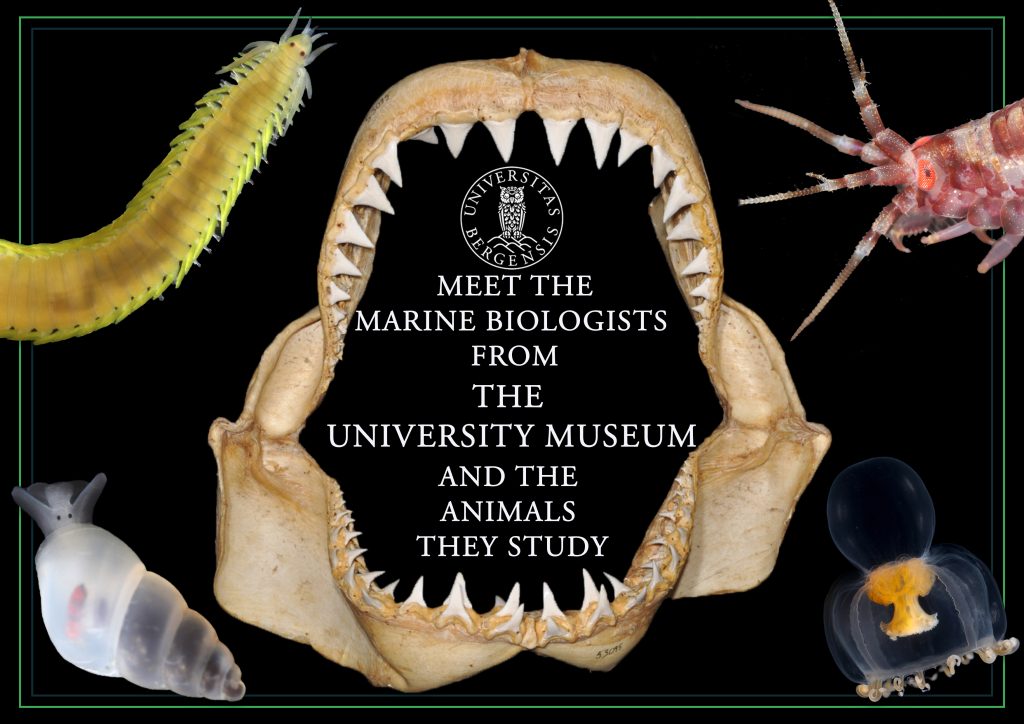
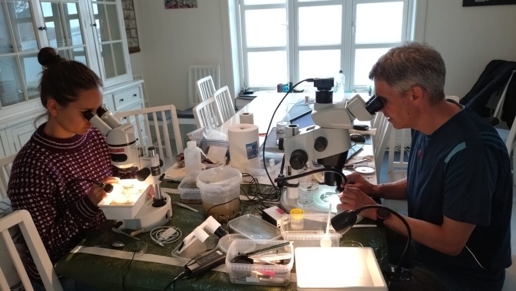
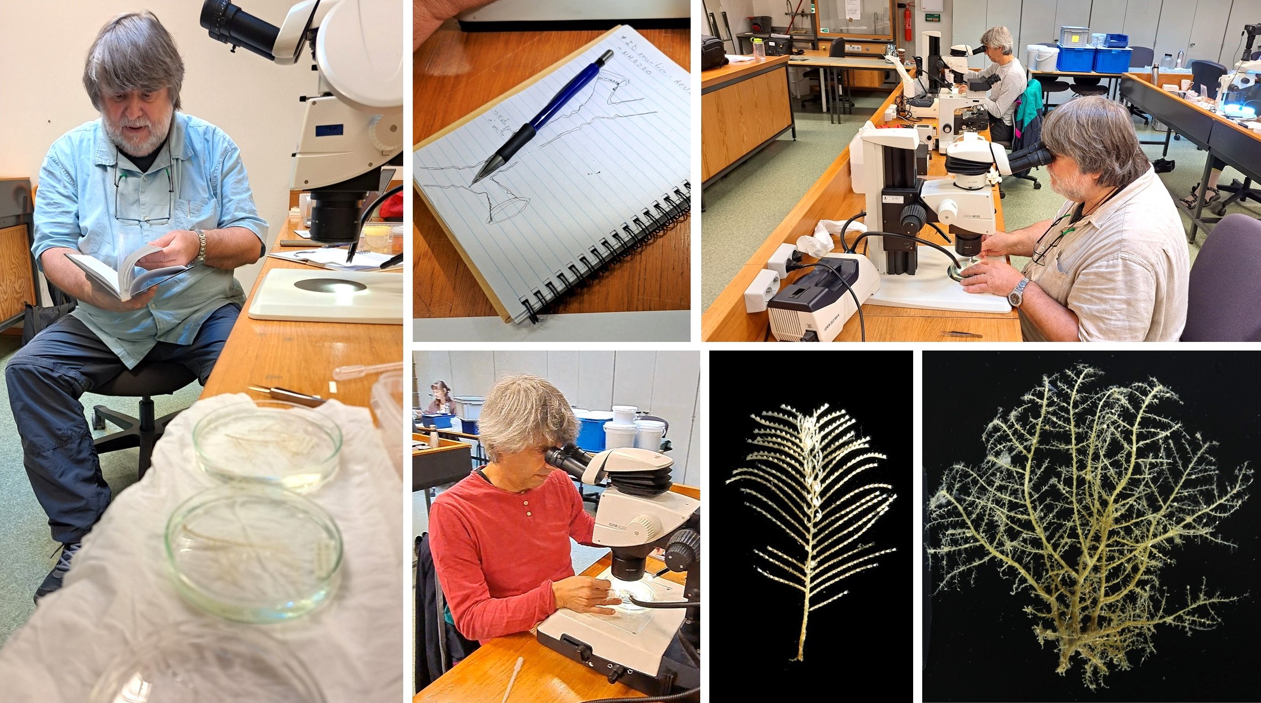
In a world full of administrative duties, unanswered emails, scary deadlines and project reports, immersing ourselves in a room full of samples of our favourite animals for an entire week is certainly therapeutic. And what a better way to do this than surrounded by colleagues and friends you admire; partners-in-crime with whom to play detective, solving challenging cases about species’ true identity.
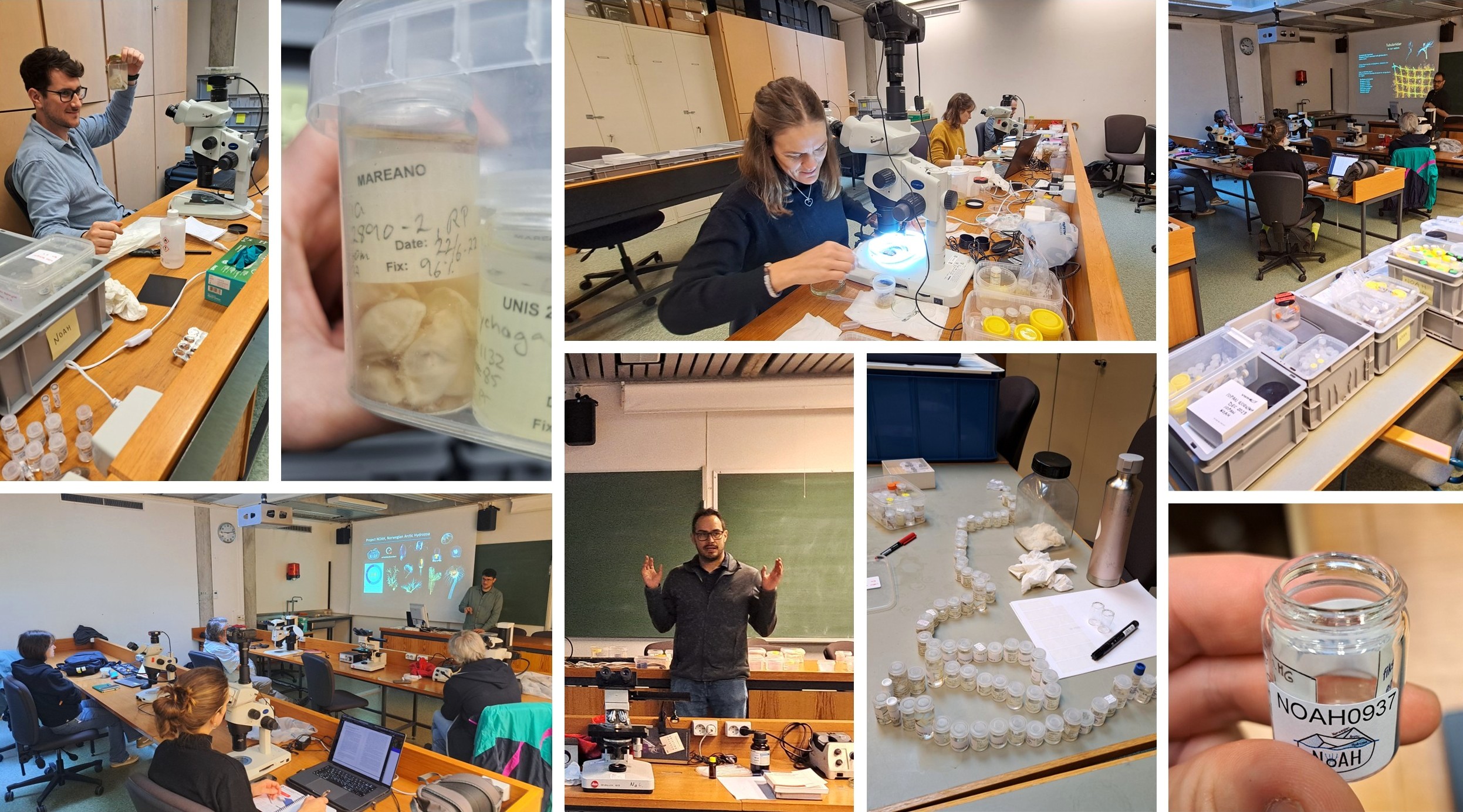
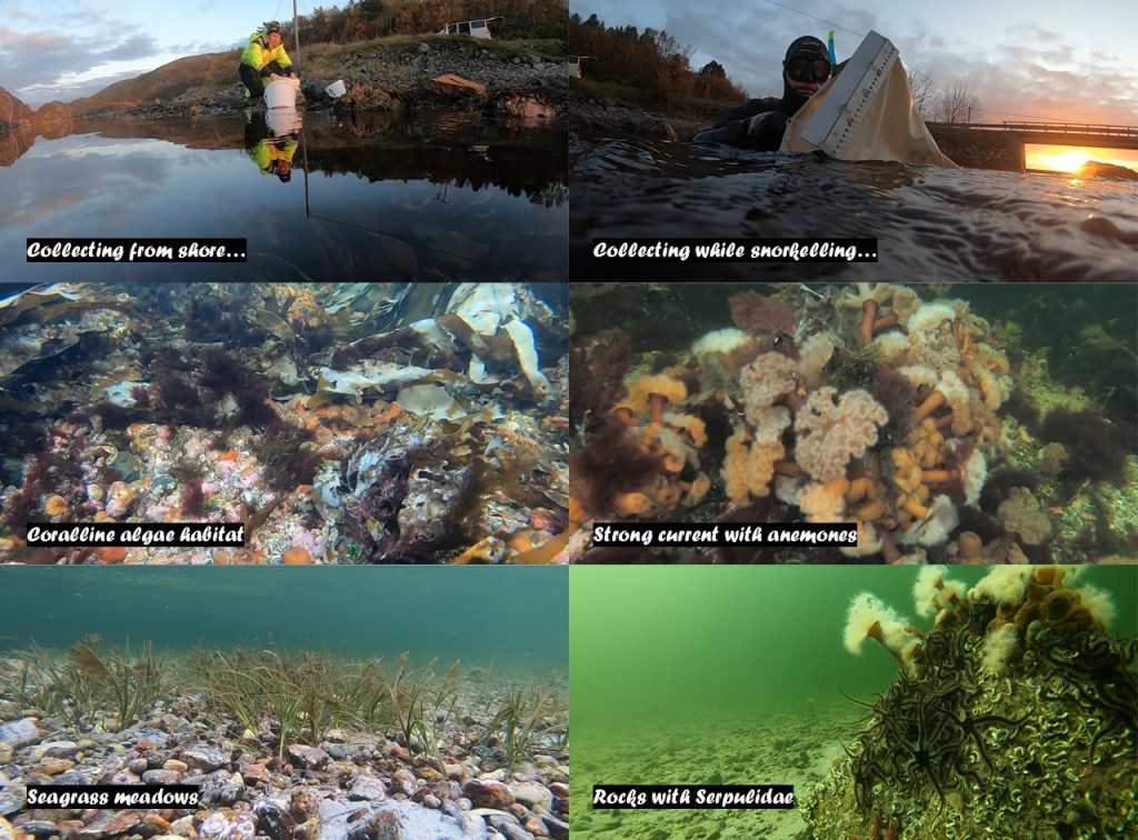
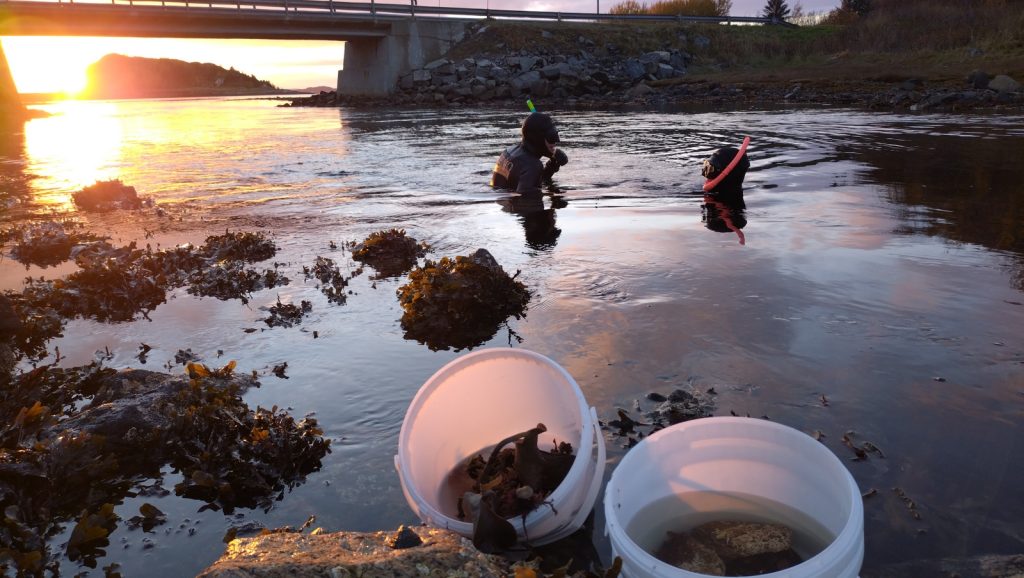
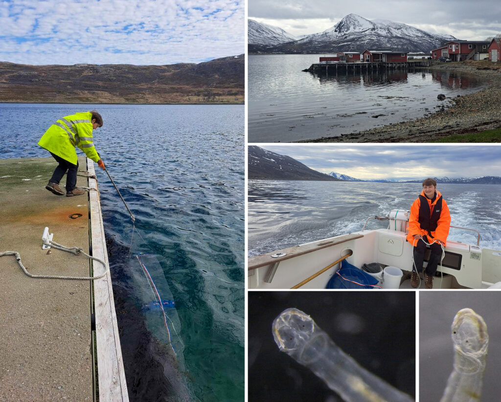
The latest NOAH Workshop on Arctic hydrozoans focused on a vast collection of samples obtained during several MAREANO surveys by the Institute of Marine Research (IMR). As hosts, it was a joy to organize and implement an event like this. The workshop participants represented all career stages (from MSc to consolidated professors), five different nationalities and four different institutions, including University of Vigo (Spain), IMR (Norway) and University of Gothenburg (Sweden), in addition to ourselves at UMB. We inventoried over 400 samples. Indeed, we surpassed NOAH sample number 1200! Yaay! Of those, a set of 95 tissue samples are now on their way to the sequencing facility at the Canadian Centre for DNA Barcoding, as part of our agreement with the Norwegian Barcode of Life (NorBOL, BOLD). Despite (or perhaps thanks to) having very long days of nearly 10.5 hours of work, we had a very rewarding and productive week, instrumental to define our next efforts on species delimitation in Arctic Hydrozoa. It was really wonderful seeing people with different backgrounds, expertise and skills joining efforts to put some order in the messy hydrozoan systematics. We leave you with some impressions from each of the workshop participants. Thank you ALL for joining us!
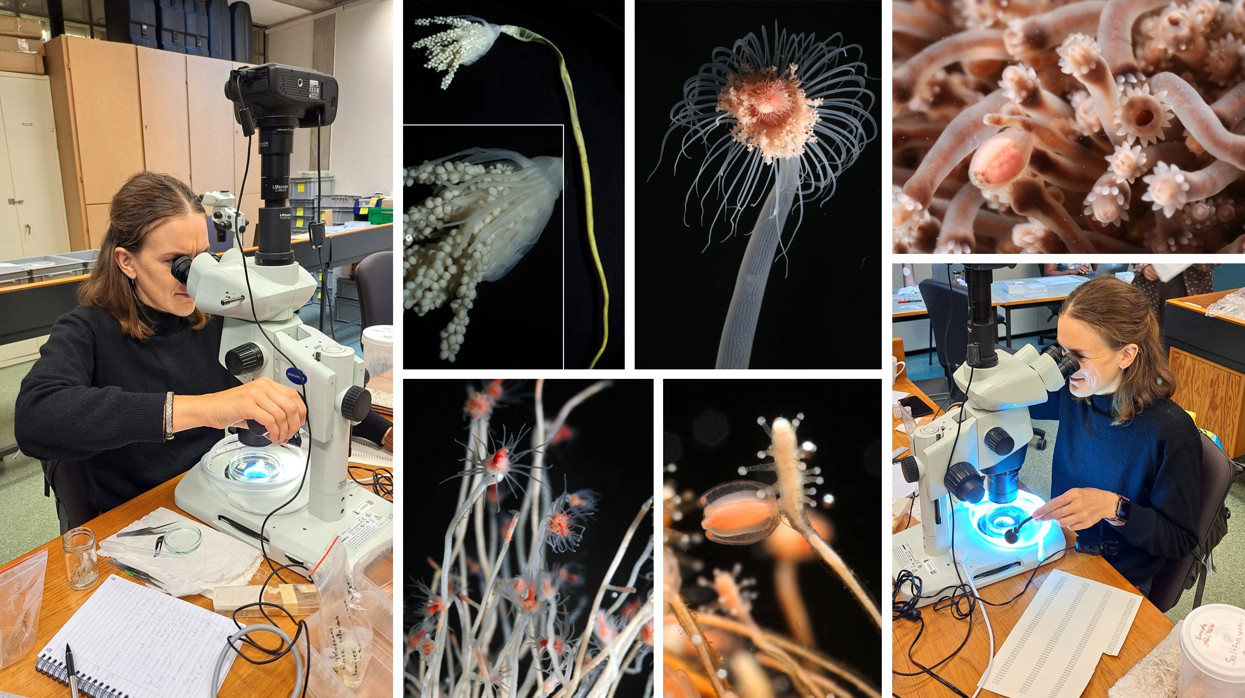
From Marta Gil, Senior Engineer, Institute of Marine Research:
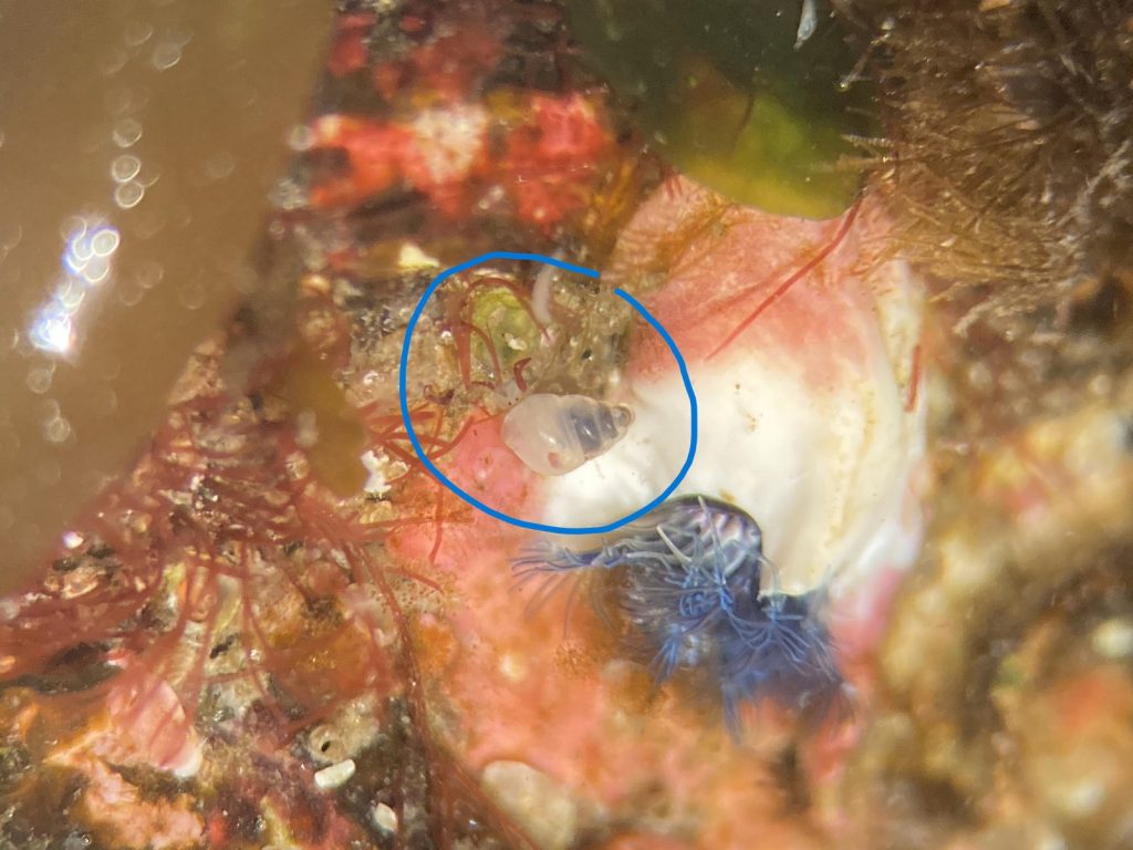
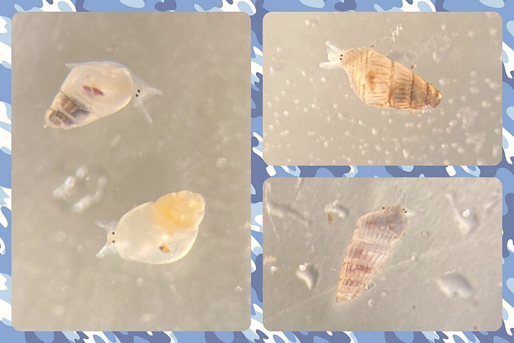
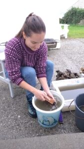
This was another incredible week collaborating with NOAH project, surrounded by hydroids and close friends! Despite being an intense week, I had so much fun in the lab checking samples, taking photos, chatting with Doris about athecate hydroids, discussing our identifications, and realizing that the new data and records we are generating will certainly shape our upcoming research efforts for the years to come. What I enjoyed the most was matching identifications made in the laboratory with the available molecular evidence. It’s very exciting to see how everything fits together. It’s like doing a puzzle!
I’m pleased that Mareano (IMR) supported and encouraged my participation in this workshop and that IMR is actively contributing with physical samples, some of which will be used to generate the first DNA barcodes for a bunch of species without prior molecular data. Collaboration between projects and institutions while sharing expertise and skills is essential: NOAH is a good example of how joining efforts is not only more efficient and productive, but also funnier and motivating. It will be exciting to see the results obtained altogether during the project (including “resurrecting” some species that were pooled as synonyms of supposedly largely distributed species). Stay tuned for updates!
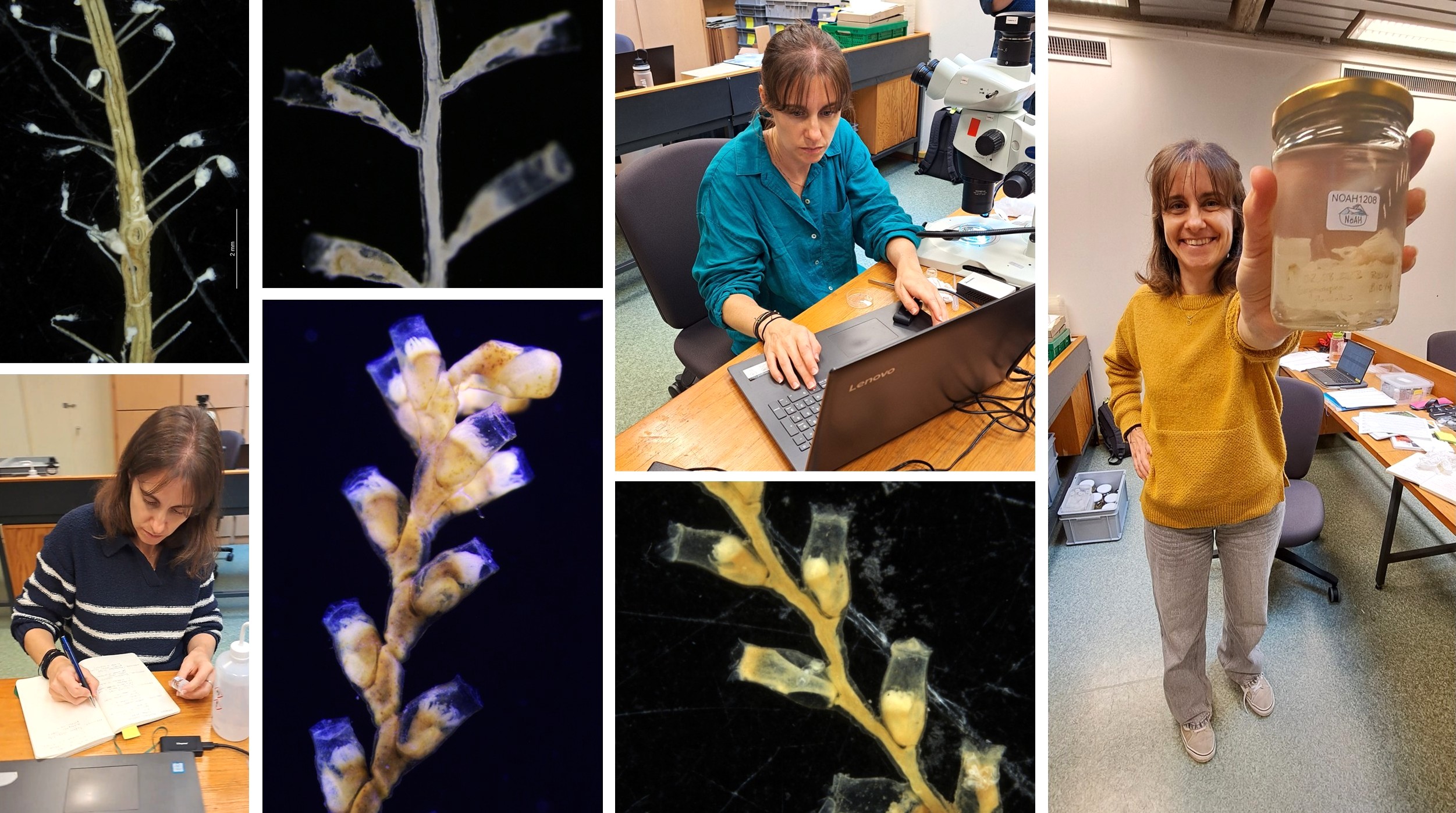
From Doris Björling, PhD student, University of Gothenburg
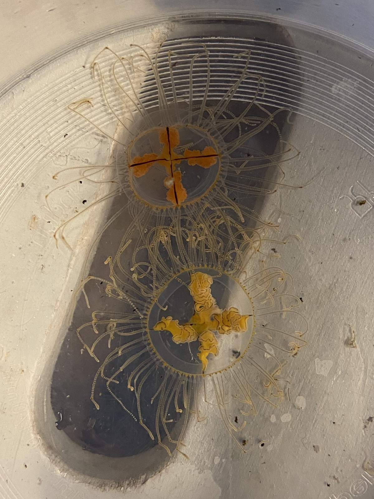
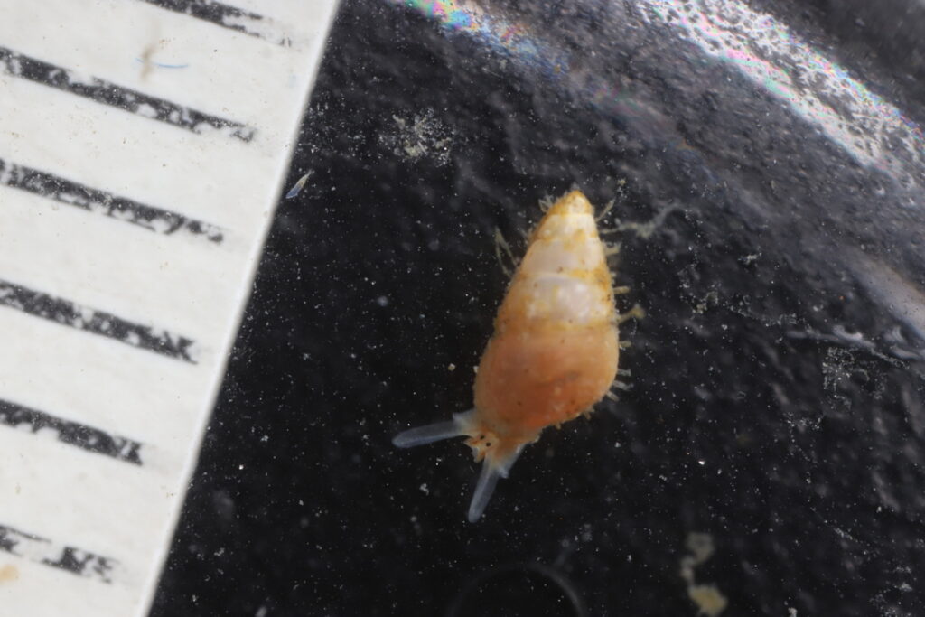
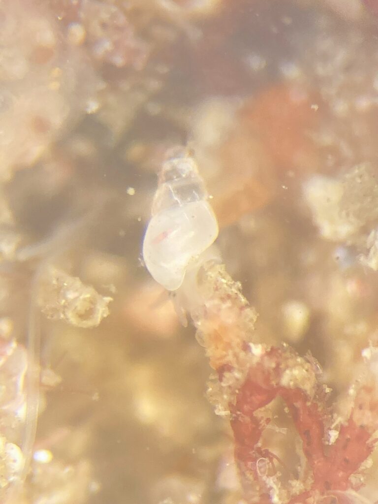
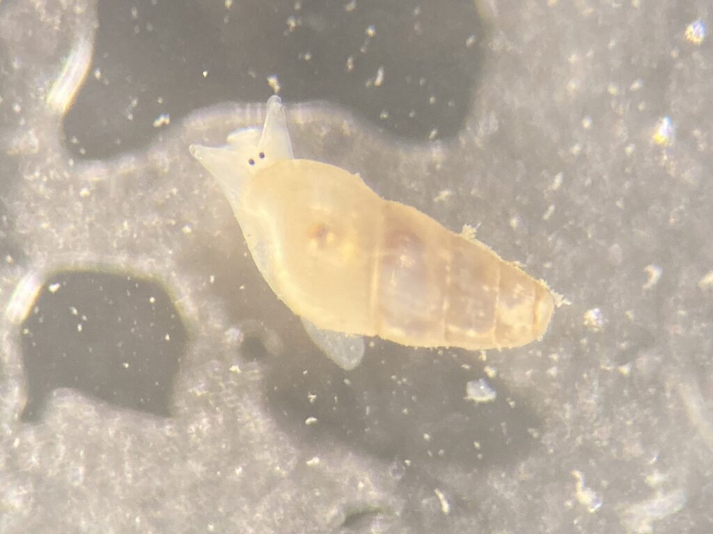
I’m a PhD student in the project HYDROINS (Hydrozoa In Sweden) looking into the diversity of Anthoathecata in Swedish and adjacent waters. With limited resources, the opportunity to explore arctic samples from NOAH, the diverse collection of NorHydro and other samples from the Manet Team collection at the University Museum of Bergen is priceless. During the workshop I’ve gotten access to samples that feed into and expand several of my upcoming projects. While representatives from at least seven anthoathecate families were examined, my main focus has been family Tubulariidae. HYDROINS and NOAH are making a collaborative effort to explore and describe this family in our waters, including the large charismatic species Tubularia regalis in the Arctic.
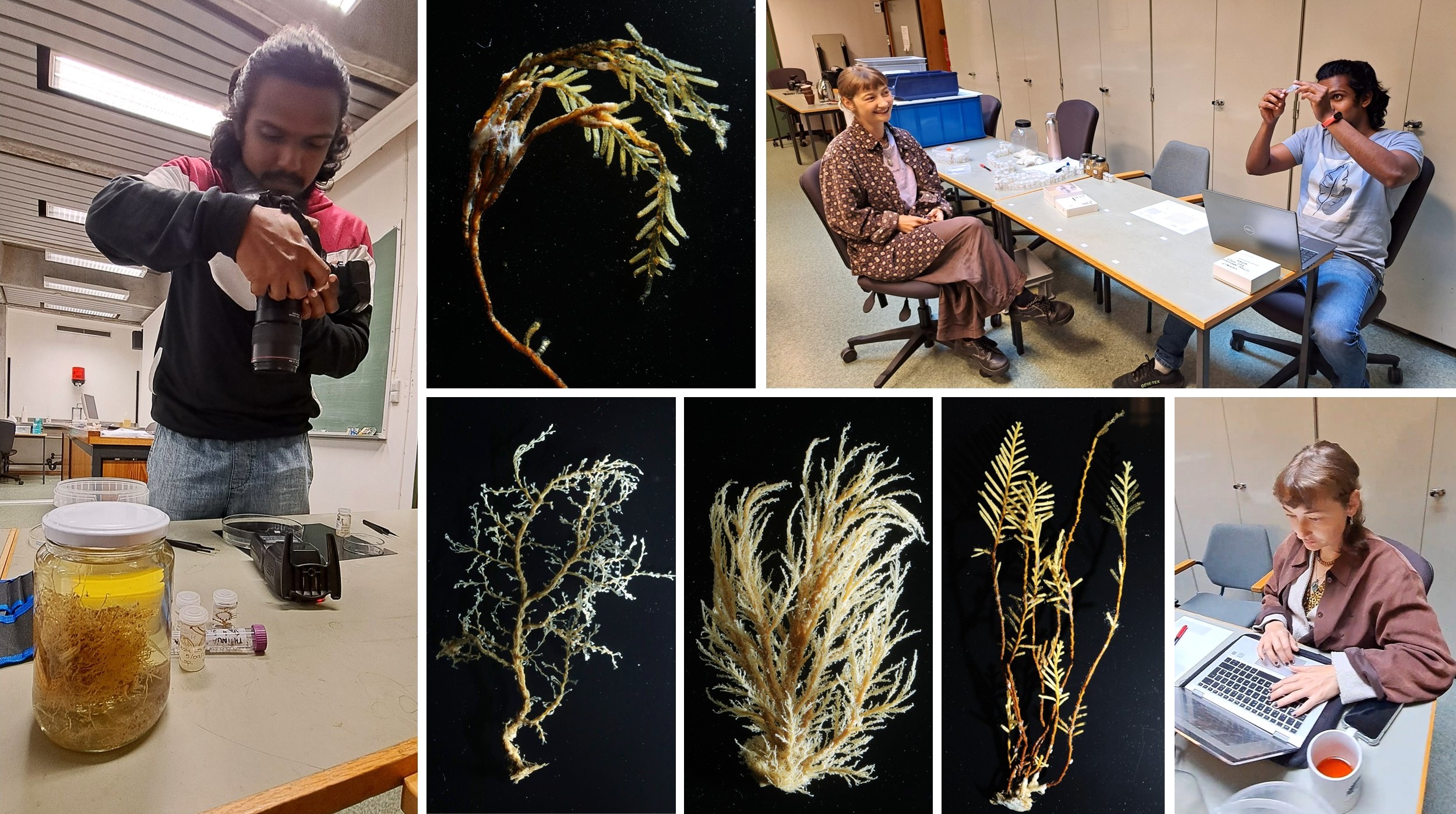
From Praveen Raj, PhD student, University Museum of Bergen
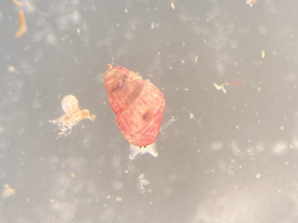
I had a fantastic time at the NOAH workshop coordinated by Joan and Luis. During the week, I helped organizing the NOAH hydrozoan collection: over 1200 samples of benthic hydroids, hydromedusae and siphonophores that has been collected and/or examined over the past 2.5 years. In addition, I also gave a hand photographing larger specimens that would not fit under the microscope. I thoroughly enjoyed these two tasks I was entrusted with. The photography part was especially rewarding, as Joan shared his macro photography tips, which greatly improved my skills. Meeting passionate researchers working on hydrozoan diversity and being part of this vibrant hydrozoan community at the University Museum of Bergen is truly inspiring.
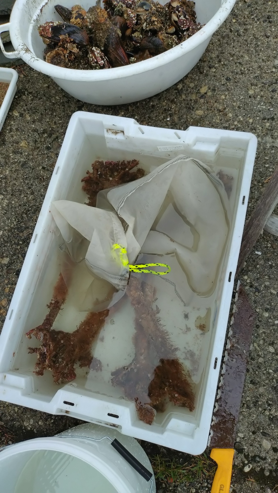
From Lea Dober, Staff Engineer, University Museum of Bergen
In my first week as technician at the University Museum, I was introduced to hydrozoans; quite different to my usual chaetognath suspects I have been working with during my Master thesis. Although hydrozoans are small and delicate creatures, they sure take up a large space in the museums’ collection! So that week we faced one big challenge: Sorting and organizing the entire NOAH hydrozoan collection. Quite some work, but the spirits and motivation were high – how could it be any different with the lovely company of experts from Spain, Mexico and Sweden which were super focused and busy with identifying the most difficult hydrozoans. Not even the call for lunch could lure them away from their microscopes! In the end we managed to sort everything and were rewarded with delicious food and the knowledge that we moved multiple steps further with the NOAH project in only one (intense) week.
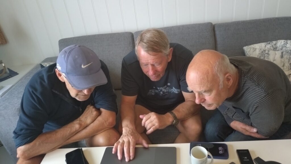
From Fran Ramil and Jose Ansín Agís, Professor and Researcher (resp.), University of Vigo.
The traditional taxonomy, based on morphological features, together with molecular approaches for hydroid identification posed an exciting challenge during the workshop, mainly within controversial taxa, like Haleciids, Aglaopheniids and “Anthoathecata”. The preliminary results are very promising and may lead to new advances in hydroid taxonomy.
Thanks Joan, thanks Luis for inviting us and organizing such a productive and wonderful meeting. Great work!

Joan and Luis, Manet Team