Category Archives: 2016 december calendar

Door #14: Where the sun doesn’t shine. Lucifer, luciferin and luciferase

Yesterday’s blog was about about Lucia, the mythical virgin who is celebrated with lights produced from combustion energy in candle lights. Lucia’s name is derived from the latin lux, meaning light. Similarly, Lucifer was one who was carrying light. We will not dwell with the ridiculous lore about Lucifer in old folk beliefs. Instead we will briefly look at real carriers of light from the organic world. Light can also be produced with other processes than fire and a range of organisms are particularly skilled in “letting there be light” in their environments. This is what we call bioluminescence. Bioluminescent substance can be used by potential prey animals to scare away predators. (Hover over picture with your mouse to see the gif.)

“Glow worm” beetles or “fire flies” of the families Lampyridae and Phengodidae are familiar to most people who have been out-door on a dark summer night. Studies of fire fly behavior have revealed how different species of these beetles communicate with their kinds using flash signals with variable frequency, intensity and duration. The biochemical mechanisms at work during bioluminescence are also relatively well studied in these beetle groups.

In the marine environments bioluminescence is known from many unrelated organisms and because different molecular reactions are involved in light production it is likely that bioluminescence must have evolved independently many times. A glowing sea may be experienced when massive densities of dinoflagellates are flashing their lights on the coast at night. Particularly Noctiluca scintillans, whose body is big enough to be visible with the naked eye, is frequently referred to in field guides to marine shore life. But many dinoflagellate species are involved in the bioluminescence that Norwegians call “morild”, – the “fire in the sea”. This phenomenon has also been called phosphorescence, however this is the process where light energy is absorbed by a substance and emitted on a different wave length. Special fluorescent proteins are responsible for glow-stick effects in organisms. (Incidentally the Greek light-carrier Phosphorus has been equated with the Latin Lucifer, and those of us who have seen white phosphorous in action understand what gave the name to this very reactive version of the element.) Studies of dinoflagellates have associated the light production with molecular bodies named scintillons and demonstrated that the biochemical activity in these objects in diatoms is governed by diurnal rhythms. This appears to make sense, because what is the point of flashing lights at day time? It may not be quite clear what use the diatoms have of producing light at night time either. However, it seems more obvious that there are functional advantages of bioluminescence in the depth of the ocean, where light does not penetrate. And it is below the so-called euphotic zone, from approximately 200 m on that bioluminescence is effective in different sorts of interactions among various animal groups. Deep water angler fish even use lit lures to attract prey.

Light emission in some animals is based on symbiosis with bacteria such as Aliivibrio fischeri.  In other cases, the animal itself may produce the active proteins. Different biochemical systems are at work in bioluminescence and the light-producing molecules are not the same in all systems. Still, as a group of oxidizing and light emitting molecules they all go by the name of luciferin.  To produce a light flash a catalyzing agent is also needed. This is provided by a group of different enzymes called luciferase.  Other active components and free ions may be involved in the reaction which may be triggered in different ways, either mechanically as with a set of oars and a rowboat when dinoflagellates are near the surface water, or by some biochemical trigger. Sometimes it must happen by some neural response in the animal.

Light organs in the lantern fish Benthosema glaciale. (from Paulsen et al. 2013

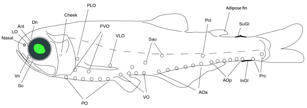
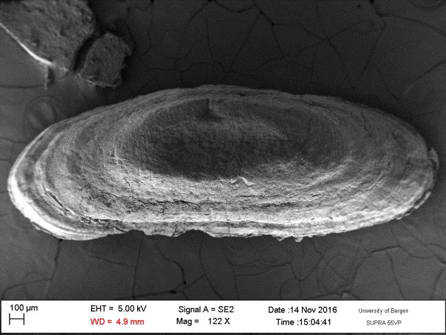
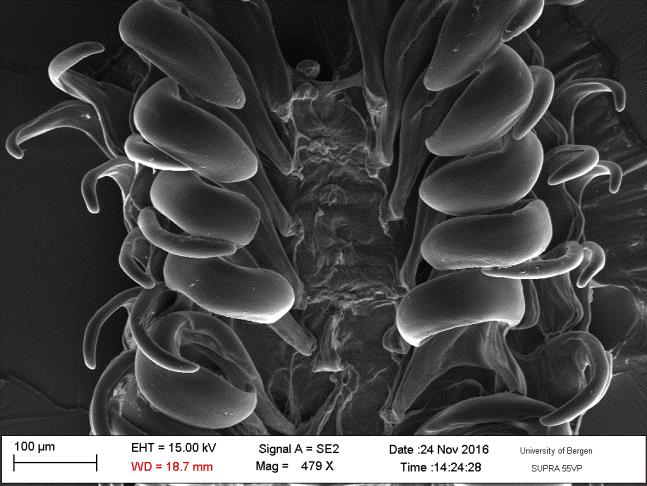
Light organs in the lantern fish Benthosema glaciale. (from Paulsen et al. 2013)

The lantern fishes are known for their photophores, – series of small organs that can produce yellow, green or blue light. Because the arrangement of photophores is different in different species, the organs are thought to play a role in communication between con-specific individuals. This may be the case for other animals as well, such as squids. It is also believed that the light organs can have a camouflaging effect by visually breaking up the silhouette of the fish, when the fish is viewed against a lighter back-ground higher up in the water column. The fish thus obtains protection from predators below by means of counter-illumination.

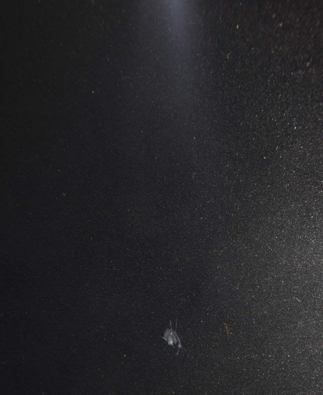
Several deep water animals are confusing potential predators by ejecting a luminescent substance towards the predator. Shrimps of the family Oplophoridae are particularly known to exercise this defensive technique.  The luciferine in shrimps is called coelenterazine and is presumably produced in the digestive gland called haepatopancreas. When the shrimp spews the glow through the mouth, the effect is somewhat similar to the one that cephalopods use when they disappear in a cloud of ink.  May we call it a “lucifer smoke-screen”?

Our museum collections have a rich material of these shrimps. Several species were collected during the MAR-ECO cruises over the Mid-Atlantic Ridge. One of them was Oplophorus spinosus shown in the picture below. While all of the oplophorids appear to be able to use a “lucifer smoke”, some of the species, including O. spinosus, also have light producing organs along their sides, somewhat similar to what we see in the lantern fishes. These photophores are complicated light generating organs with lenses and reflectors. They may be able to filter the wave length, and also the intensity and direction of the emitted light. In oplophorids, such organs are only found in three of the most closely related genera of the family, according to a recent study by Wong et al. (2015).  Interestingly, these animals also have two types of eye pigments. One type is shared with other oplophorids and is sensitive to the blue-green part of the light specter.  The other pigment is also sensitive to the shorter wave length in UV light. Because of the special abilities of the eyes it is tempting to think that these shrimps somehow are using the photophores in communication with individuals of their species. If so, Oplophorus spinosus and similar light talk would be a perfect case for biosemiotics. “Please shrimp, tell us about the world view from your perspective!” However, it is possible that the use of the photophores is only for counter-illumination when the shrimps are performing vertical migrations in the water column.

Oplophorus spinosus - a bioluminescent mid water shrimp (Photo: David Shale, MAR-ECO)

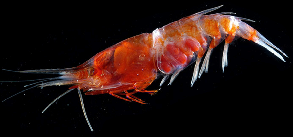
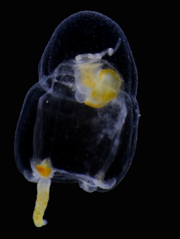
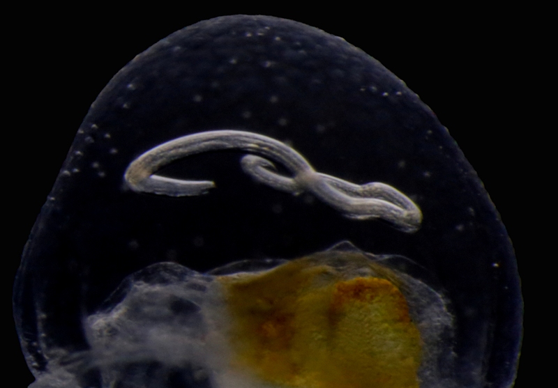
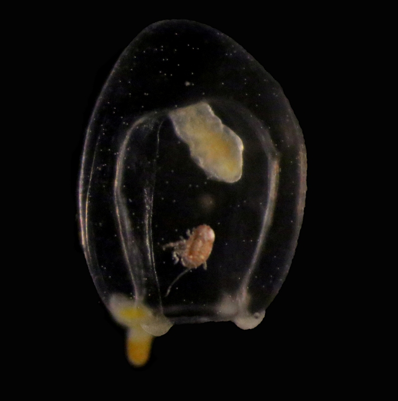
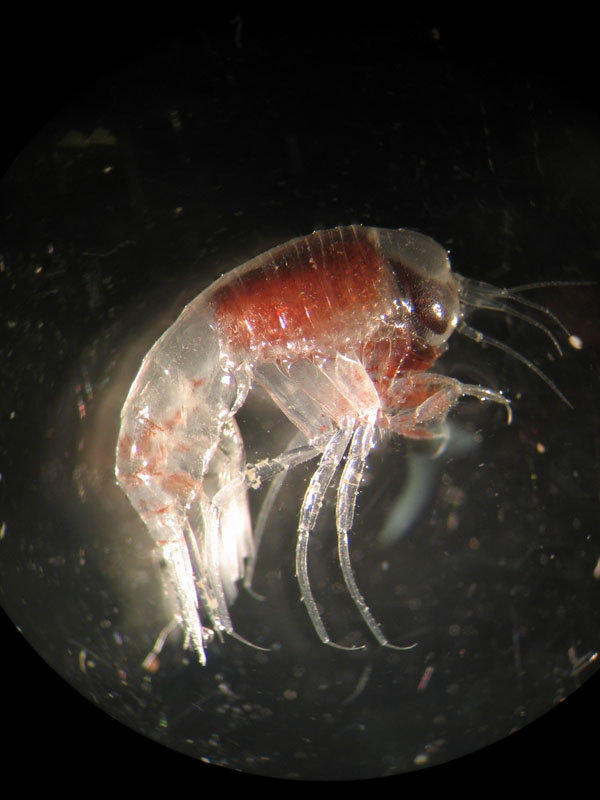
Oplophorus spinosus – a bioluminescent mid-water shrimp carrying large eggs (Photo: David Shale, MAR-ECO).

The “signalling abilities” of bioluminescent compounds are exploited in biotechnology and cell research. Luciferase from Oplophorus has been exploited as a so-called reporter gene in visualization of cell activities and gene transcription. May be it is not too far-fetched to see the shrimps as some kind of “light-carriers”.


Papers

Inoue S, Kakoi H, Goto T. (1976) Oplophorus luciferin, Bioluminescent substance of the Decapod shrimps, Oplophorus spinosus and Heterocarpus laevigatus. J.C.S. Chem. Comm. 966:1056-1057.

Poulsen JY, Byrkjedal I, Willassen E, Rees DJ, Takeshima H, Satoh TP, Shinohara G, Nishida M, Miya M. (2013).Mitogenomic sequences and evidence from unique gene rearrangements corroborate evolutionary relationships of Myctophiformes (Neoteleostei). BMC Evolutionary Biology 13:111.

Shimomura O, Masugi T, Johnson FH, Hanedal Y. (1978) Properties and reaction mechanism of the bioluminescence system of the deep-sea shrimp Oplophorus gracilorostris. Biochemistry 17:994-998.

Wong JM, Pérez-Moreno JL, Chan T.-Y, Frank TM, Bracken-Grissom HD. (2015) Phylogenetic and transcriptomic analyses reveal the evolution of bioluminescence and light detection in marine deep-sea shrimps of the family Oplophoridae (Crustacea: Decapoda). Molecular Phylogenetics and Evolution 83:278–292

Door #13: Lucia – with a ray of enlightenment?

Many kids will appear as “lucifers” today – light bearers. Celebration of Santa Lucia’s day seems to have won increasing popularity in Norway over the recent decades. Much of this cultural practice has clearly spread from Sweden, where the tradition is so deeply rooted since early 1900 that Swedish racists seem to believe that Lucia was an Arian blonde from remote mythical times of Astrid Lindgren’s Småland? But according to legend, Lucia was a christian martyr from Sicily in the fourth century. The story goes that she was tortured because of her conviction. She obtained her post mortem status as a heroine because she sacrificed her eyes for the “true faith” and was thus expelled to the darkness.

"Lussekatt" is a pastry made for Santa Lucia day. Is it supposed to express the track of the sun through the year?

“Lussekatt” is a pastry made for Santa Lucia day. Is it supposed to express the track of the sun through the year?

Norwegian Wikipedia (of “Luciadagen”) claims that Lucia day has no actual association with Advent. “It is simply a celebration in commemoration of Sta. Lucia’s death on the 13th of December”, it is stated. But wait a minute! Take some time to look at the two calendars from the year 1700 that I reconstructed with the Timeanddate facility. This was the year when the Gregorian calendar revision was finally introduced in Denmark-Norway. Until that time Scandinavians had used the calendar that Julian Caesar’s government introduced 45 BC.

Reconstructed calendars for Denmark-Norway and Sweden from the year 1700. Notice that Norwegian Christmas Eve and Swedish Santa Lucia day are on the same day,

Reconstructed calendars for Denmark-Norway and Sweden from the year 1700. Notice that Norwegian Christmas Eve and Swedish Santa Lucia day are on the same day,

The Julian calendar is assuming a mean year length of 365.25 days, so the Julian clock was ticking about 20 minutes too fast compared to the mean tropical year. The Earth moves around the sun with a speed of about 30 km per second. As observed from the Earth, the mean time it takes for the Sun to return to the same point in the annual revolution is the tropical year. Revolution time is slightly variable, but since it is closer to 365.24 than to 365.25 the mismatch between the calendar and the season was increasing one day about every 128 years. In order to correct for the 10 days accumulated difference, the new Gregorian calendar was introduced in the catholic world in 1582.

Protestant Scandinavian countries may not have been able to discriminate between science and catholic religious practices. They delayed any action with the calendar until 1700 when the seasonal anomaly had increased to 11 days. In Denmark-Norway it was decided to delete those 11 days from the last part of February so that March 1st would follow immediately after February 18. The Swedes decided to go for a more gradual procedure. They reckoned that the calendar would be in order with time if they just neglected some successive leap years and skipped February 29 for some time. So they took this first small step by deleting February 29 in 1700. You can see how that worked out for February in the two calendars.

Travelling across the Norwegian-Swedish border in 1700 would imply the crossing of “multiple date lines”. Although it is questionable whether Lucia was celebrated to any extent in Sweden at the time, we could imagine that we wanted to travel there to take part in the feast for this catholic saint on the 13th of December. The Swedish calendar would show that Lucia is the fourth day in week number 50, which was a Thursday. However, the fourth day of week 50 in Norway was the 23rd. If we remember that the adjustment to the Gregorian calendar in 1700 should imply deletion of 11 days and that Sweden only got rid of one of those, we should rather consider the fifth day of week 50, namely Friday the 24th as the one that compares to Lucia day in Sweden. So with a couple of not particularly magic calculations we have matched Lucia’s deathday with Christmas Eve!
Could it be that these two mythical celebrations are actually rooted in fancy human imagination spun around the same astronomical event? Certainly so because in the course of time the Julian date for the winter solstice changed from 23rd to 22nd in the first centuries and in the 13th century the happening was down to 13th of December. This is the simple explanation to why some are confused by the fact that Lucia’s day is also called the darkest day of the year. Well, dear Swedes and sweet-hearts. Not any more.
We know that winter solstice, the day when the track of the sun has reached its southernmost point at the moving Tropic of Capricorn, was measured already 5000 years ago by stone age people at Newgrange in Ireland. During the regime of Julius Caesar the winter solstice was dated by Roman astronomers to December 25th and this became the celebration of the sun god Sol Invictus in the Roman Empire. However, the list of solar deities in pre-Cristian cultures is a long one and celebrations of winter solstice were significant in many pagan traditions before the customs were absorbed by Christian ways. Various brands of mystics still seem to recognize winter solstice as a moment of great spiritual significance. But maybe it would be sobering to think of the phenomenon in terms of solar energy influx and the amazing cascading biological effects of the seasons. In biology is where the mysteries are. Indeed the solstice marks an ecological and existential turning point for life on the northern hemisphere. At times it was dated to the 13th of December. This year it is happening on December 21st at 11:44.

EW

Door #12: All aboard the jelly cruise!

Travelling alone through the water column may be a dangerous business: reaching the final destination is not always guaranteed, the risk of being eaten is high, and even finding food may prove a difficult task… which is why several animals choose to travel comfortably on or inside jellyfish and siphonophores!

Jellyfish are commonly involved in relationships of parasitism and phoresis (i. e., when one organism is mechanically transported by another without any further physiological dependence), and many examples have been observed of these interactions around the world. For instance, it’s not unusual to find hyperiid amphipods and sea-spiders –as well as lobster and crab larvae – piggybacking on the surface of large and tiny jellyfish, and while it’s still not clear whether or not all these passengers feed on their means of transportation, real parasitism and jelly-feeding has been confirmed for at least some of them. Jellyfish may also transport parasitic worms to their final hosts (like the nematode you see in the pictures), acting as carriers of parasites towards fish and mammals, and sometimes, eventually reaching humans as well!

Euphysa aurata medusa with parasitic nematode larva. Korsfjord, February 2016. Credit: Aino Hosia.

Euphysa aurata medusa with parasitic nematode larva. Korsfjord, February 2016. Credit: Aino Hosia.

A close-up of 2 showing the parasite embedded in the mesoglea (jelly) of the host. Credit: Aino Hosia.

A close-up of 2 showing the parasite embedded in the mesoglea (jelly) of the host. Credit: Aino Hosia.

Euphysa aurata medusa with crustacean ectosymbiont. Raunefjord, December 2016. Credit: Luis Martell

Euphysa aurata medusa with crustacean ectosymbiont. Raunefjord, December 2016. Credit: Luis Martell

These two hydromedusae of Euphysa aurata were collected this year in the fjords south of Bergen, and are only an example of jellyfish harboring other animals in this area. The species is a common and widespread jellyfish around here, but its role in the transmission of parasites and transportation of small crustaceans has never been explored. It might well be that, together with its gelatinous relatives, E. aurata will prove to be involved in many more biological interactions than we previously thought!

Luis Martell

Door #11 Invertebrately inspired art?

Scientific illustrations today are usually formed within quite strict limits. We use photographs or drawings of small details, and these are all connected to one specific specimen that preferably is to be found in a scientific collection.

But can other approaches also help us? The artist Pippip Ferner has long found her inspiration in nature, and especially the (marine) invertebrates. Maybe her pictures can inspire us to examine other details in our study-animals? Maybe a picture can inspire you to think more about nature, the sea, or invertebrates – their lives and lores? These are not pictures that are meant to be scientifically accurate, but rather fabulations inspired by the wild things that happen when evolution gets to do as it pleases…

Some of Pippis drawings are inspired from scientific drawings, both old and new, some are from animals we have looked at together.

Here are some of Pippips pictures from this year, and the animals that inspired them. These three pictures were chosen to be part of the Evolution and Art section of the international science conference Evolution this summer in Austin, TX.

"Tunicate anatomy" (c) Pippip Ferner

“Tunicate anatomy” (c) Pippip Ferner

Pippip says about this first picture:

“A scientific illustration of a TUNICATE is the inspiration for this work. Tunicates are sort of last stage before vertebrates. Clues for this is found in the larva that has a notochord, comparable to the spine of vertebrates. It has cerebral vesicle equivalent to a vertebrate’s brain, sensory organs that includes an eyespot to detect light and an otolith, which helps the animal orient to the gravity.
Fascinated by the thought of this “slimy blob” having many features similar to humans resulted in this quite complex outcome. The overload of insistent lines has given the tunicate quite a sophisticated system.”

Komodo National Park sea squirt (Polycarpa aurata). Photo: Nick Hobgood (wikipedia)

Komodo National Park sea squirt (Polycarpa aurata). Photo: Nick Hobgood (wikipedia)

On the left is a photo of a live tunicate. This photo is from Indonesia, but tunicates are common to find also in our colder waters. They can be solitary as this one, or colonial – where several tunicates form a colony together by budding, so that one large colony basically has the exact same DNA. Most tunicates are sessile (they sit attached to one place), but some live floating around in the water. The best known of these pelagic tunicates are the salps of the southern oceans.


Internal anatomy of a tunicate (Urochordata). Adapted, with permission, from an outline drawing available on BIODIDAC. (Wikipedia)

Internal anatomy of a tunicate (Urochordata). Adapted, with permission, from an outline drawing available on BIODIDAC. (Wikipedia)

A scientific illustration of a tunicate in a Biology textbook will look something like this:


Moving to other invertebrates, Pippip has worked with clams:

"Bivalve anatomy" (c) Pippip Ferner

“Bivalve anatomy” (c) Pippip Ferner

“In this image I question how the clam lives in symbiosis with other species as its shell gets weaker due to climate changes. The drawing might resemble the results of some kind of scientific inquiry with references to the anatomy of a clam (bivalve).
In my work I let my own artistic evolutionary process make the clam into something more abstract.”

(If you wait until door # 22, there might be a story that relates to bivalves that live with others…)

This is a Ctenophore, a comb jelly:

"Comb jelly anatomy" (c) Pippip Ferner

“Comb jelly anatomy” (c) Pippip Ferner

“The starting point of this work was a detailed illustration from biologist Ernst Haeckel’s (Artforms in Nature) of a comb jelly/ctenophorae. The comb jelly differs from other jellyfish with more sophisticated nervous system with both synapses and individual muscle cells.
The outcome of this drawing is a tribute to the beauty of the structure of this organism.”

Jelly fishes anf Comb jelly fishes. Illustration: Ernst Haeckel, Kunstformen der Natur 1904, plate 27

Jelly fishes anf Comb jelly fishes. Illustration: Ernst Haeckel, Kunstformen der Natur 1904, plate 27

Ctenophores are predatory planktonic jellies. The special thing about them, according to our Jelly-specialist Aino, is that they have a rotational symmetry. The diagnostic feature of comb jellies are their comb-rows that they use for swimming. The photos above represent the three groups of comb jellies – all of them are present in Norway.


To the right is the Haeckel-picture she started from, and here is a film of Comb jellies from the Chicago Shedd Aquarium.


Pippip, Anne Helene and Aino


Door #10: Siphonophores

Today, I thought I’d introduce to you to a cool group of animals that is ubiquitous in the oceans (including the Norwegian seas), but unfamiliar to most people. Siphonophores (“kolonimaneter” in Norwegian) belong to cnidarians, a group that includes corals, anemones, hydroids and jellyfish, and is characterized by the presence of stinging cells used in prey capture. All siphonophores are predatory, and use their stinging tentacles to catch small crustaceans or, in the case of some species, even small fish.

The most (or only) familiar siphonophore for the majority of people is probably the highly venomous Portuguese Man O’War (Physalia physalis), which can be spotted floating on the surface of the ocean or stranded on beaches. However, it is not really representative of the group as a whole, as most siphonophores live in the water column of the open ocean rather than its surface. There are around 200 described species of siphonophores.

The most fascinating feature of siphonophores is their peculiar body plan. While siphonophores may appear to be a single animal, they are in fact a colony of physiologically connected and genetically identical but morphologically diverse individuals called zooids that have specialized to carry out different tasks for the colony. Siphonophores belong to the class Hydrozoa (“polyppdyr” in Norwegian), which covers two basic body plans: the polyp/hydroid and the medusa.

Schematic of a physonect siphonophore. From http://www.siphonophores.org (CC-by-nc-sa)

Schematic of a physonect siphonophore. From http://www.siphonophores.org (CC-by-nc-sa)

The various zooids comprising a siphonophore colony can also be divided into these main groups. For example, the zooids used for swimming, called nectophores, are medusoid, while the feeding zooids, or gastrozooids, are polyp-like. The siphonophore colony can also include specialized defensive, protective and reproductive zooids. All the zooids forming a colony arise by budding from a single fertilized egg. The different zooids are specialized to the degree that they cannot function as individual animals any more, and are only able to perform their specific tasks as parts of the siphonophore colony.

Anterior nectophore, posterior nectophore and eudoxid of the calycophoran siphonophore Dimophyes arctica – a common species in Norwegian waters. Photos by Aino Hosia (cc-by-sa)

Anterior nectophore, posterior nectophore and eudoxid of the calycophoran siphonophore Dimophyes arctica – a common species in Norwegian waters. Photos by Aino Hosia (cc-by-sa)

The zooids, for example the swimming nectophores, vary in appearance between species, and can be used for species identification. In addition, the various types of zooids in the colony are arranged in a strict species specific pattern, providing the intact colonies of each species with their particular appearance. While the individual zooids are generally small, millimeters to centimeters in size, some siphonophore species, like Praya dubia, may have colonies that reach 40 m in length! Siphonophore colonies generally have a zone of one or more (up to several dozen) swimming nectophores at the front, used to pull the colony through water. Behind this nectosome is the siphosome, which contains the feeding, reproductive and other zooids in a repeating pattern, each iteration of which is called a cormidium. In some species (suborder Calycophorae), these cormidia are released as small free-living reproductive colonies called eudoxids. Unfortunately, siphonophore colonies are extremely fragile and tend to fall apart during standard plankton sampling with nets, leaving behind a bewildering array of small bits and pieces – part of the reason they are relatively poorly known to most people.

Colony of physonect siphonophore Physophora hydrostatica, aka hula skirt siphonophore. Photo by Aino Hosia (cc-by-sa)

Colony of physonect siphonophore Physophora hydrostatica, aka hula skirt siphonophore. Photo by Aino Hosia (cc-by-sa)

Intact siphonophore colonies are beautiful, but often utterly alien in appearance. It is interesting to consider where to draw the line between an individual and a colony. While we as individuals have specialized organs to carry out our various bodily functions, siphonophore colonies are made up of specialized interdependent individuals or zooids similarly carrying out their specific tasks.

As part of project HYPNO we are charting the diversity of pelagic hydrozoans, including siphonophores, in Norway. There are ~15 species observed in Norwegian waters, and some, particularly Dimophyes arctica, Lensia conoidea and Nanomia sp. are extremely common components of marine plankton. However, siphonophores are primarily noticed when they become a nuisance: For example, mass occurrences of Muggiaea atlantica and Apolemia uvaria have in the past killed large numbers of farmed fish in Norway, with resulting losses to aquaculture companies.

– Aino (HYPNO)

Intrigued by siphonophores? For more information, visit e.g. http://www.siphonophores.org/  by Casey Dunn.

Door #9: Research stay of Juan Moles

Juan working at the Museum

Juan working at the Museum

During my stay at the University Museum of Bergen I have been working on the diversity and systematics of Antarctic philine snails. Most of the samples were collected during different cruises on board of the RV Polarstern in the Eastern Weddell Sea, Bouvet Island, and South Shetland Islands (West Antarctica). I photographed all specimens and then clipped them for the DNA analysis (see pictures).


I was able to work at the DNA lab with excellent resources for DNA extraction, amplification, purification, and sequencing.

I am indebted to Louise Lindblom who helped me at the beginning of my crusade there. After a first barcoding of all the material we identified six clades, from which we selected a maximum of three specimens to further sequence the ribosomal genes 16S and 28S and the nuclear gene codifying for the Histone 3.

The first phylogenetic tree with all partitions resulted in the finding of novel clades that now deserve further investigation.

Prof. Manuel António E. Malaquias and his PhD Student Trond Oskars helped me dissecting the material for anatomical analyses. Important taxonomical characters were those related to the male reproductive system, the digestive tract as well, and the shell. After the dissections and drawings of the main parts I prepared the hard structures such as the radula, the shell, and the gizzard plates for Scanning Electron Microscopy (SEM) as well as some soft structures after critical point drying. I could photograph all these material at the same facilities of the museum being helped by Irene and Katrine. After the two months of work, I ended up having huge amount of anatomical and molecular data that deserves further processing. See a picture of the radula and a gizzard plate:

Moreover, I was able to join the student diving club and make several dives to get to know the local flora and fauna. I could even collect some other heterobranch slugs for the barcoding project of the museum. See a couple of pictures of the nudibranch Limacia clavigera and Onchidoris muricata.

Overall, Bergen is a nice city to visit surrounded by nice mountains, good (but not cheap) beers, beautiful fjords, and nice people. I hope I can come back with a postdoctoral position to further enjoy the country and meet more Viking descendants.

-Juan

Door #8: the ups and downs of a marine werewolf?

When we think about what drives the ecosystems, much of the initial responsibility is put on the sunlight. This is mainly because of the photosynthesis, and thus the basic pieces of almost all food-webs, but light is also important for the animals. Many animals use visual cues to find food, and whether you search for food or do not want to become food, the presence (or absence) of light will help you.

Themisto sp swims up into the dark night. Photo: Geir Johnsen, NTNU

Themisto sp swims up into the dark night. Photo: Geir Johnsen, NTNU

Seawater is a pretty good stopper of light. We don’t need to dive far down before we are in what we consider a dark place, and less and less light finds its way the deeper we come. We tend to call the depths between 200 and 1000 m “the twilight zone”: most light stops way before 200m and the last straggling lumens give up at 1000m.

Most places on earth has a daily division between a dark and a light period: night and day. This is the ultimate reason for what is often called “the largest motion on earth”: Millions of zooplankton hide out in the darker parts of the water column during the day, and then move up to feed on the plants living in the light-affected parts of the water during the night (when predators will have a hard time seeing them). This daily commute up and down is called Diel Vertical Migration (DVM).

Themisto sp among the many smaller particles. (The light in this picture is from a flash). Photo: Geir Johnsen, NTNU

Themisto sp among the many smaller particles. (The light in this picture is from a flash). Photo: Geir Johnsen, NTNU

But what about the waters north of the polar circle? These areas will for some time during the winter have days when the sun stays under the horizon the entire day – this is “the Dark time” (Mørketid). At higher latitudes, there will be several days, or even weeks or months when the sun is so far below the horizon that not even the slightest sunset-glow is visible at any time. In this region, we have long thought that the Dark time must be a dead or dormant time.


The acoustic signals that gave the first indications of LVM. Figure 2 from Last et al 2016.

The acoustic signals that gave the first indications of LVM. Figure 2 from Last et al 2016.

We could not have been more wrong! It turns out that during the polar night, the DVM moves from being on a 24 hr cycle (sunlight-induced), to a 24.8 hour cycle! What is now the driver? The moon !(The lunar day is 24.8 hrs). Another thing that shows us that the moon must give strong enough light that predators can hunt by it, is that every 29.5 days most of the zooplankton sinks down to a depth of 50m: this falls together with the moon being full. Researchers have started to call this LVM (Lunar-day Vertical Migration) to show the difference to the “normal” DVM. There are of course lots of complicated details such as the moons altitude above the horizon and its phase that influences the LVM, but in general we can say that during the polar night (the Very Dark time), the “day” as decided by light has become slightly longer than normal.

The full moon, photographed by the Apollo 11 crew after their visit. Photo: NASA, 1969

The full moon, photographed by the Apollo 11 crew after their visit. Photo: NASA, 1969

Themisto - the werewolf. Note that the whole head is dominated by eyes - this is a visual hunter! Photo: Geir Johnsen, NTNU

Themisto – the werewolf. Note that the whole head is dominated by eyes – this is a visual hunter! Photo: Geir Johnsen, NTNU

Some of the larger animals taking part in the LVM are the amphipods Themisto abyssorum and Themisto libellula. They are hunters – so their reason to migrate up in the water column is not the plants, but the animals eating the plants; their favourite food are copepods of the genus Calanus. These are nice and quite energy-rich small crustaceans that eat the microscopic plants in the upper water column. We have sampled both Themisto-species in the middle of the winter (january), and their guts were filled to the brim with Calanus, so we know that they continue hunting by moon-light. They are such voracious hunters that some researchers have started to call them marine werewolves: the moonlight transforms them from sedate crustaceans to scary killers…


But, if they are the hunters, why do they spend so much time in the deep and dark during the lighter parts of the day? The hunters are of course also hunted. Fish such as polar cod (Boreogadus saida),  birds such as little auk (Alle alle) and various seals like to have their fill of the Themisto species. So – life has its ups and downs, and the dance of hunter and hunted continues into the dark polar night…

Anne Helene


Literature:

Berge J, Cottier F, Last KS et al (2009) Diel vertical migration of Arctic zooplankton during the polar night. Biology Letters 5, 69-72.

Berge J, Renaud PE, Darnis G et al (2015) In the dark: A review of ecosystem processes during the Arctic polar night. Progress in Oceanography 139, 258-271.

Kintisch E (2016)  Voyage into darkness. Science 351, 1254-1257

Kraft A, Berge J, Varpe Ø, Falk-Petersen S (2013) Feeding in Arctic darkness: mid-winter diet of the pelagic amphipods Themisto abyssorum and T. libellula. Marine Biology 160, 241-248.

Last KS, Hobbs L, Berge J, Brierley AS, Cottier F (2016) Moonlight Drives Ocean-Scale Mass Vertical Migration of Zooplankton during the Arctic Winter. Current Biology 26, 244-251.

Door # 7: Always on my mind…?

Today is #WormWednesday on Twitter, and we figured that it was a good day to introduce you to this rather unlucky fellow and his sidekick…

The orange (coloured in Photoshop) is the parasite. The two long sacks are filled with eggs.

The orange part (coloured in Photoshop) is the parasite. The two long sacks are filled with eggs.

They were collected during our field work in Sletvik in October. The worm is a polychaete in the genus Terebellides, whilst the parasite is a Copepod. This species rich group of small crustaceans have many modes of life, but parasitism is a common one, with about half of the ~13 000 species being parasites.

headache2_zerene HEADACHE3

Door # 6: Stuffed Syllid

Todays calendar critter is a Trypanosyllis sp. – a undescribed species from the genera Trypanosyllis in the family Syllidae. It most closely resembles a species described from the Mediterranean Sea. The Norwegian species is common in coral rubble, and has been assumed to be the same species as the one described from the Mediterranean. Genetic work reveals that these two are in fact separate species, and thus the Norwegian one is a new species awaiting formal description and naming. (If you read Norwegian, you can learn more about how species are described and named here: Slik gir vi navn til nye arter).

A new species of Trypanosyllis, collected in Sletvik, Norway. Photo by Arne Nygren. CC-by-sa

A new species of Trypanosyllis, collected in Sletvik, Norway. Photo by Arne Nygren. CC-by-sa

This specimen was collected, identified and photographed by Arne Nygren during our field work in Sletvik as part of his work on cryptic polychate species in Norway.

Syllids have opted for a rather fascinating way of ensuring high fertilization rates; something called epitoky: they asexually produce a special individual – the epitokous individual – from their bodies, and release this to go swimming in search of a mate. In the photo you can see that the female reproductive body (epitoke) is filled with orange eggs and has its own set of eyes, close to the middle of the animal. This section will break away from the mother animal and swim away in search of a male reproductive body to reproduce with. The mother animal will then grow a new female reproductive body.

-Arne & Katrine

Door #5: A visit from Mario

The collections have many guest researchers come here to work on our material, and one of our most frequent guests of lately has been Mario, who makes the long trip from Colombia to study both the West African material that we have from the MIWA-project, and to work on Nordic material. We asked him to make a contribution to the blog, and got the folllowing:

Mario in the Lab

Mario in the lab

For October – November visit.

For my third time in the Museum, I have found, as always, very good company from my colleagues in the lab: Katrine, Nataliya, Jon and Tom. Deep morphology and molecular method discussions over very good coffee were the “breaks” between periods of hard work at the microscope.

This time, I take to my home two papers close to completion; one about species of the genus Pista (Terebellidae) with additional information to what I found during my last visit in January. The second paper is about species in the subfamily Polycirrinae (Terebellide) from the West coast of Africa.

The idea is combine drawings, digital photos of specimens with methyl-green staining pattern and SEM pictures, as well as molecular information that will hopefully help us separate species and make better estimates of the region’s biodiversity.

Field work - somewhat cold and windy

Field work – somewhat cold and windy


The visit – which was without snow and with only a few showers of rain in Bergen (!), though with some very cold and windy moments at the Marine Station of the University of Trondheim – and sharing time with recognized polychaetologist as Fred Pleijel, Torkild Bakken, Eivind Oug, and Arne Nygren, was as spectacular as to know the Aurora Borealis.

Aurora borealis and a hooded tropical visitor. Photo: K.Kongshavn

Aurora borealis and a (hooded) tropical visitor. Photo: K.Kongshavn