Category Archives: 2015 december calendar

Door #14: A world of colour and slime

Welcome to the world of Nudibranchs!

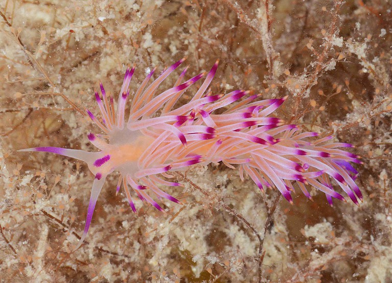
Flabellina rubrolineata (Mozambique) Photo: M. Malaquias

Flabellina rubrolineata (Mozambique) Photo: M. Malaquias

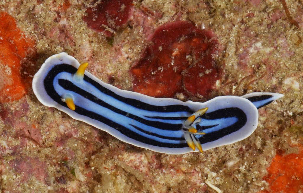
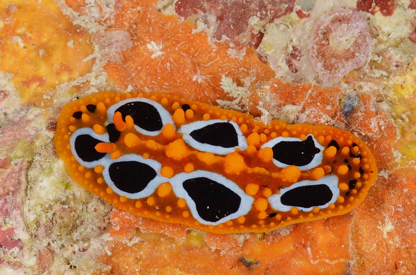
The nudibranchs are among the most beautiful animals in our seas. The palette of colours, shapes, and adaptations depicted by this group of gastropod molluscs has no parallel. Some species have no more than few millimetres where others can reach nearly half a meter. Some have a smooth skin, others are covered with long and delicate appendices.

Chromodoris boucheti (Mozambique) Photo: M. Malaquias

Chromodoris boucheti (Mozambique) Photo: M. Malaquias

Phillidia ocellata (Mozambique) Photo: M. Malaquias

Phillidia ocellata (Mozambique) Photo: M. Malaquias

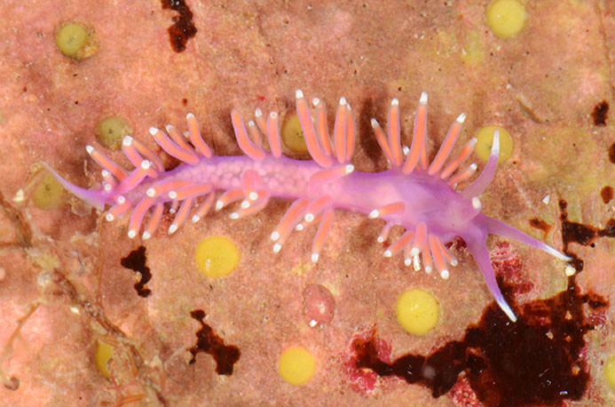
Most are benthic, but some are pelagic drifting with the oceanic currents. Nudibranchs feed on sponges, bryozoans, crustaceans, and cnidarians and even can incorporate in their tissues nematocysts sequestered from their prey which they use in self-defence. Probably, the most striking feature of these gastropods is the lack of a shell and presence of bright colours. These colours are usually a warning signal indicating the presence of deterrent chemicals some of them with pH values as low as 1 or 2. Some of these chemicals are biologically active and have been investigated for the treatment of several types of cancer diseases.

Flabellina pedata (Norway) Photo: M. Malaquias

Flabellina pedata (Norway) Photo: M. Malaquias

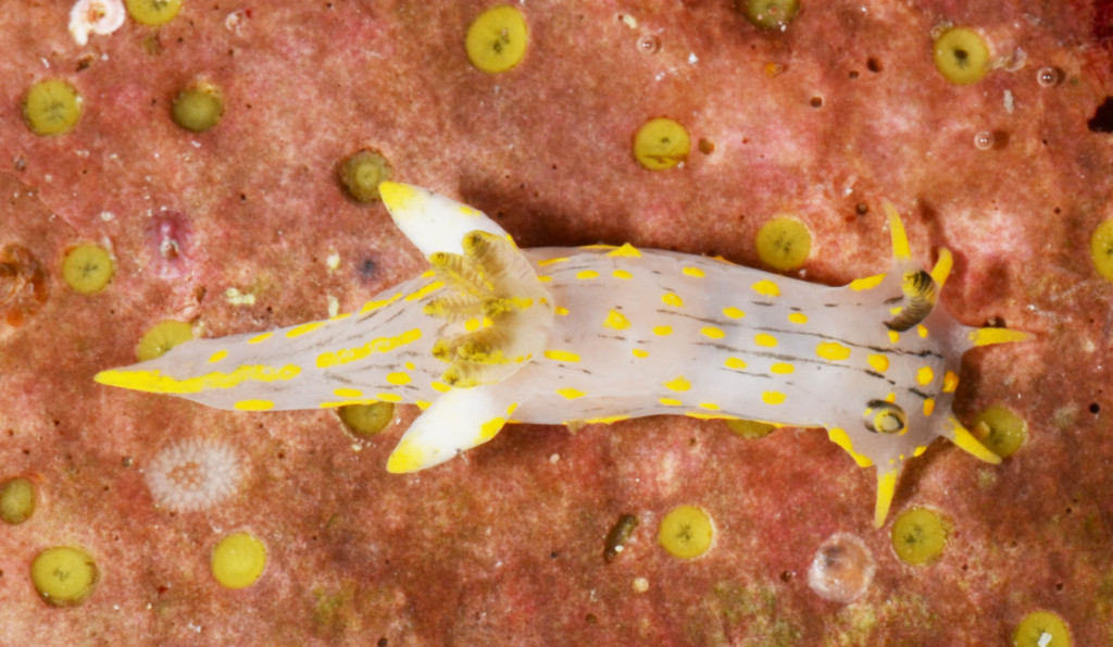
Polycera quadrilineata (Norway) Photo: M. Malaquias

Polycera quadrilineata (Norway) Photo: M. Malaquias

-Manuel

Door #13: Time for rejuvenation

Some of the fundamental existential impacts of the solar cycle were certainly understood by the Neolithic people who built Newgrange and were able to align the gigantic construction with the position of the sun rise at winter solstice. It was a point of return in “the wheel of time”, the annual cycle of “ageing, rebirth, and rejuvenation of Nature”. But how living individuals reproduce and come into being was a mystery right up to modern times. The Roman writer in natural history, Pliny (ca 70 AD), for instance stated that: “…after six months’ duration , frogs melt away into slime, though no one ever sees how it is done; after which they come to life again in the water during the spring, just as they were before. This is affected by some occult operation of Nature, and happens regularly every year. Mussels, also, and scallops are produced in the sand by the spontaneous operations of nature.”

Although the famous experiments by Francesco Redi had refuted some ideas about “spontaneous generation” in the mid 16-hundreds, the concept was still an important part of Lamarck’s theory of evolution that was opposed by his colleague Cuvier. Birth, of course, has also been a subject of discussions when pondering the mysteries of the Mary cult: was it really a case of parthenogenesis? What is really going on in the making of a body – the “process of incarnation”?

IMG_2861

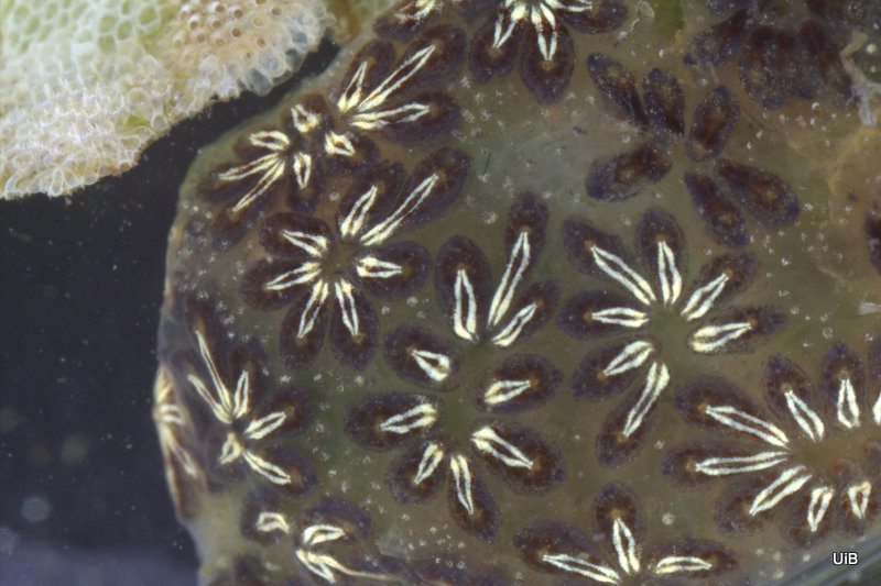
Botryllus schlosseri (photo: K. Kongshavn)

Botryllus schlosseri, the “golden star tunicate”, is a common species on Atlantic coasts and recently has expanded its distributions to other seas as a result of human marine travelling. Researchers at the University of Bergen (Delsuc et al 2006) found that the tunicates belong to an evolutionary lineage that is the closest to vertebrates (including humans). B. schlosseri is relatively easy to keep in aquaria and has taught us a lot about reproduction and life cycles.

The similarity between the tunicates and the vertebrates are only apparent in the early stages of tunicate life. The larvae have a body with a tail containing the “chorda”, and a dorsal nerve tube, – both unique characteristic features of the Chordate animals (see figure 1A in in Voskoboynik el al. 2013). But these similarities disappear within a few hours when the free swimming larva has settled on some surface substrate and started the metamorphosis into the sack like body of an adult tunicate with a filter feeding gut. The larva was the result of sexual reproduction, the merged genetic material from sperm and egg. However, the metamorphosed individual will soon begin to reproduce asexually by budding off a copy of itself in a neighbouring position. The results of such multiplications are clusters of two to 12 genetically identical individuals in a star like pattern. These individuals, called zooids, are active for relatively short time, about a week at 19 oC, until they become inactive and gradually are reabsorbed by other cells in the colony while being replaced by new zooids. This sort of programmed cell death is called apoptosis and researches believe that studies of B. schlosseri can reveal some of what is going on with ageing and death of cells. It has been estimated that in an adult human body there is apoptosis of about 50 to 70 billion cells per day. Fortunately there is also renewal of cells, like in the growing colony of Botryllus. Very interesting things may happen if the zooids from different larvae are meeting up at the margins of two colonies with the so-called ampullae. Botryllus has a self-recognition system that is controlled by just one gene, but the gene occurs in many variants (alleles). If the alleles from two colonies are compatible, the blood vessel systems of the two colonies may grow together so that one colony is actually formed by zooids with different genetics. This is somewhat analogous to what happens between mother and child in the mammalian placenta. If the compatibility of two colonies is bad, they will “fight” each other in an inflammatory immune reaction. Such processes have special interest with respect to understanding immune systems and the outcome of organ transplantation.

It takes about 3-4 weeks for a colony to become sexually mature so that egg and sperm may be released in turn, avoiding self-fertilization. The duration of a colony is believed to be about 12 to 18 months in Norwegian waters (Moen & Svendsen 2008).

The reproduction system of B. schlosseri is just one of many different reproduction systems of animals. Where does individuality begin and stop? Would a zooid greet its neighbour with “Merry Christmas, I!”?

Suggested reading:

Delsuc et al. (2006). Tunicates and not cephalochordates are the closest living relatives of vertebrates. Nature: 439:965-968.

Manni et al (2007). Botryllus schlosseri: A model ascidian for the study of asexual reproduction. Developmental Dynamics 236(2): 335-352.

Moen & Svendsen (2008) Dyreliv i havet. KOM Forlag.

Tiozzo et al. (2006). Programmed cell death in vegetative development: Apoptosis during the colonial life cycle of the ascidian Botryllus schlosseri. Tissue and Cell 38 (3): 193-201

Voskoboynik et al. (2013) The genome sequence of the colonial chordate, Botryllus schlosseri Elife. DOI: 10.7554/eLife.00569.001

-Endre

Door #12: Plankton sampling with a vertebrate view!

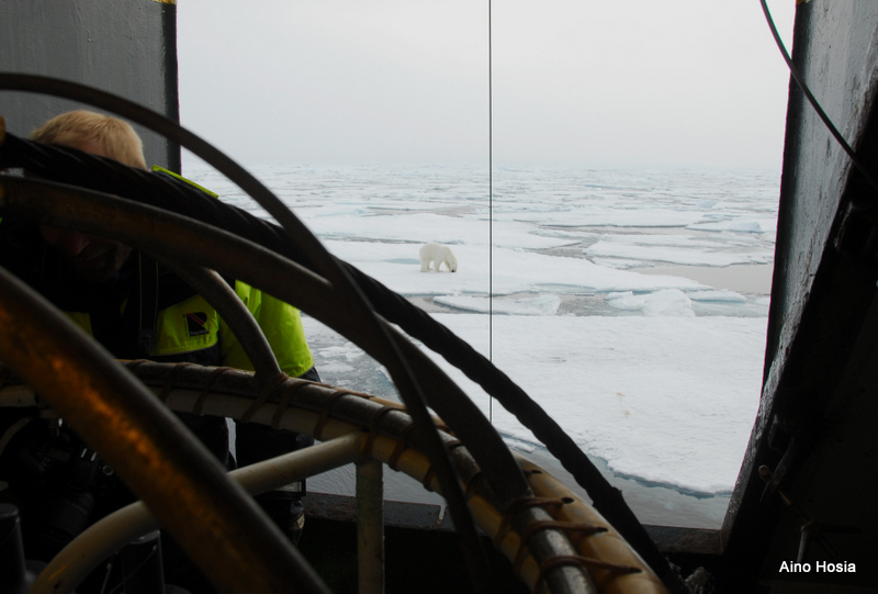
HYPNO participating on an Arctic cruise by the Institute of Marine Research on RV Helmer Hanssen 17 Aug – 7 Sep 2015.

Julekalender Aino 2-001Most of the pelagic hydrozoans for HYPNO are collected with simple plankton nets, in the case of this Arctic cruise the double one you see in the picture. The net is towed vertically from above the bottom to the surface, bringing with it a representative sample of plankton – inclusive hydromedusae and siphonophores – from the entire water column. Standard plankton nets are generally lowered and retrieved at a speed of ~0.5 ms-1.

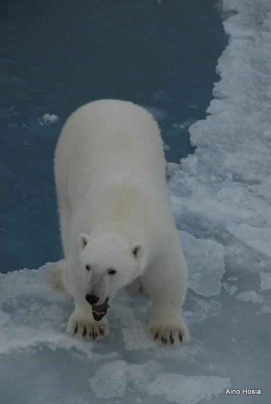
This particular station in the Arctic basin was over 2000 m deep, which means that a single tow takes more than an hour to complete. Sometimes waiting for the sample to come up can get a bit tedious – not at this station, though! With this beauty turning up right outside the hangar opening, the wait didn’t feel long at all!

SI_Arctic 24-8-2017 SI_Arctic 24-8-2016-Aino

Door #11: Just a white blob?

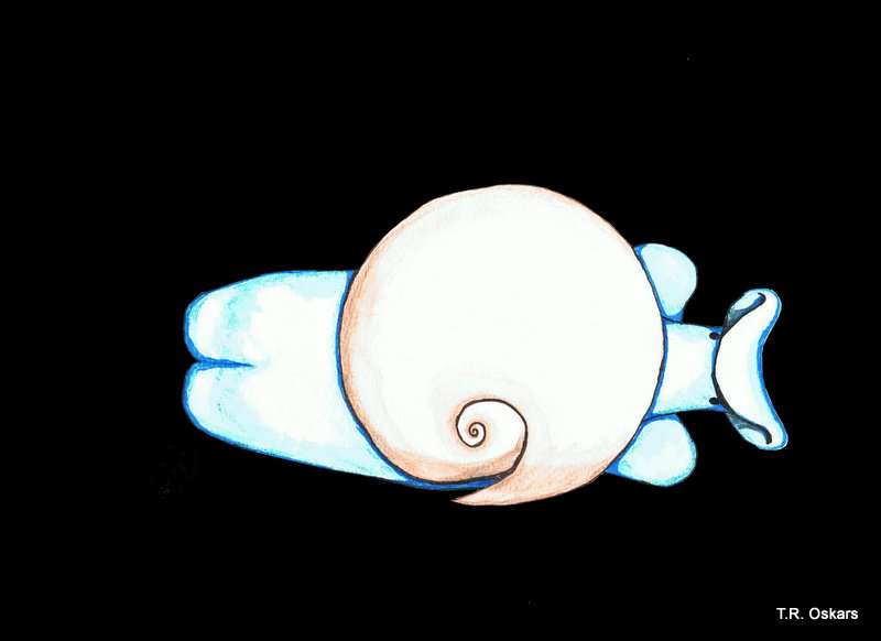
Colobocephalus costellatus repainted from M. Sars (T.R. Oskars)

Colobocephalus costellatus repainted from M. Sars (T.R. Oskars)

When researching small, obscure sea slugs you are bound to run into surprises. Partly because it often takes a long time between discovery and identification, and also because a lot of the really interesting stuff is first revealed when new methods become widely available.

In 2011 a team of researchers from the Invertebrates collection were sampling specimens in Aurlandsfjorden for the Invertebrate collections and range data for the Norwegian Biodiversity Information Centre (Artsdatabanken). Among other interesting critters they found a 2 mm long white blob. While not initially impressive this small blob turned out to be the enigmatic cephalaspidean sea slug Colobocephalus costellatus (Cephalaspidea: Heterobranchia) described by Michael Sars from Drøbak in 1870. At the time of its re-discovery it was thought that this species, which is unique for Norway, had not been seen or collected since M. Sars first laid hands on it 145 years ago (more (in Norwegian) here). However, you continuously discover more information in the course of scientific work. During their work on the enigmatic slug Lena Ohnheiser and Manuel Malaquias found in the literature that the species had in fact been discovered a couple of times since 1870, first by Georg Ossian Sars in Haugesund some years after his father, and more recently by Tore Høisæter of Bio UIB in Korsfjorden outside Bergen.

Still, no in-depth analyses have been done on this species since M. Sars until Nils Hjalmar Odhner of the Swedish Natural History Museum drew the animal from the side showing some of the organs of the mantle cavity.

Most authors have had real difficulties to place this slug within the cephalaspids, and M. Sars even thought is possible that the slug might not be an opisthobranch. Some placed it within Diaphanidae based only on the globular shell, a family that has been poorly defined and often used as a “dump taxon” for species that hare hard to place. Yet others thought it might even be the same as the equally enigmatic Colpodaspis pusilla, which has been suggested to be a philinid sea slug (flat slugs digging around in mud and sand).

What was unique about the most recent find was that this was the first time it was collected alive and photographed with high magnification. The material was also so fresh that Lena and Manuel could dissect the animal and study its internal organs. In their 2014 paper “The family Diaphanidae (Gastropoda: Heterobranchia: Cephalaspidea) in Europe, with a redescription of the enigmatic species Colobocephalus costellatus M. Sars, 1870” they tried to resolve the relationships between these globe shelled slugs. What they found was that Diaphanidae was likely not a real grouping of species, containing at least three distinct groups, where one group was Colobocephalus and Colpodaspis, which were closely related to each other, but also quite distinct.
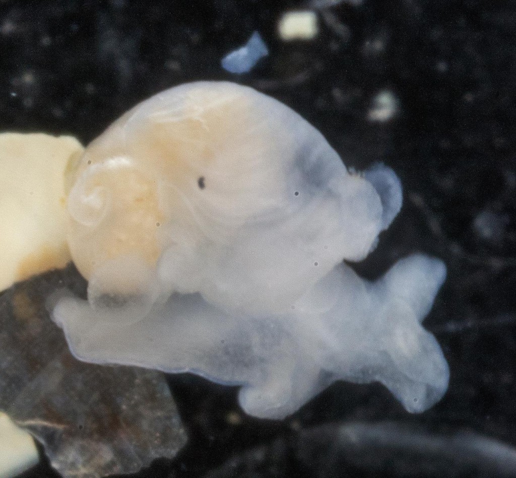
Colobocephalus costellatus M. Sars, 1870. Photo Lena Ohnheiser, CC-BY-SA. Also featured on http://www.artsdatabanken.no/File/1292

Colobocephalus costellatus M. Sars, 1870. Photo: Lena Ohnheiser, CC-BY-SA. Also featured on http://www.artsdatabanken.no/File/1292

Another new development with the sampling in Aurlandsfjorden was that the slugs were preserved in alcohol rather than formalin. Formalin is good for preserving the morphology of animals, but it destroys DNA. On the other hand, alcohol is perfect for preserving DNA. This lead to C. costellatus to be included in a 2015 DNA based phylogenetic analysis of cephalaspidean sea slugs.
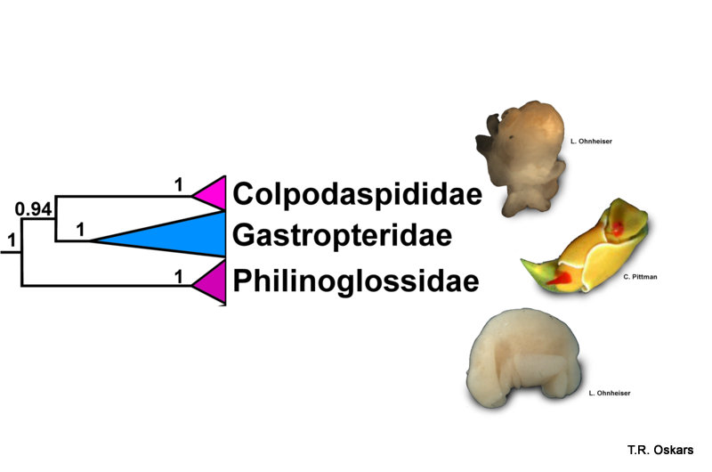
Modified Tree from Oskars et al. (2015)

Modified Tree from Oskars et al. (2015)

This resulted in that the slug was found to be indeed an Opisthobranchia, and as Lena and Manuel thought, Colobocephalus and Colpodaspis were placed in their own family, Colpodaspididae. Whereas the traditional “Diaphanidae” was split apart. Even weirder was the sea slugs that were shown to be the closest relatives of Colpodaspididae, which were neither the philinids or the diaphanids. The closest relatives turned out to be slugs that are equally as weird and unique as Colpodaspididae, namely the swimming and brightly colored Gastropteridae (sometimes called Flapping dingbats) and the Philinoglossidae, which are tiny wormlike slugs that live in between sand grains.
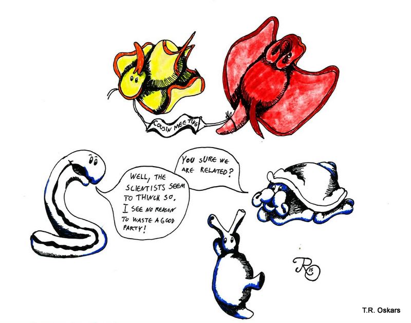
*Cousin Meeting*  - "You sure we are related?"  - "Well, the scientists seem to think so. I see no reason to waste a good party!"

*Cousin Meeting*
– “You sure we are related?”
– “Well, the scientists seem to think so. I see no reason to waste a good party!”

So it took 145 years from its discovery before Colobocephalus became properly studied and its family ties revealed, but it is still mysterious as we do not know much about their ecology or diet.

Suggested reading:

Colobocephalus costellatus: http://www.biodiversity.no/Pages/149747

Colpodaspis pusilla: http://www.biodiversity.no/Pages/149766

Philinoglossa helgolandica: http://www.biodiversity.no/Pages/149915

Høisæter, T. (2009). Distribution of marine, benthic, shell bearing gastropods along the Norwegian coast. Fauna norvegica, 28.

Gosliner, T. M. (1989). Revision of the Gastropteridae (Opisthobranchia: Cephalaspidea) with descriptions of a new genus and six new species. The Veliger, 32(4), 333-381.

Odhner, N.H. (1939) Opisthobranchiate Mollusca from the western and northern coasts of Norway. Kongelige Norske Videnskabers Selskabs Skrifter, 1939, 1–92.

Ohnheiser, L. T., & Malaquias, M. A. E. (2014). The family Diaphanidae (Gastropoda: Heterobranchia: Cephalaspidea) in Europe, with a redescription of the enigmatic species Colobocephalus costellatus M. Sars, 1870. Zootaxa, 3774(6), 501-522.

Oskars, T. R., Bouchet, P., & Malaquias, M. A. E. (2015). A new phylogeny of the Cephalaspidea (Gastropoda: Heterobranchia) based on expanded taxon sampling and gene markers. Molecular phylogenetics and evolution, 89, 130-150.

Sars, M. (1870) Bidrag til Kundskab om Christianiafjordens fauna. II. Nyt Magazin for Naturvidenkaberne, 172–225.

-Trond

Door #10: Old Stoneface

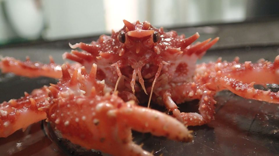
Today’s critter is a Lithodes maja, or Northern stone crab (Trollkrabbe in Norwegian). They live in depths between 80-500 meters, where they feed on algae, bottom dwelling animals, and of scavenging. They are much smaller than their relatives the King crab (Paralithdodes camtschaticus), reaching up to 150 mm across the carapace.

Despite the name, they are not true crabs – Brachyura, but rather Anomurans: “As decapods (meaning ten-legged), anomurans have ten pereiopods (legs), but the last pair of these is reduced in size, and often hidden inside the gill chamber (under the carapace) to be used for cleaning the gills. Since this arrangement is very rare in true crabs (for example, the small family Hexapodidae), a “crab” with only eight visible pereiopods is generally an anomuran.”  (Wikipedia)

Hello, there!

Hello, there! Shake hands? Photo: H. Hektoen

Martin encountered this one when participating on this year’s final MAREANO survey in the Barents Sea. MAREANO has been working on mapping the depth and topography, sediment composition, contaminants, biotopes and habitats through a combination of video stations and physical sampling of sediments and animals in Norwegian waters since 2006.

A cruise typically lasts between 10 and 20 days, and for most years MAREANO has had 2-3 cruises. The amount of stations and collected material is staggering!

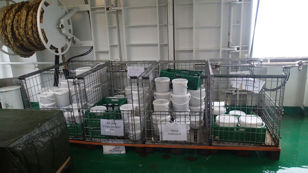
The pile of samples halfway through the cruise Photo: M. Hektoen

The pile of samples halfway through the cruise Photo: M. Hektoen

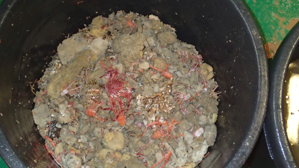
A bucket of beam trawl collected material - sponges and Munida (squat lobsters) are dominant, together with our friend from the picture above. Photo: M. Hektoen

A bucket of beam trawl collected material – sponges and Munida (squat lobsters) are dominant, together with our friend from the picture above. Photo: M. Hektoen

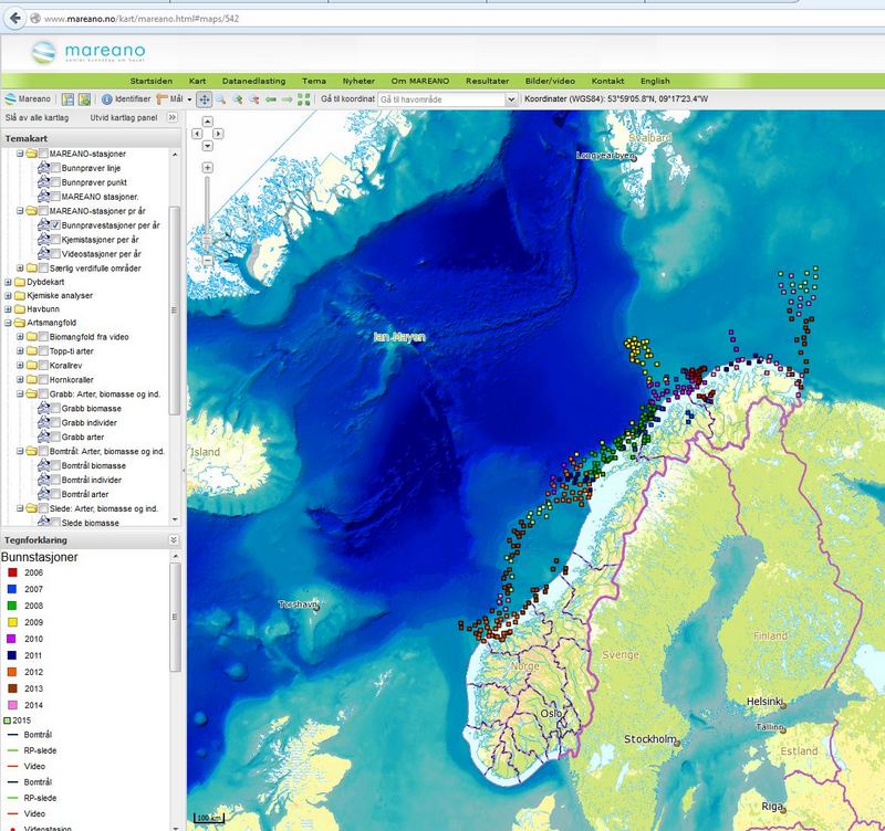
Below is a map over the “full stations”, the stations that also include physical samples of biological material from grab, sled and trawl. These samples are split into fractions, some to be further processed by MAREANO, whilst others are bulk fixated without further analysis. The MAREANO-identified animals and unsorted fractions from these stations are deposited at the University Museum once MAREANO is done with them. We then continue to process them; decide which samples are significant, sort the unsorted fractions, implement material into the museum collections, and make it available for further research. For the interactive maps, go here.

Screenshot from mareano.no showing the bottom stations per year.

Screenshot from mareano.no showing the bottom stations per year.

-Martin & Katrine

Door #9: Delving into the DNA

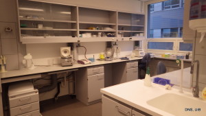
From the pre-PCR lab

From the pre-PCR lab

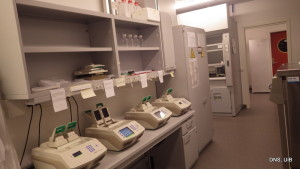
The four PCR-machines lined up

The four PCR-machines lined up

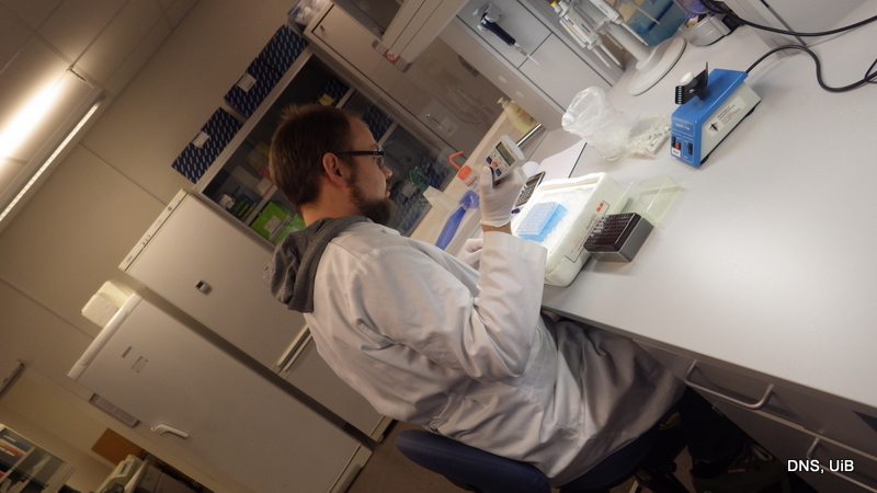
We are very fortunate in that we have a modern DNA lab available «just down the street» from us, as the University Museum is part of the shared Biodiversity laboratories (BDL) structure.

The BDL is a formalized cooperation between three research groups at Dept. of Biology (Marine biodiversity, Geomicrobiology and the EECRG), and two of the research groups at the University Museum. One of the senior engineers if this lab is a Museum employee, and from time to time we are also able to hire in other collaborators for specific projects.


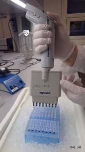
Pipetting

Pipetting samples onto one of the plates that we fill with DNA-extracts


For the past couple of months we’ve had a technician – Morten – working on resolving some of the challenges that we run into when we work on COI barcoding of marine invertebrates.

Unlike many of the other groups that this method works exceedingly well for (like the Diptera), we are experiencing difficulties in obtaining DNA barcodes from a significant proportion of our samples.

IMGP0775-001We are currently focussing particularly on the Polychaeta (bristle worms), as this is the group we have submitted the majority of samples from in both our major barcoding projects: MIWA (Marine Invertebrates of Western Africa) and NorBOL (Norwegian Barcode of Life).


Morten has been working on obtaining DNA from the more problematic species, by troubleshooting and tinkering on various aspects of the ways we extract and amplify genes.

Basically there are more or less standardized ways of obtaining DNA, and these methods normally works well. Unfortunately (for various reasons) this is not always the case, and this is where we have to alter the protocols to see if we can find a way to retrieve the sample DNA from the specimens.

So far it looks quite promising; we’ve been able to fill in some of the most important “blanks” in our datasets – and we’re not done yet!

– Morten & Katrine

Door #8: One jar –> many, many vials

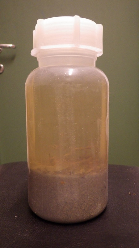
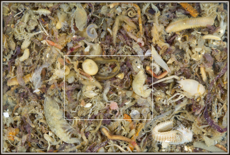
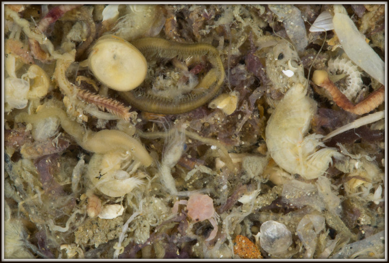
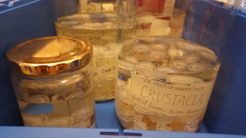
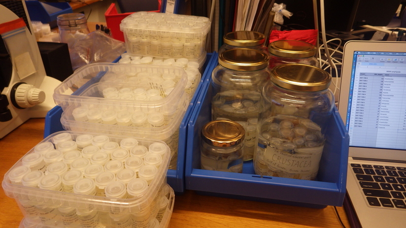
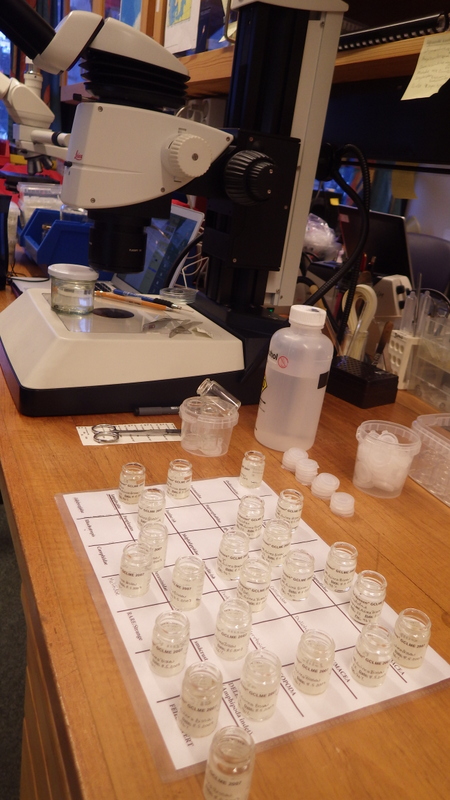
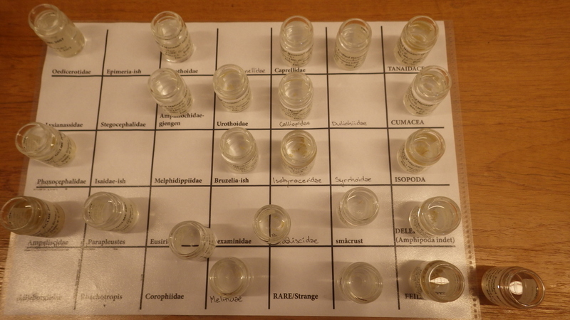
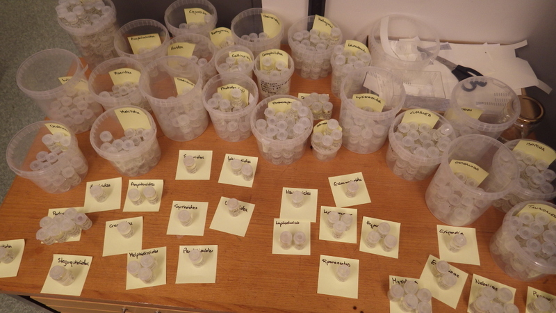
Sorting the Crustacean samples from our

Marine Invertebrates of Western Africa Project (MIWA)

Most work is more fun when working together. It also makes for better science to cooperate – and the easiest way to cooperate on taxonomy is to sit at the same lab for some time – to be able to look at the same specimens and see the same details that should be examined. This is the plan for the amphipods from the MIWA-project. Ania Jażdżewska from the University of Łódź in Poland is visiting our lab for an extended week of collaboration with me in a mini-workshop on the amphipod samples.

But before a visitor can come, preparations are necessary. So for the last 6 weeks I have been sorting all the ethanol-samples of our west-african crustaceans into separate orders (isopods, tanaidaceans, cumaceans, decapoda), and the amphipods (also an order) have been sorted to family.

98 samples have been split into 629 smaller vials – ready to be further examined when Ania comes.

We promise a follow-up on what this brings of fun science!

 -Anne Helene

Door #7: Shrimp and salad

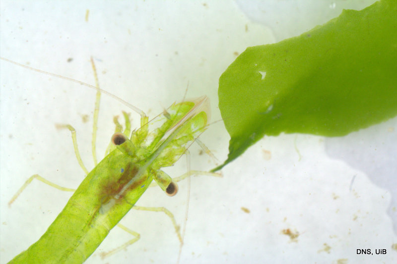
Hippolyte varians Leach, 1814 in sea lettuce, Ulva lactuca

Hippolyte varians Leach, 1814 in sea lettuce, Ulva lactuca

This small shrimp can be found on the shore in small ponds at low tide. The species was recorded from Western Norway already in the early 19 hundreds by research curators in Bergens Museum (Appellöf, 1906; Grieg, 1927). Interestingly, as also observed by Appellöf (1906:page 124) individuals of this the species appear in many contrasting colour variants due to the ability of the the body to mimic the colours in the environment. The Norwegian name of Hippolyte varians is «sjøgressreke», which refers to its association with sea grass meadows (Zostera marina), in which a green body colour would seem to be an appropriate camouflage appearance. The specimen on this picture was caught in the littoral at the island Turøy amongst sea lettuce, Ulva lactuca. We see that there is a good colour match between the shrimp and the lettuce.

However, reddish, pink, brownish, black or even white colours have been are observed in other environments and it seems that at night time the shrimp may take a rest from the camouflage by attaining a transparent whitish blue appearance as shown by Moen’s picture here.

Knowledge about the distribution and biology of Hippolyte varians is summarised by C. d’Udekem d’Acoz. 

References

Appellöf, A. 1906. Die Dekapoden Crustaceen. Meeresfauna von Bergen 2(3): 113-238.

Grieg, J.A. 1927. Decapoda Crustacea from the west coast of Norway and the North Atlantic. Bergens Museums Aarbok 7:1-53.

Udekem d’Acoz, C. (1996). The genus Hippolyte Leach, 1814 (Crustacea: Decapoda: Caridea: Hippolytidae) in the east Atlantic Ocean and the Mediterranean Sea, with a checklist of all species in the genus. Zoologische Verhandelingen (Leiden) 303: 1-133.

-Endre

Door #6: Associated Amphipods

Amphipods are a group of small crustaceans where most of the species we know are benthic (bottom dwelling) and marine. But within the benthic habitat there are many niches, and one of the more intriguing is the many ways of living on or inside another benthic animal. A few species become parasitic (feeding on their host), but for the most species living like this, it does not look like eating the host is the main objective. In these cases we term the amphipods as “associated with a host”.

To document some of these associations, I have had a wonderful cooperation with an amazing underwater photographer this year. Lill Haugen has photographed amphipods associated with hydroids, and sampled the amphipods afterwards for us. Documenting this kind of association is almost impossible without the help of divers – if we are lucky enough to sample a hydroid with our normal sampling gear, the amphipods fall off. It is not easy spotting these small animals for a diver either, but Lill says that it becomes easier with practice.

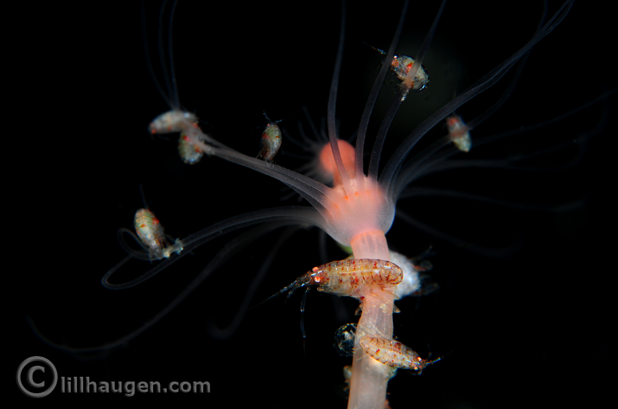
An amphipod family at home. Photo by Lill Haugen, all rights reserved

An amphipod family at home. Photo by Lill Haugen, all rights reserved

This photo is from the Oslofjord, at 25 m depth. With the photos from Lill we are able to say that this particular amphipod (from the family Stenothoidae) looks like it keeps the hydroid as a family home. The parents sit on the “stem” of the hydroid, and their children sit on the tentacles of the “flower”. This might be both to provide extra protection and food for the amphipod-children. The adult amphipods are 5mm long, their children 3mm.

Earlier studies have shown that amphipods of the family Stenothoidae often associate with molluscs – we have found several different species living inside bivalves (shells). Other amphipods might associate with other crustaceans such as crabs, or with sponges, anemones or snails (gastropoda).

For most amphipod species we know nothing about their life-history and possible associations. But the more we examine them, the more we learn..

Suggested reading:

Tandberg, A.H., Schander, C., Pleijel, F. (2010) First record of the association between the amphipod Metopa alderii and the bivalve Musculus Marine Biodiversity Records, 3, e5, doi:10.1017/S1755267209991102

Tandberg, A.H., Vader, W., Berge, J. (2010) Studies on the association of Metopa glacialis (Amphipoda, Crustacea) and Musculus discors (Mollusca, Mytilidae). Polar Biology, 33, 1407-1418

Vader, W., Tandberg, A.H. (2013) A survey of amphipods associated with mollusks. Crustaceana 86(7-8), 1038-1049

Vader, W., Tandberg, A.H. (2015) Amphipods as associates of other crustacea: a survey. Journal of Crustacean Biology 35(4), 522-532

-Anne Helene

Door #5: A (so far) undescribed species of bristle worm

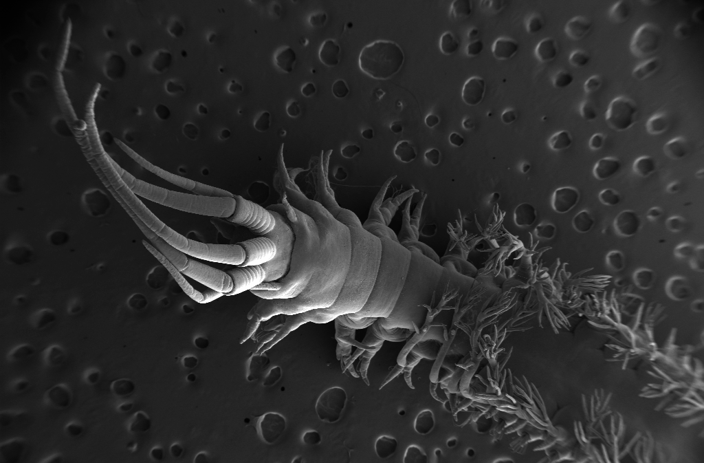
Diopatra sp

Diopatra sp. Photo: M. Hektoen

Pictured above is a cute polychaete (bristle worm) from the genus Diopatra. It was collected in Mauritania, and has been photographed using Scanning Electon Microscopy (SEM). Although I ended up describing 9 new species of Diopatra worms in my master’s thesis, many worms were still left undescribed, this is one of those.

-Martin