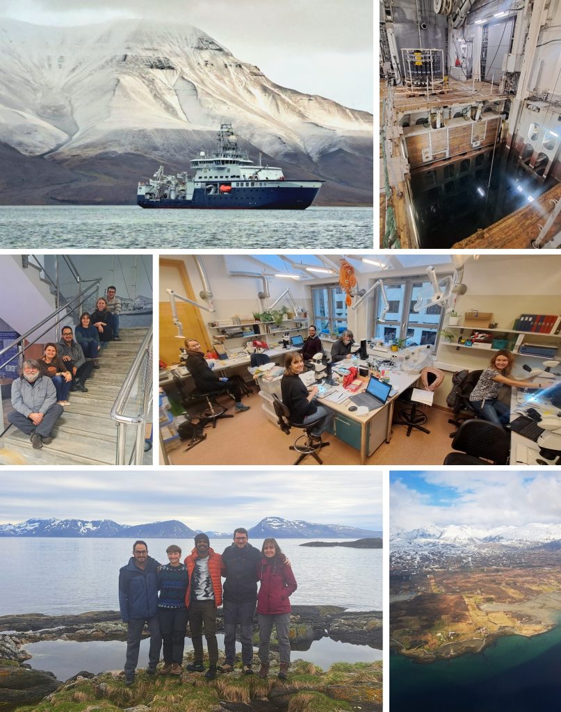
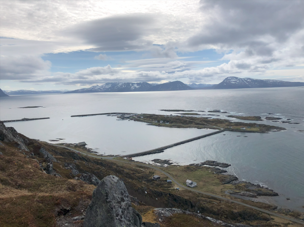
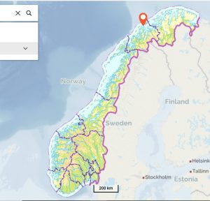
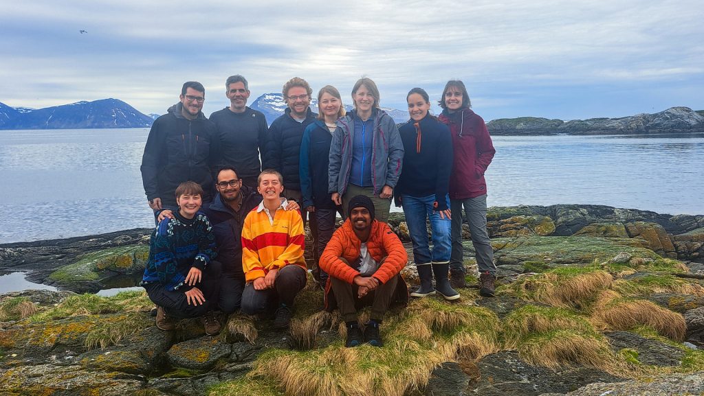
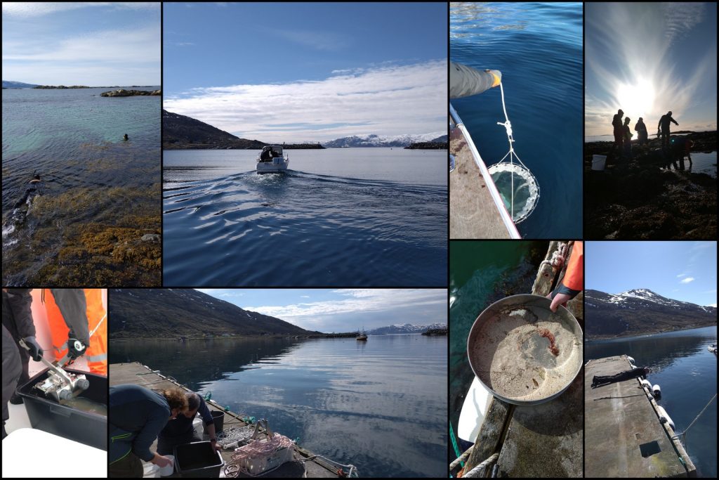
From 21 till 28th of May, researchers, technicians, and students of the University Museum’s marine section, travelled up North to Torsvåg, close to Tromsø, for joint fieldwork. The participants represented several Artsdatabanken projects that cover marine fungi, hydrozoans, polychaetes, parasites of jellyfish, comb jellies and chaetognaths, bryozoans, marine amphipods and finally the Lower Heterobranchia and Pyramidellidae gastropods. In this blog you can read about the general experience of the fieldwork and more details about the different projects. And here you can read about the fieldwork through the eyes of two master students who joined. And if you want to read more adventures and see more pictures check out this blog post! It was a large group of young and more experienced scientists which created the perfect opportunity for a lot of knowledge transfer.
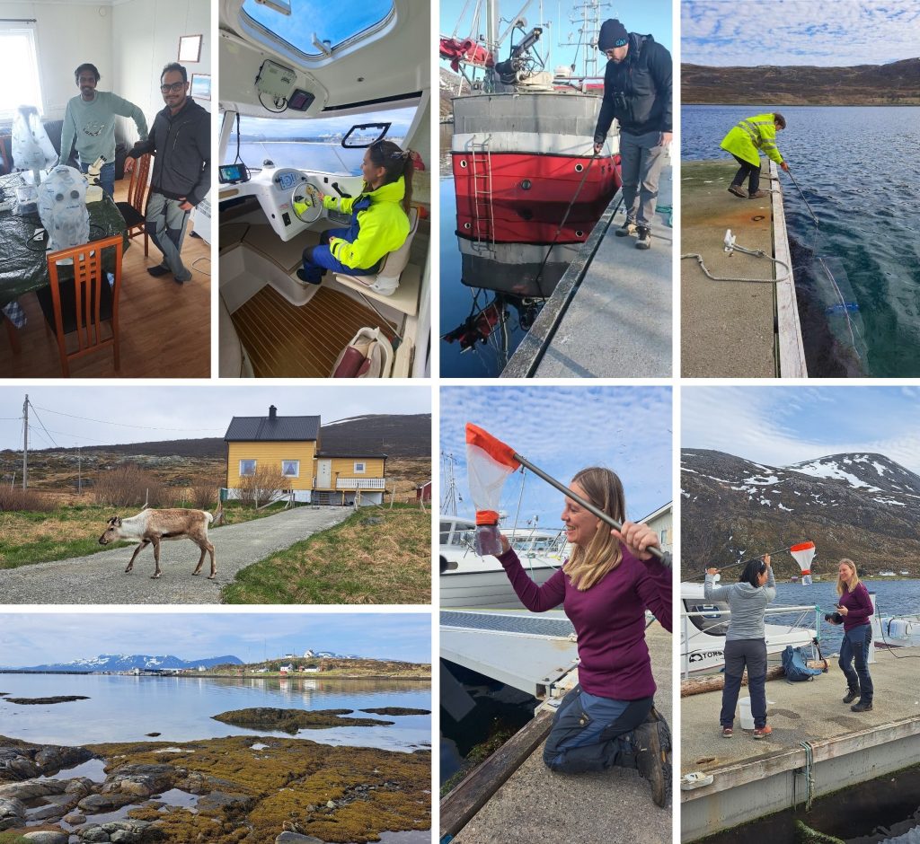
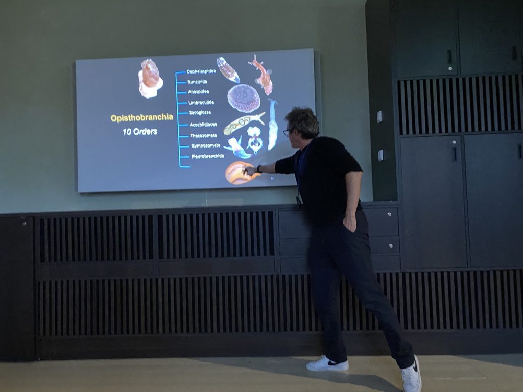
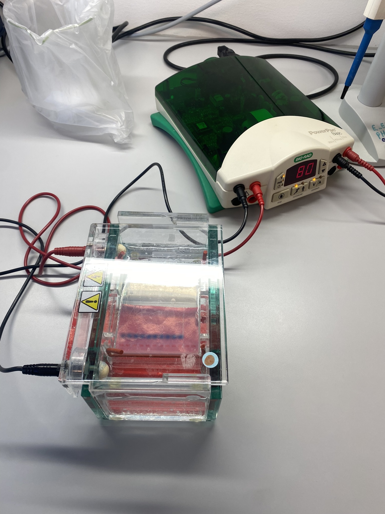
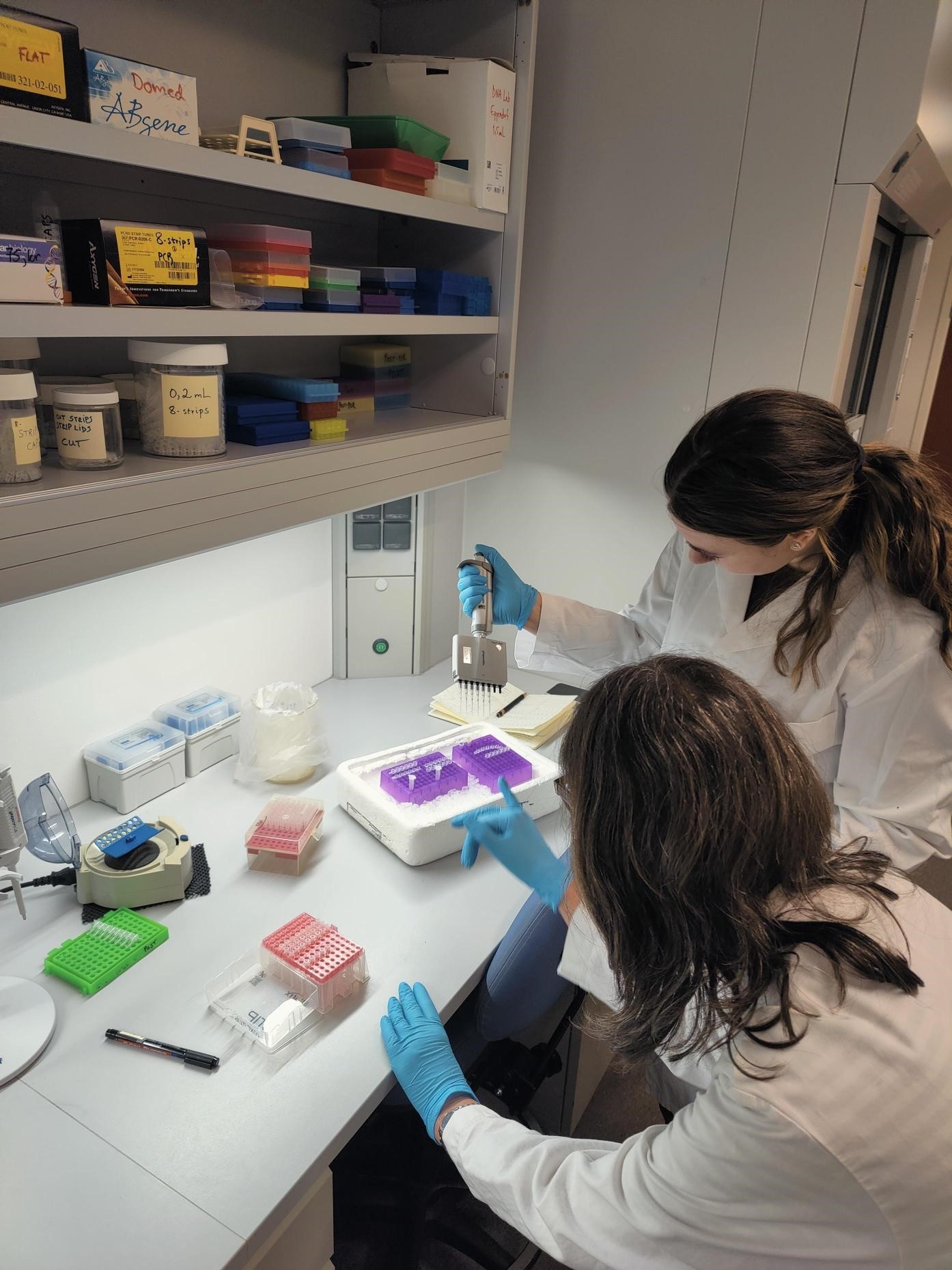
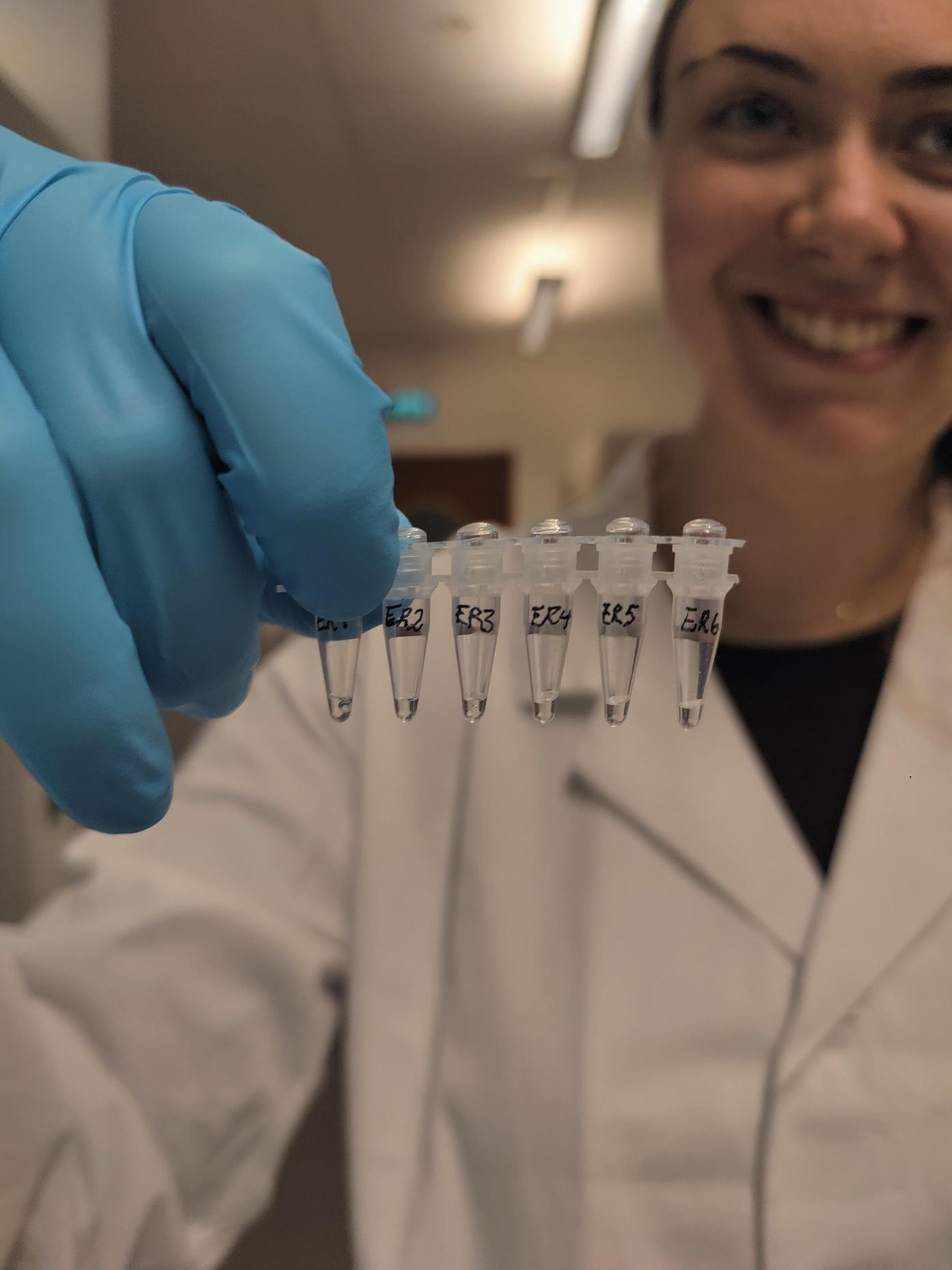
This was also the first “big” fieldwork trip for the Lower Heterobranchia and Pyramidellidae project, after several sampling events in the Bergen area, which you can read here. Both Lower Heterobranchia and Pyramidellidae include small snails, just a few millimetres in length that are hard to identify (they can resemble other small species of gastropods). The diversity of these tiny sea snails is poorly understood in Norway, and thus, during this project these sea snails will be studied by combining DNA barcoding and shell characters. Sampling will be based on the use of dredges, grabs, and snorkelling.
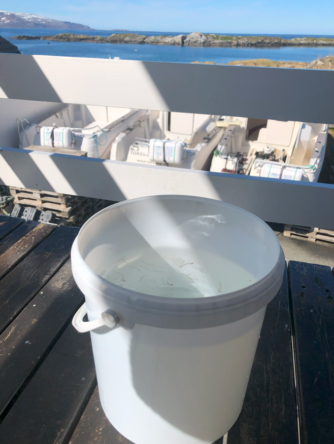
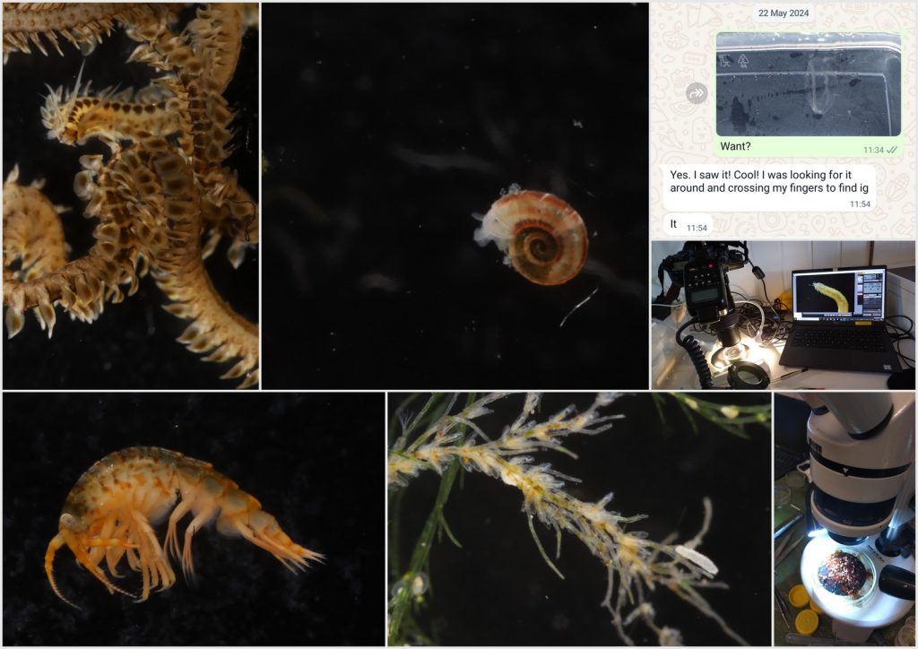
Pyramidellidae are regarded as ectoparasites and are often found living on other molluscs or other marine invertebrates, but also free in soft sediments. The Lower Heterobranchia are often found on algae, for example on the stipes and in between the holdfasts of large kelp. Because of the small size of these snails the best way to collect them is by sampling the substrate they live on. So, this is what we did, we went snorkelling several times in the cold waters of Northern Norway, but thanks to good neoprene layers we were able to keep warm and simultaneously looking like seals!
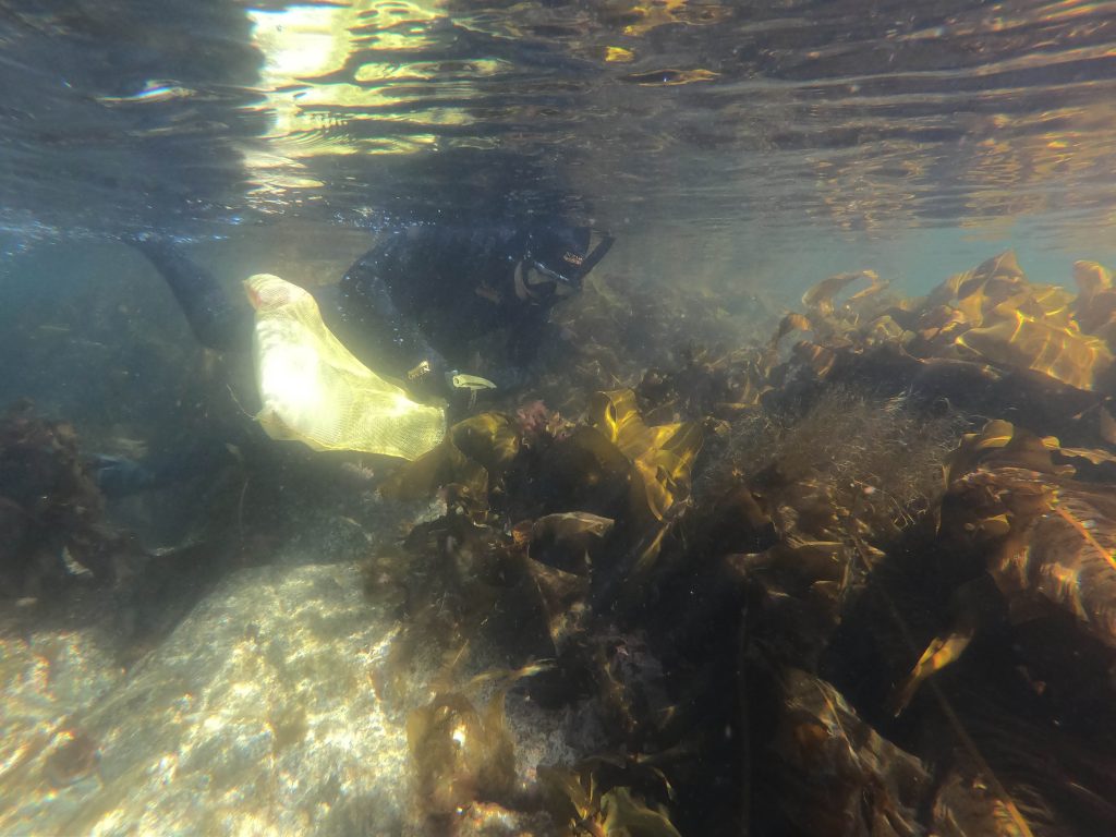
Collecting the right substrates for the Lower Heterobranchs and Pyramidellidae while snorkeling. Photo by Eva Charlotte Samson, UiB
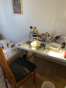
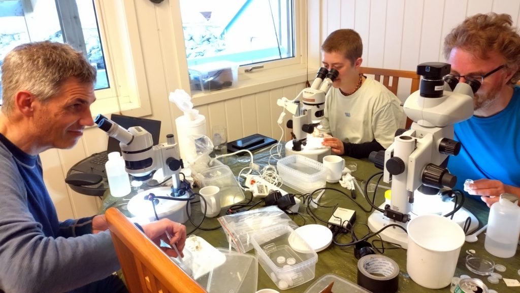
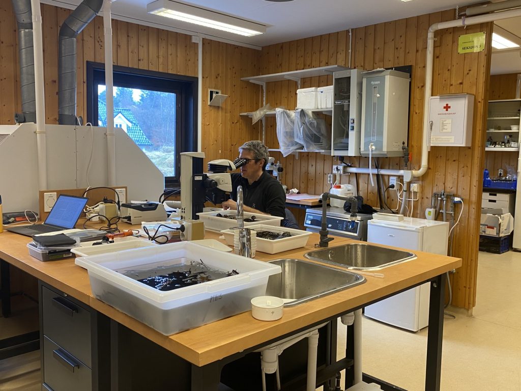
Little lab set-up in the kitchen to sort through the samples. Photo by Cessa Rauch, UiB.
It was challenging to find our snails; there was plenty of kelp and high diversity of many other taxonomic groups, but the conditions were not exactly right especially for the pyramidellids that seem to prefer areas with strong currents.
So, even though we sampled many different habitats, we often ended up not finding our snails when back in the lab sorting under the microscope.
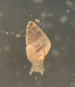
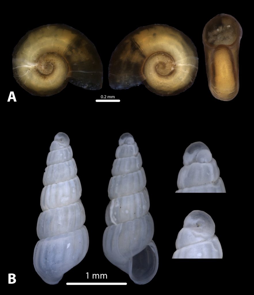
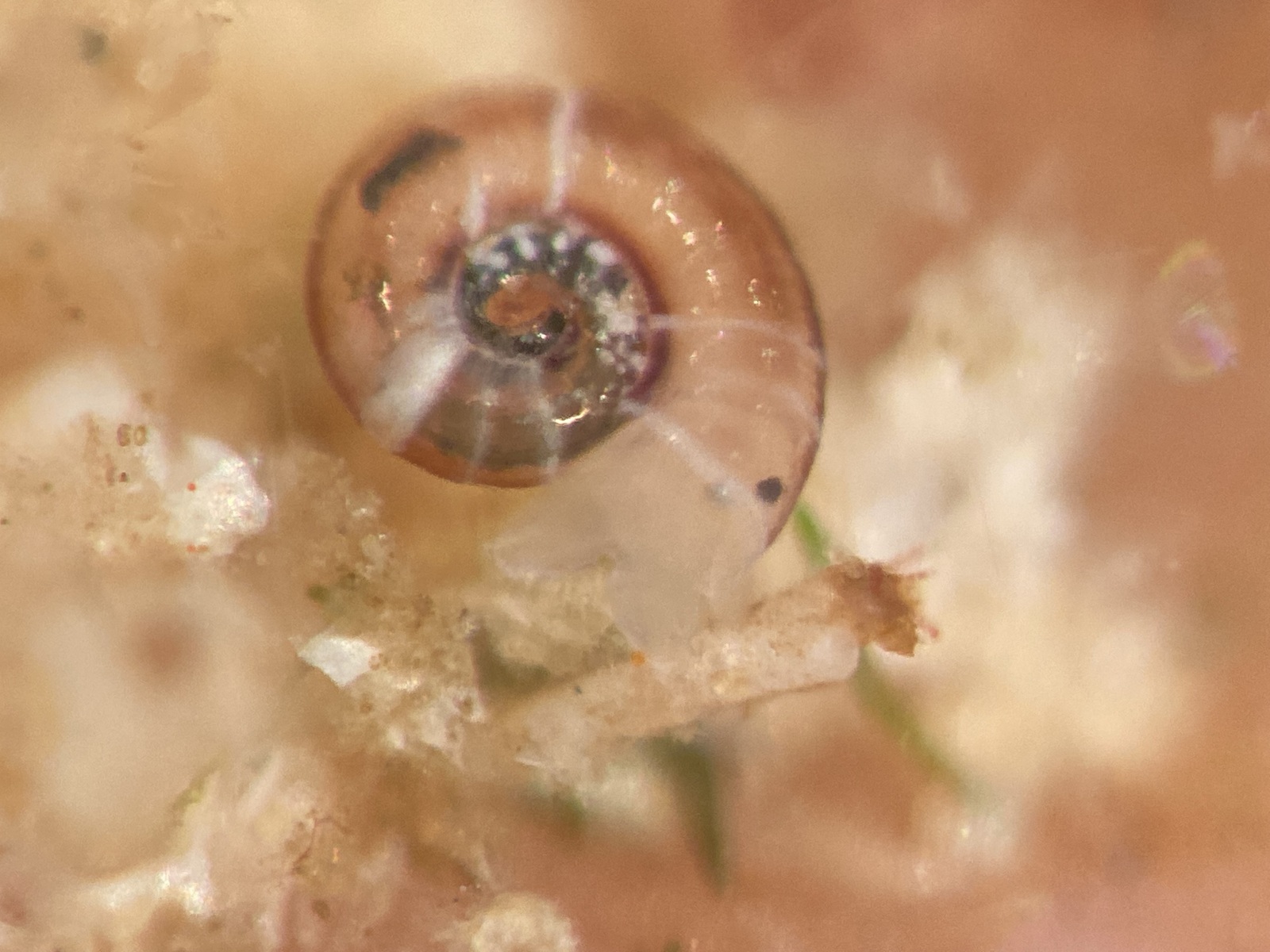
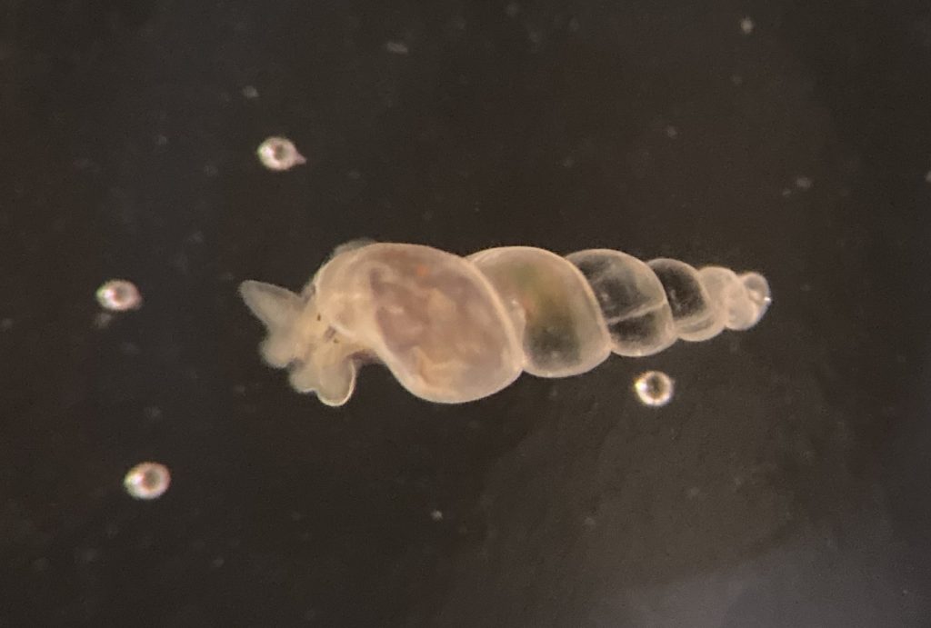
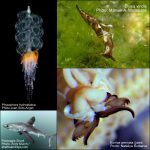
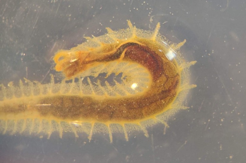
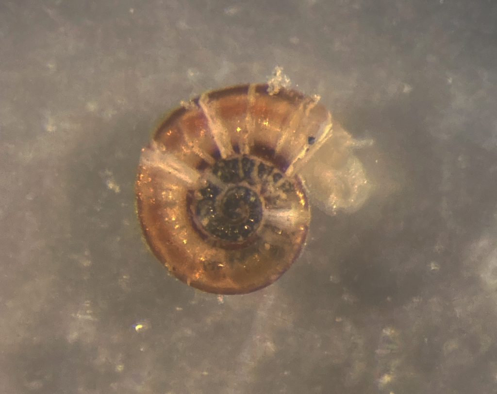
Species of Pyramidellidae; Odostomia turrita. Photo by Cessa Rauch, UiB.
Yet, after collecting a ton of material and spending many hours sorting, we finally found one pyramidellid!
In this case Odostomia turrita.
Odostomia are neat little pyramidellids that have glands with distinct colours, which makes somewhat easier the identification of species.
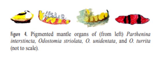
The different colour patterns distinguish different Odostomia species. Source from Høisæter 2014. (Høisæter, T. (2014). The Pyramidellidae (Gastropoda, Heterobranchia) of Norway and adjacent waters. A taxonomic review).
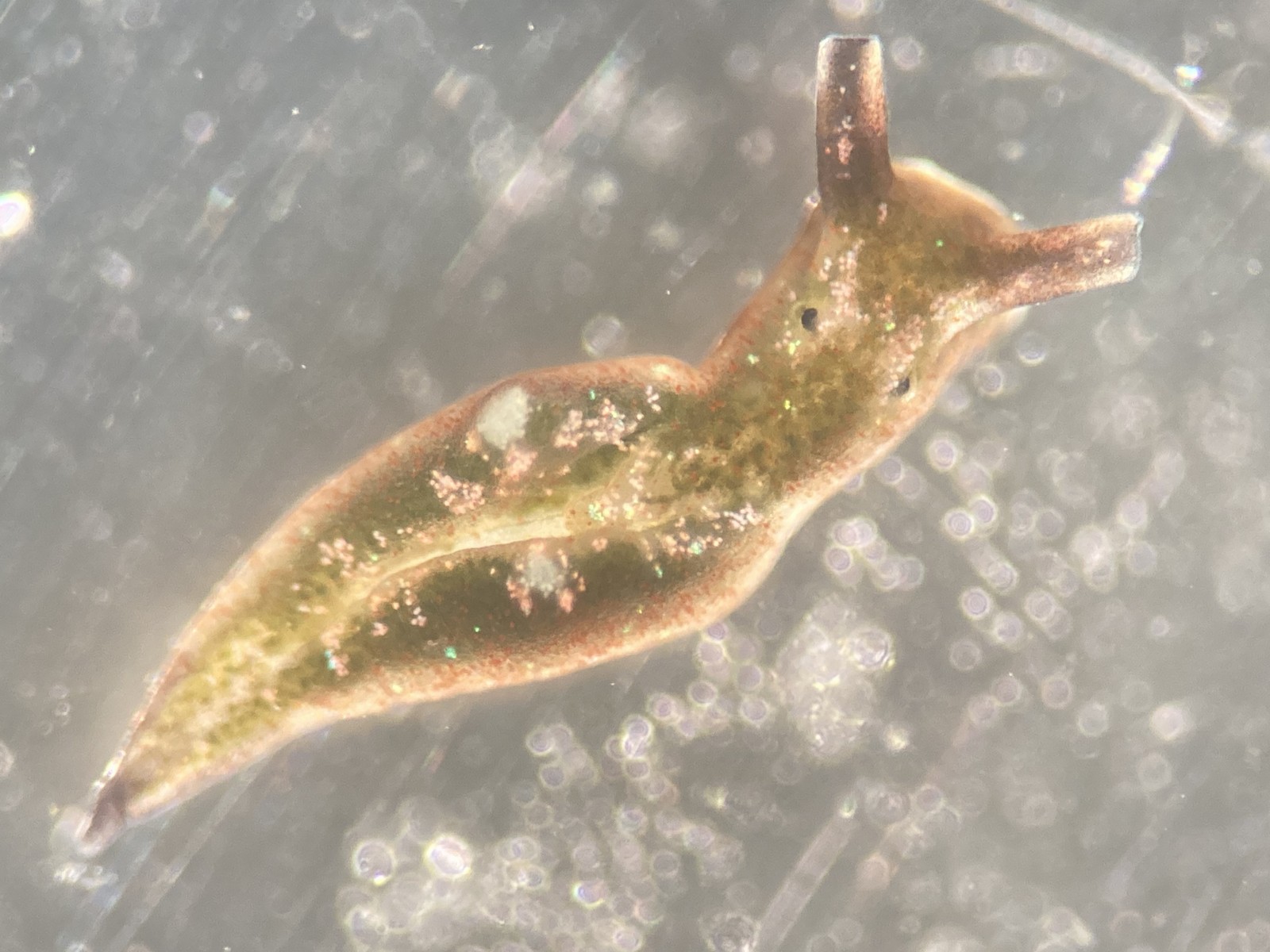
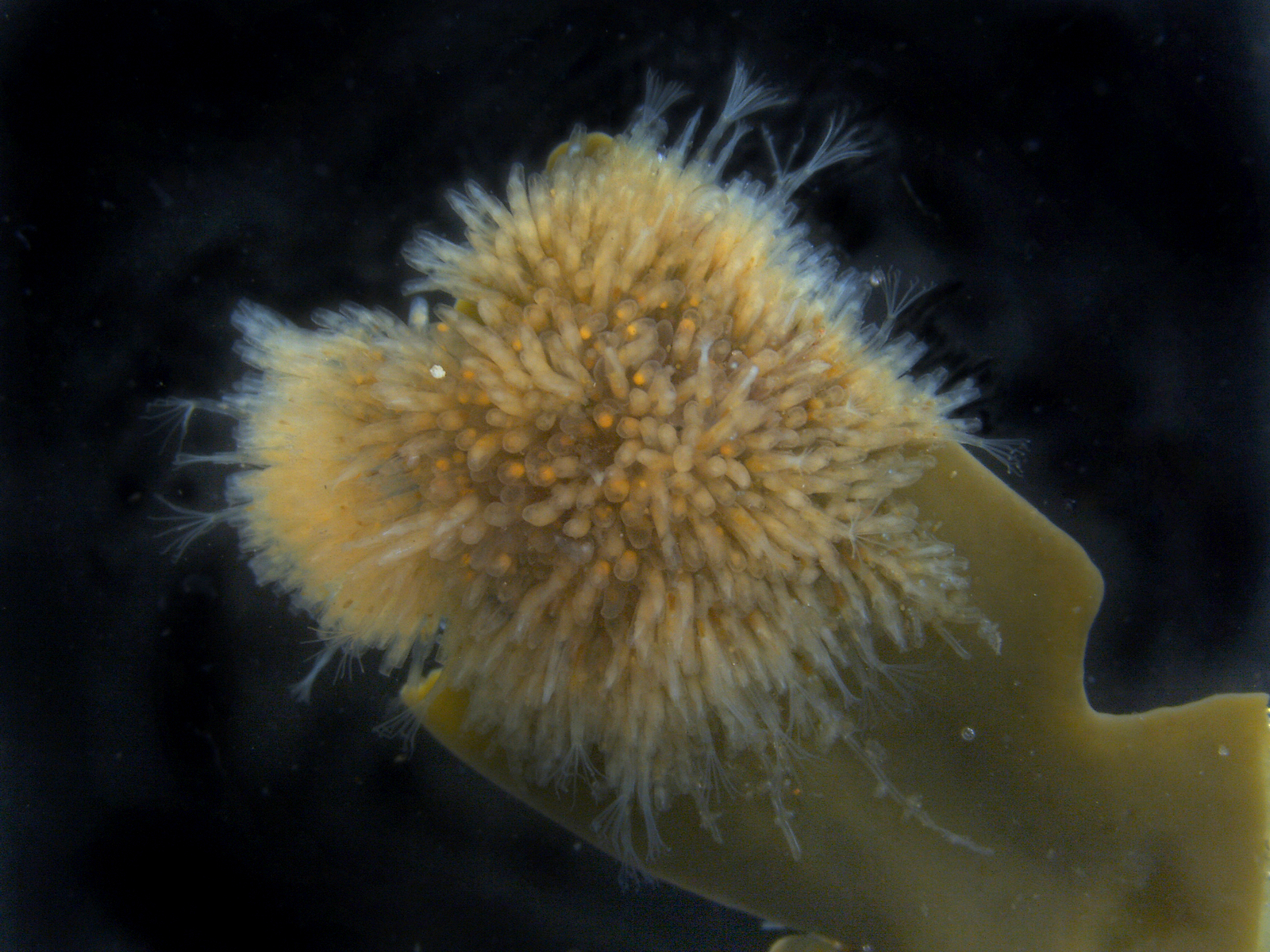
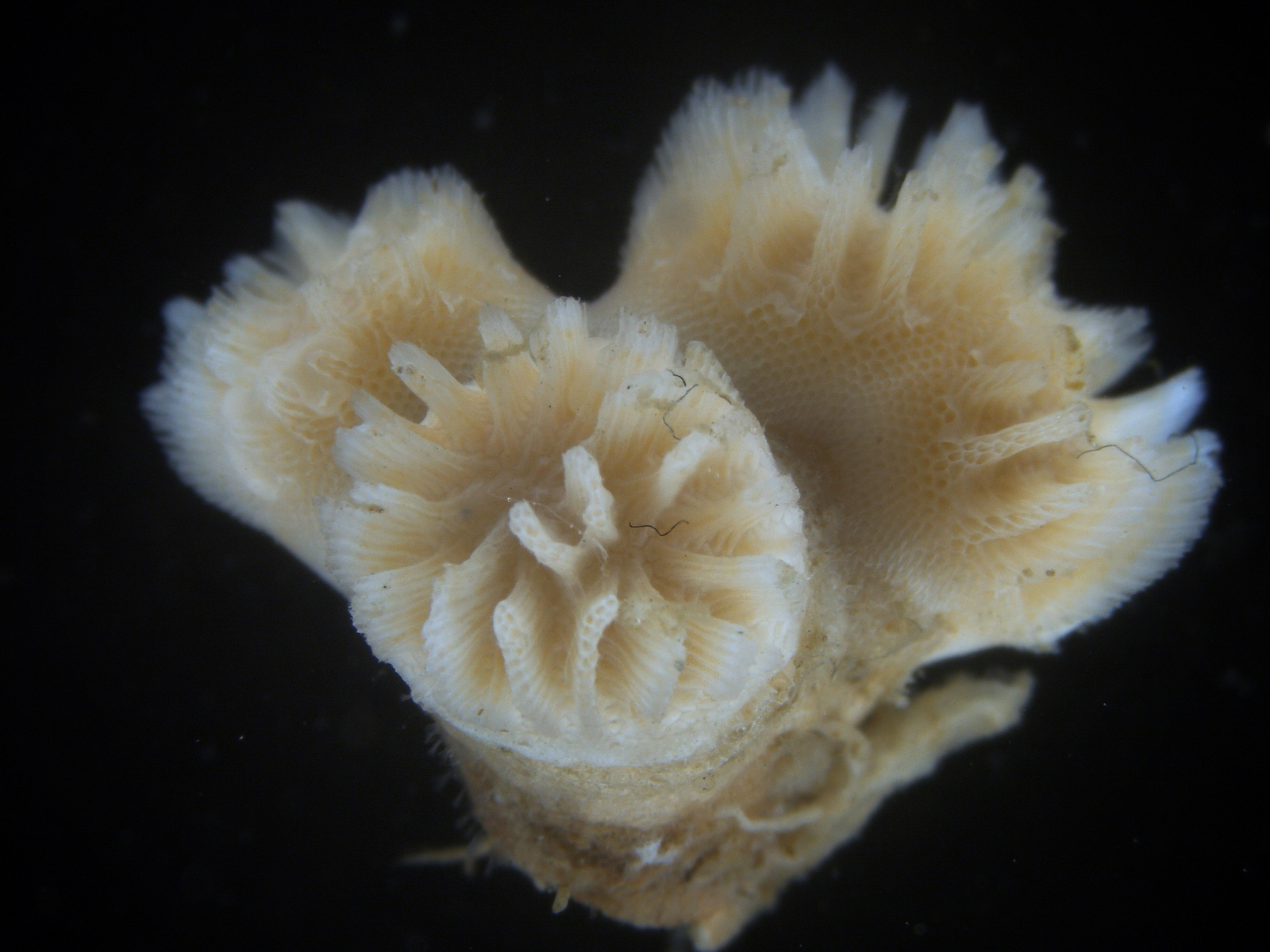
Although minute, the lower heterobranch were “easier” to find… They seem to be less picky with the environment, and on kelp and sand we managed to sample a few different species, amongst others two very similar ones: Ammonicera rota and Omalogyra atomus. In addition, we found in the sand and gravel small snails of the genus Rissoella – R. globularis.
-
- One species of Lower Heterobranch; Ammonicera rota. Photo by Cessa Rauch, UiB.
-

- Another Lower Heterobranch species; Omalogyra atomus. Photo by Cessa Rauch, UiB.
-
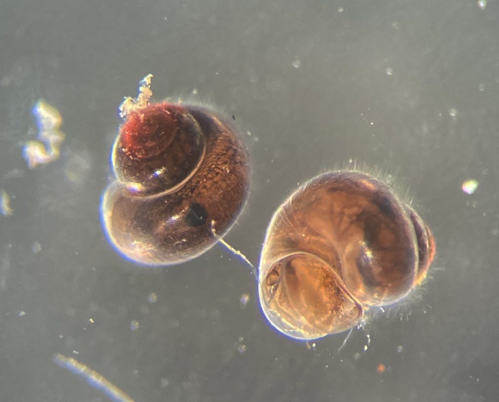
- Species of Rissoella globularis that belong to the Lower Heterobranchs. Photo by Cessa Rauch, UiB.
Field season has just started, this was a good beginning for a busy Summer with many more blogs to come!
-Cessa & Manuel